[level-membership-for-basic-science-category]
Intestinal Trematodes
1. List the clinically significant intestinal trematodes.
2. Describe the general life cycle of the intestinal trematodes and identify the life cycle stage infective for humans.
3. Describe the diagnostic methods used to identify intestinal trematodes.
4. Explain the pathogenesis of intestinal trematode infections.
5. List the drugs of choice for intestinal trematode infections.
6. Describe the environment where intestinal trematodes are found, the route of transmission, and preventive measures.
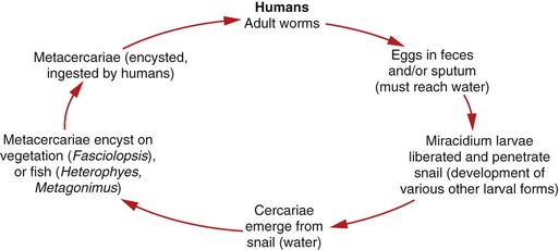
The adult worms are located in the small intestine, where they lay eggs that may be embryonated or remain unembryonated until they are shed from the body via feces. The egg continues developing after reaching the water, and a ciliated, free-swimming miracidium larva is released. The miracidium enters a snail host and develops into a redia (cylindrical larvae), followed by development into tailed cercariae. The cercariae emerge from the snail and encyst as a metacercariae (encrusted larvae) on water plants or fish. A human host ingests raw or undercooked plants (Fasciolopsis buski) or fish (Heterophyes heterophyes, Metagonimus yokogawai) containing the metacercariae, which exycyst in the intestinal tract, attach, and mature into adults (Figure 56-1).
Fasciolopsis Buski
General Characteristics
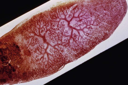
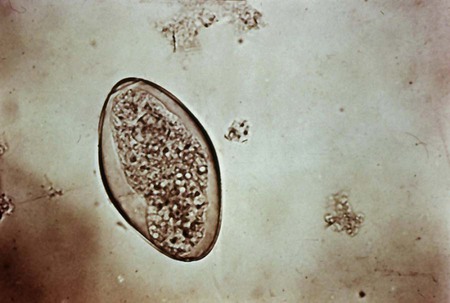
The adults of F. buski have an elongated shape and range from 20 to 75 mm long to approximately 8 to 20 mm wide (Figure 56-2). They have an oral sucker at the anterior end and a ventral sucker located about midway to the posterior end. The eggs, which are indistinguishable from those of Fasciola hepatica (Figure 56-3), are oval and elongated, transparent, and yellow-brown with an operculum (lid) at one end, and they range in size from 130 to 140 µm long to 80 to 85 µm wide and may be unembryonated.
Heterophyes Heterophyes and Metagonimus Yokogawai
General Characteristics
Laboratory Diagnosis
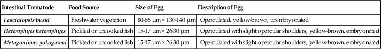
Identification of the intestinal trematodes is made by recovery of eggs, or in rare cases adults, from stool specimens using a sedimentation method such as formalin-ethyl acetate. The sediment may be examined in a wet mount with or without iodine. Because the eggs of Fasciolopsis buski are identical to those of Fasciola hepatica, and those of Heterophyes heterophyes and Metagonimus yokogawai are very similar, diagnosis may also require assessment of symptoms, obtaining a travel history, and/or recovery of adult worms. Table 56-1 shows the diagnostic characteristics of the intestinal trematodes.
TABLE 56-1
Diagnostic Characteristics of Intestinal Trematodes
| Intestinal Trematode | Food Source | Size of Egg | Description of Egg |
| Fasciolopsis buski | Freshwater vegetation | 80-85 µm × 130-140 µm | Operculated, yellow-brown, unembryonated |
| Heterophyes heterophyes | Pickled or uncooked fish | 15-17 µm × 26-30 µm | Operculated with slight opercular shoulders, yellow-brown, embryonated |
| Metagonimus yokogawai | Pickled or uncooked fish | 15-17 µm × 26-30 µm | Operculated with slight opercular shoulders, yellow-brown, embryonated |
1. Fluke eggs are equipped with a lid at the top of the shell called a/an:
2. The infective life cycle stage of a fluke is the:
3. What intermediate host is required in the life cycle of all trematodes?
4. Which two of the following are small flukes whose eggs are generally undistinguishable?
5. What laboratory finding is common in F. buski infections?
a. Increased levels of vitamin B12
b. Increased serum bilirubin levels
6. The drug of choice for an intestinal trematode infection is:
[/level-membership-for-basic-science-category][not-level-membership-for-basic-science-category]
Intestinal Trematodes
1. List the clinically significant intestinal trematodes.
2. Describe the general life cycle of the intestinal trematodes and identify the life cycle stage infective for humans.
3. Describe the diagnostic methods used to identify intestinal trematodes.
4. Explain the pathogenesis of intestinal trematode infections.
5. List the drugs of choice for intestinal trematode infections.
6. Describe the environment where intestinal trematodes are found, the route of transmission, and preventive measures.
The adult worms are located in the small intestine, where they lay eggs that may be embryonated or remain unembryonated until they are shed from the body via feces. The egg continues developing after reaching the water, and a ciliated, free-swimming miracidium larva is released. The miracidium enters a snail host and develops into a redia (cylindrical larvae), followed by development into tailed cercariae. The cercariae emerge from the snail and encyst as a metacercariae (encrusted larvae) on water plants or fish. A human host ingests raw or undercooked plants (Fasciolopsis buski) or fish (Heterophyes heterophyes, Metagonimus yokogawai) containing the metacercariae, which exycyst in the intestinal tract, attach, and mature into adults (Figure 56-1).
Fasciolopsis Buski
General Characteristics
The adults of F. buski have an elongated shape and range from 20 to 75 mm long to approximately 8 to 20 mm wide (Figure 56-2). They have an oral sucker at the anterior end and a ventral sucker located about midway to the posterior end. The eggs, which are indistinguishable from those of Fasciola hepatica (Figure 56-3), are oval and elongated, transparent, and yellow-brown with an operculum (lid) at one end, and they range in size from 130 to 140 µm long to 80 to 85 µm wide and may be unembryonated.
