Intestinal Cestodes
1. Describe the distinguishing morphologic characteristics, clinical disease, basic life cycle (vectors, hosts, and stages of infectivity), and laboratory diagnosis for the intestinal cestodes included in this chapter.
2. Define and identify (where appropriate) the following parasitic structures: scolex, proglottids, rostellum, hermaphroditic, oncosphere, hexacanth embryo, strobila, bothria, and coracidium.
3. Compare and contrast autoinfection and hyperinfection.
4. List several methods of control and prevention of tapeworm infection.
5. Correlate the life cycles with the specific diagnostic stage(s) for each organism.
The intestinal cestodes are commonly referred to as tapeworms. Tapeworms have a long, segmented, ribbonlike body with a specialized structure for attachment, or scolex, at the anterior end. The adult tapeworm consists of a chain of egg-producing units called proglottids, which develop posteriorly from the neck region of the scolex. The crown of the scolex, rostellum, may be smooth or armed with hooks. The body of the worm (proglottids) varies in the geometric characteristics or number of segments according to the genus and species of the cestode. The mature cestode is hermaphroditic. In other words, the organism contains both male and female reproductive organs. Food is absorbed from the host through the worm’s integument, the outer covering or skin of the organism. Adult worms typically inhabit the small intestine; however, humans may be host to either the adult or the larval forms, depending on the infecting species. Humans infected with a cestode pass the eggs in the feces. The embryo may be visible within the tapeworm egg as an oncosphere (larva tapeworm within an embryonic envelope, infective stage) or hexacanth embryo. The intermediate host ingests feces containing the adult tapeworm eggs, which further develop into the larva of the cestode. Cestodes generally require one or more intermediate hosts for the completion of their life cycle. Intestinal tapeworm infections are generally asymptomatic. However, if the larval stage develops in human organs outside the intestine, they may cause additional life-threatening complications. Serologic tests are not available for the diagnosis of tapeworm infections, therefore requiring skilled laboratorians for proper morphologic identification of the organism. Fresh or preserved stools are the specimen of choice for ova and parasites (O&P) examination and cestode identification. Preserved stool containing adult worm segments (strobila) or the scolex may also be used for diagnosis. Chapter 47 describes the methods and specimen requirements in more detail as they relate to parasitology. This chapter describes the intestinal cestodes of public health importance.
Diphyllobothrium Latum
General Characteristics
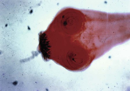
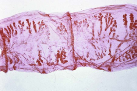
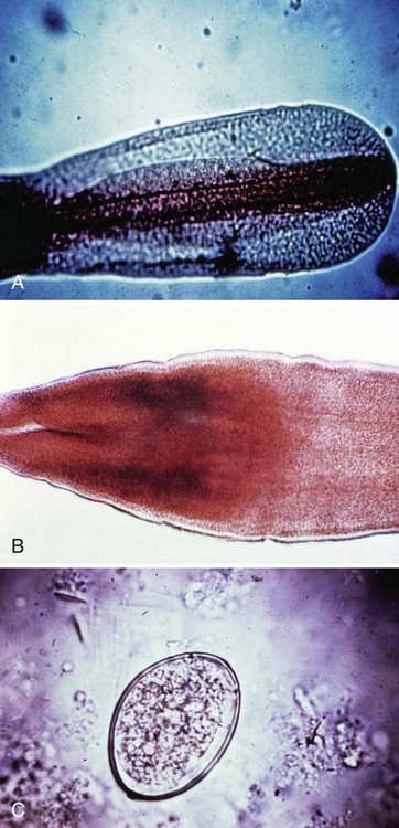
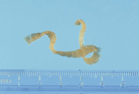
Diphyllobothrium latum, the freshwater broad fish tapeworm, is the largest human tapeworm. Adults have been known to reach up to 10 m in length, with more than 3000 to 4000 proglottids, and reside within a host for 30 years or more. The proglottids are characteristically wider than long with a central rosette-shaped uterine structure (Figure 54-1). The scolex is spatulate and contains two shallow sucking grooves referred to as bothria (Figure 54-1, B). D. latum has unembryonated eggs. The eggs are operculated with a terminal knob, similar to trematode eggs (Figure 54-1, C). The intermediate hosts include crustaceans and freshwater fish.
Pathogenesis and Spectrum of Disease
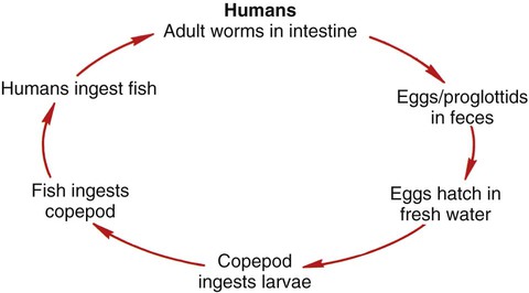
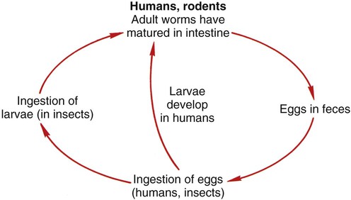
Diphyllobothrium latum is the only cestode to have an aquatic life cycle (Figure 54-2). Fish serve as the reservoir host, with humans serving as the definitive host. D. latum eggs are found in the feces of infected humans and other fish-eating mammals. Once passed into a water source, such as a lake, the life cycle requires two intermediate hosts. After incubation in freshwater for approximately 2 weeks, the mature eggs release the first larval stage (coracidium). The coracidia are ingested by copepods. The fish feed upon the small crustaceans ingesting the procercoid larvae. Within the freshwater fish, the larvae develop into the infective larvae. D. latum infection occurs through the ingestion of poorly cooked freshwater fish containing the plerocercoid larval form. D. latum matures to an adult tapeworm within the human small intestine. Infection is usually asymptomatic, but mild gastrointestinal symptoms may occur such as diarrhea, abdominal pain, fatigue, vomiting, or dizziness. Symptoms vary depending on the worm burden and the host’s immune response to the organism. The tapeworm nutritional requirements may decrease the host’s vitamin B12 level, resulting in megaloblastic anemia (Table 54-1).
TABLE 54-1
Epidemiology of the Intestinal Cestodes That Cause Disease in Humans
| Parasite | Habitat (Reservoir) | Mode of Transmission |
| Diphyllobothrium latum | Adult tapeworms can be found in a number of wild animals, the most important being dogs, bears, seals, and walrus, that serve as reservoir hosts; the human serves as the definitive host. | Occurs via ingestion of poorly cooked freshwater fish containing the sparganum or plerocercoid larval form. |
| Dipylidium caninum | Humans serve as an accidental host for the dog tapeworm. Dogs, cats, and wild animals serve as a reservoir host. Arthropods such as the dog and cat flea serve as an intermediate host. | Transmitted to humans through the ingestion of dog/cat fleas. |
| Hymenolepis nana | The dwarf tapeworm can also occur in rodents; the human can serve as both intermediate and definitive host, with development from the egg to adult worm occurring in the human intestine. | Primarily acquired from accidental ingestion of eggs from an adult tapeworm, most commonly via fecal-oral exposure. |
| Hymenolepis diminuta | The rat tapeworm frequently infects rodents, and rarely infects humans. | Infects humans after infected mice and rats contaminate food with their feces. |
| Taenia solium | Humans serve as the definitive host for the pork tapeworm, whereas pigs and humans serve as intermediate hosts. | Humans become infected when cyst-infected pork is ingested. |
| Taenia saginata | Humans serve as the definitive host for the beef tapeworm, whereas cows/camels serve as intermediate hosts. | Humans become infected when cyst-infected beef is ingested. |
Laboratory Diagnosis
Both eggs and proglottids may be found in patient’s feces. Visualization of the eggs is enhanced using a wet preparation of the patient’s stool sample. Diagnosis is made by identification of the ovoid, operculated, yellow-brown eggs (58 to 75 µm by 40 to 50 µm) passed in abundance in the stool. They are sometimes confused with the eggs of Paragonimus. The mature gravid proglottids are wider than long (3 × 11 mm), often in chains, and contain a rosette-shaped central uterus (Figure 54-3). Species identification is through assessment of the morphologic characteristics of the proglottids as previously described (Tables 54-2 and 54-3).
TABLE 54-2
Common Human Parasites, Diagnostic Specimens, Tests, and Positive Findings
| Organism | Acquire Infection from: | Location in Host | Diagnostic Specimen | Diagnostic Test* | Positive Specimen | Comments |
| Intestinal cestodes Taenia saginata (beef) |
Ingestion of raw beef | Intestine | Stool and/or proglottids | O&P, India ink proglottids | Eggs, proglottid branches | Both Taenia eggs look alike; need gravid proglottid or scolex for definitive identification. |
| Taenia solium (pork) | Ingestion of raw pork | Stool and/or proglottids | O&P, India ink proglottids | Eggs, proglottid branches | ||
| Diphyllobothrium latum | Ingestion of raw freshwater fish | Stool and/or proglottids | O&P | Eggs, proglottid shape | ||
| Hymenolepis nana | Tapeworm eggs | Stool | O&P | Eggs | ||
| Hymenolepis diminuta | Grain beetles | Stool | O&P | Eggs | ||
| Dipylidium caninum | Fleas from dogs/cats | Stool and/or proglottids | O&P | Eggs, proglottid shape |
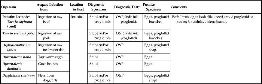
*Although serologic tests are not always mentioned, they are available for a number of parasitic infections. Unfortunately, most are not routinely available. Contact your state public health laboratory or the Centers for Disease Control and Prevention in Atlanta, Georgia.
TABLE 54-3
Cestode Parasites of Humans (Intestinal)
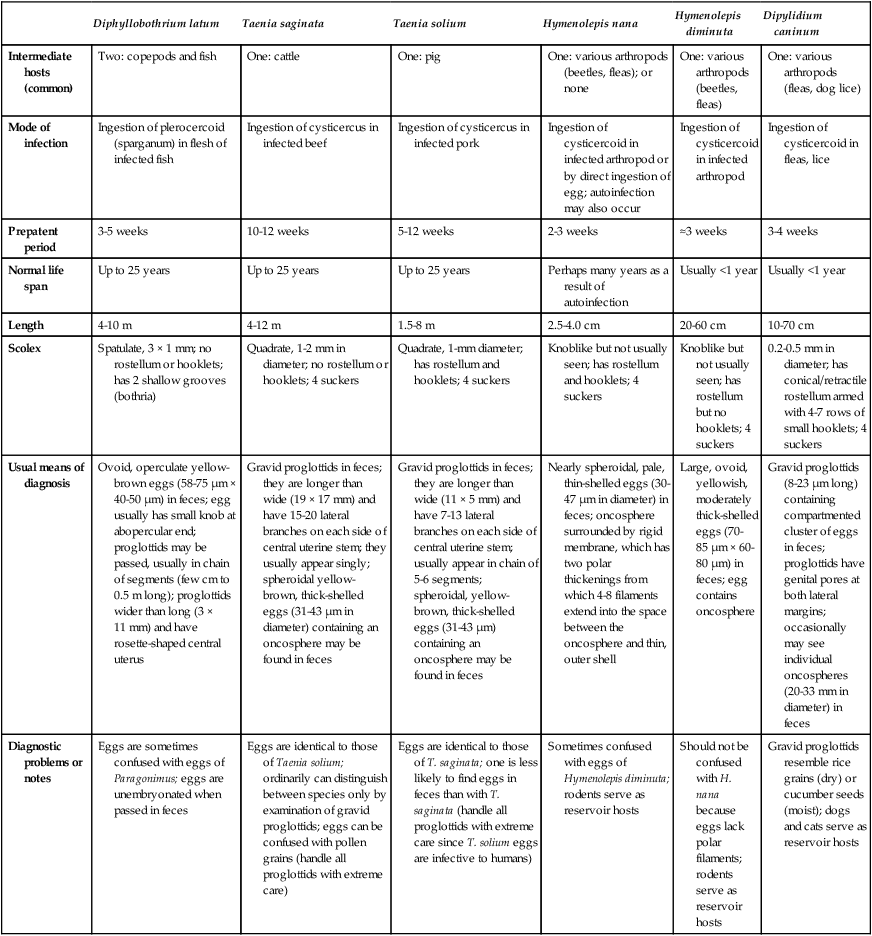
| Diphyllobothrium latum | Taenia saginata | Taenia solium | Hymenolepis nana | Hymenolepis diminuta | Dipylidium caninum | |
| Intermediate hosts (common) | Two: copepods and fish | One: cattle | One: pig | One: various arthropods (beetles, fleas); or none | One: various arthropods (beetles, fleas) | One: various arthropods (fleas, dog lice) |
| Mode of infection | Ingestion of plerocercoid (sparganum) in flesh of infected fish | Ingestion of cysticercus in infected beef | Ingestion of cysticercus in infected pork | Ingestion of cysticercoid in infected arthropod or by direct ingestion of egg; autoinfection may also occur | Ingestion of cysticercoid in infected arthropod | Ingestion of cysticercoid in fleas, lice |
| Prepatent period | 3-5 weeks | 10-12 weeks | 5-12 weeks | 2-3 weeks | ≈3 weeks | 3-4 weeks |
| Normal life span | Up to 25 years | Up to 25 years | Up to 25 years | Perhaps many years as a result of autoinfection | Usually <1 year | Usually <1 year |
| Length | 4-10 m | 4-12 m | 1.5-8 m | 2.5-4.0 cm | 20-60 cm | 10-70 cm |
| Scolex | Spatulate, 3 × 1 mm; no rostellum or hooklets; has 2 shallow grooves (bothria) | Quadrate, 1-2 mm in diameter; no rostellum or hooklets; 4 suckers | Quadrate, 1-mm diameter; has rostellum and hooklets; 4 suckers | Knoblike but not usually seen; has rostellum and hooklets; 4 suckers | Knoblike but not usually seen; has rostellum but no hooklets; 4 suckers | 0.2-0.5 mm in diameter; has conical/retractile rostellum armed with 4-7 rows of small hooklets; 4 suckers |
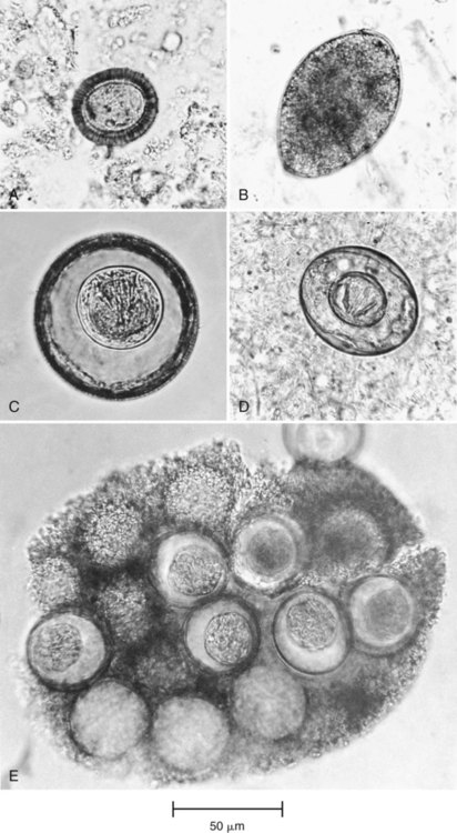
| Usual means of diagnosis | Ovoid, operculate yellow-brown eggs (58-75 µm × 40-50 µm) in feces; egg usually has small knob at abopercular end; proglottids may be passed, usually in chain of segments (few cm to 0.5 m long); proglottids wider than long (3 × 11 mm) and have rosette-shaped central uterus | Gravid proglottids in feces; they are longer than wide (19 × 17 mm) and have 15-20 lateral branches on each side of central uterine stem; they usually appear singly; spheroidal yellow-brown, thick-shelled eggs (31-43 µm in diameter) containing an oncosphere may be found in feces | Gravid proglottids in feces; they are longer than wide (11 × 5 mm) and have 7-13 lateral branches on each side of central uterine stem; usually appear in chain of 5-6 segments; spheroidal, yellow-brown, thick-shelled eggs (31-43 µm) containing an oncosphere may be found in feces | Nearly spheroidal, pale, thin-shelled eggs (30-47 µm in diameter) in feces; oncosphere surrounded by rigid membrane, which has two polar thickenings from which 4-8 filaments extend into the space between the oncosphere and thin, outer shell | Large, ovoid, yellowish, moderately thick-shelled eggs (70-85 µm × 60-80 µm) in feces; egg contains oncosphere | Gravid proglottids (8-23 µm long) containing compartmented cluster of eggs in feces; proglottids have genital pores at both lateral margins; occasionally may see individual oncospheres (20-33 mm in diameter) in feces |
| Diagnostic problems or notes | Eggs are sometimes confused with eggs of Paragonimus; eggs are unembryonated when passed in feces | Eggs are identical to those of Taenia solium; ordinarily can distinguish between species only by examination of gravid proglottids; eggs can be confused with pollen grains (handle all proglottids with extreme care) | Eggs are identical to those of T. saginata; one is less likely to find eggs in feces than with T. saginata (handle all proglottids with extreme care since T. solium eggs are infective to humans) | Sometimes confused with eggs of Hymenolepis diminuta; rodents serve as reservoir hosts | Should not be confused with H. nana because eggs lack polar filaments; rodents serve as reservoir hosts | Gravid proglottids resemble rice grains (dry) or cucumber seeds (moist); dogs and cats serve as reservoir hosts |
Dipylidium Caninum
General Characteristics
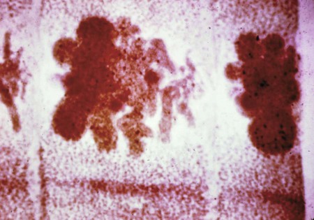
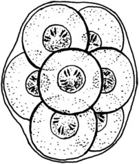
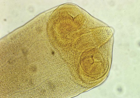
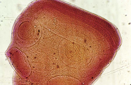
D. caninum, the cat or dog tapeworm (Figure 54-4), is a double-pored tapeworm consisting of many small proglottids. As the tapeworm matures, the proglottids separate and pass in the stool. They may be recognized on the basis of their characteristic “cucumber seed” appearance when they are wet, as well as their resemblance to a dried grain of rice. Adult tapeworms measure 10 to 70 cm in length. The scolex contains four suckers and an armed rostellum. Egg packets may also be found in the feces of the host.
Epidemiology
Laboratory Diagnosis
Symptoms of Dipylidium infection are similar to those of pinworm infection; however, the treatments are very different. The laboratory should confirm suspected infections. Proglottids (8 to 23 µm) may be seen in the stool. D. caninum is also referred to as the “cucumber seed” tapeworm as previously described (see Figure 54-3). The first sign of infection may be the appearance of seedlike particles in the stool or undergarments of the patient. These particles are the egg-bearing segments of the tapeworm. Groups of egg packets may be found in the stool (Figures 54-5 and 54-6). The adult worms have a scolex with four suckers and a conical/retractile rostellum armed with four to seven rows of small hooklets (Figure 54-7). Patients may also develop a moderately elevated eosinophilia.
Hymenolepis Nana
General Characteristics
Pathogenesis and Spectrum of Disease
H. nana has an unusual life cycle; ingestion of the egg can lead to the development of the adult worm in humans, thus bypassing the need for an intermediate host (Figure 54-8). Humans can serve as both intermediate and definitive hosts. Infection occurs by accidentally ingesting dwarf tapeworm eggs. This happens most commonly through direct fecal-oral transmission or accidental ingestion of an infected arthropod. The worm resides within the upper ileum of the intestinal tract. Once infected, the dwarf tapeworm may reproduce inside the body, thus causing autoinfection. Autoinfection is essentially a reinfection or constant reproduction of the parasite within the host. Massive infection with several thousand worms may follow autoinfection, resulting in hyperinfection. Hyperinfction refers to a large parasitic burden within the host. Autoinfection appears to initiate a cellular and humoral immune response. The immune response will provide the host with some protective immunity. Most patients are asymptomatic. Symptomatic patients may experience weight loss, nausea, weakness, loss of appetite, diarrhea, and abdominal discomfort. Young children, especially those with a heavy infection, may develop headache, itchy bottom, or difficulty sleeping. Dwarf tapeworm infection may be misdiagnosed as pinworm infection.
Laboratory Diagnosis
Adult worms and proglottids are rarely seen in stool specimens. Diagnosis is typically through the identification of eggs in stool specimens. Eggs are characterized by the presence of a thin shell enclosing an embryo (oncosphere) with six hooklets contained within two layers of membrane. The eggs are spheroidal, pale, and thin-shelled (30 to 47 µm in diameter). The eggs of H. nana and Hymenolepis diminuta are very similar. However, H. nana eggs are smaller and have polar filaments present in the space between the oncospheres and the eggshell (see Figure 54-5). The egg morphology is easily distinguishable in fresh or formalin-fixed fecal samples. It is important to note that eggs are infectious and therefore unpreserved specimens should be handled carefully. Concentration techniques and repeated examinations will increase the likelihood of detecting light infections. Some patients may demonstrate a low-grade eosinophilia.
Hymenolepis Diminuta
General Characteristics
Laboratory Diagnosis
Proglottids are rarely seen in the stool; diagnosis is made by the identification of eggs. The eggs (70 to 85 µm by 60 to 80 µm) are large, ovoid, yellowish, and moderately thick-shelled. The eggs contain a six-hooked oncosphere with the absence of polar filaments in the space between the oncosphere and the eggshell (see Figure 54-5). The eggs are clearly differentiated from H. nana because of the absence of polar filaments.
Taenia Solium
General Characteristics
Pathogenesis and Spectrum of Disease
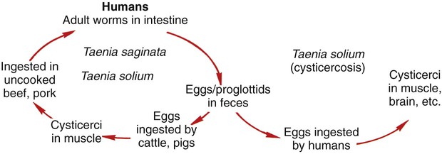
T. solium infection can result in the presence of both adult and larval stages in the human host (Figure 54-9). Infection begins when the intermediate host ingests embryonated eggs in feces. Once the egg is ingested, the hexacanth embryo is released into the intestine where the embryo penetrates the mucosa. The embryo then matures into a cyst (cysticercus) in the tissue. Humans may become infected when they eat raw or undercooked pork containing embedded cysts. Pork tapeworm infection is usually caused through the ingestion of multiple worms. During ingestion and subsequent digestion of the infected meat, the cysticercus is released and attaches to the mucosa within the small intestine of the human host. The cysticercus matures into an adult worm within approximately 5 to 12 weeks. The eggs are then released in the host’s feces. Accidental ingestion of the eggs by the human host may also result in migration of the embryo through the intestine to other areas of the body, including the eyes, brain, muscle, or bone. In addition, the proglottids are motile and may migrate out of the anus. Infection of the adult tapeworm causes few clinical symptoms, although abdominal pain, diarrhea, indigestion, and loss of appetite may be present as a result of irritation to the mucosa of the intestinal wall. The major complication with T. solium is cysticercosis (larval forms throughout the body), in which the human host becomes the intermediate host and harbors the larvae in tissues as previously described. This infection is further discussed in Chapter 76.
Laboratory Diagnosis
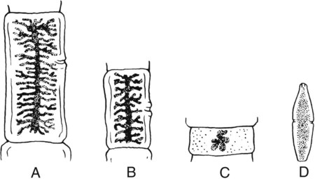
Serologic diagnosis is unreliable for infections with T. solium. Diagnosis of Taenia tapeworm infection is through the examination of stool samples. Individuals suspected of infection with T. solium should be asked if they have passed any notable tapeworm segments in their stool. Stool specimens should be collected on 3 different days and microscopically examined for the presence of Taenia eggs (see Figure 54-5). Tapeworm eggs can be detected in the stool 2 to 3 months after the tapeworm infection is established. Eggs are round or slightly oval (31 to 43 µm in diameter) and yellow-brown with a thick striated shell containing a six-hooked oncosphere. Diagnosis is based on the recovery of eggs or proglottids in stool or from the perianal area. T. solium and T. saginata cannot be differentiated on the basis of egg morphology. Speciation requires the examination of gravid proglottids or the scolices. T. solium gravid proglottids are longer than wide (19 × 17 mm) and may be distinguished from T. saginata according to the number of uterine branches. T. solium contains 7 to 13 lateral uterine branches along the proglottid (Figure 54-10), whereas T. saginata contains more than 13 branches. Uterine branches may be visualized by staining the proglottids with India ink. The scolex contains a neck region that is typically short and half the width of the scolex and differs from that of T. saginata by the presence of four suckers with hooks in a double row (Figure 54-11). The adult worm is usually 3 to 5 m long. Extreme care should be taken when handling infectious stool, since T. solium proglottids and eggs are extremely infectious. Additional laboratory findings may include a low-grade eosinophilia, increased serum IgE level, and the presence of atypical lymphocytes in the cerebrospinal fluid.
Taenia Saginata
General Characteristics
Laboratory Diagnosis
The stool should be examined for proglottids and eggs; eggs may also be present on anal swabs. The eggs of T. saginata are indistinguishable from those of T. solium. The uterus of T. saginata is longer than wide and typically contains 15 to 18 lateral branches on each side (see Figure 54-10). The scolex has four suckers and is unarmed or does not contain any hooklets (Figure 54-11). Stool specimens should be handled with care since the eggs cannot be distinguished from those of T. solium. Slight eosinophilia may develop.
Case Study 54-1
A 50-year-old man presented to the physician complaining of headaches and difficulty maintaining his balance. On physical exam the physician noticed a lump in the man’s left calf. Initial lab results including a complete blood count and differential were normal. The ESR (erythrocyte sedimentation rate) was slightly elevated, indicating generalized inflammation. The physician asked the man if he had traveled to any areas outside of the United States in the past 12 months. The man had recently returned from volunteering in Haiti. The physician prescribed niclosamide and had the patient collect three stool samples over the next 12 days. Following treatment the following parasite was recovered from the man’s stool (see Figures 54-12 and 54-13).