Increased intracranial pressure

Published on 07/02/2015 by admin

Filed under Anesthesiology

Last modified 22/04/2025

Print this page

rate 1 star rate 2 star rate 3 star rate 4 star rate 5 star
Your rating: none, Average: 0 (0 votes)

This article have been viewed 2317 times

Increased intracranial pressure

C. Thomas Wass, MD

Intracranial pressure (ICP) is determined by the relationship of the volumes of the intracranial vault (formed by the skull) and the intracranial contents. The latter is composed of three volume compartments: brain parenchyma, cerebrospinal fluid (CSF), and blood. By definition, intracranial hypertension exists when ICP is sustained above 15 mm Hg.

Intracranial elastance

Historically, the intracranial pressure-volume relationship has been termed compliance in the medical literature. Compliance is defined as unit or units of volume (e.g., intracranial volume) change per unit or units of pressure (e.g., ICP) change (i.e., ΔV/ΔP). However, the pressure-volume curve presented in Figure 132-1 and most other textbooks actually depicts the reciprocal of compliance, or elastance.

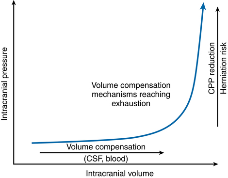
Elastance is defined as ΔP/ΔV. Under normal physiologic conditions, small volume increases in any one of the three intracranial compartments results in little or no change in ICP. The compensatory mechanisms that initially protect against an elevation in ICP are (a) translocation of intracranial CSF through the foramen magnum to the subarachnoid space surrounding the spinal cord, (b) increased CSF absorption through the arachnoid granulations, and (c) translocation of blood out of the intracranial vault. Once these mechanisms are exhausted, abrupt increases in ICP occur in association with small increases in intracranial volume (see Figure 132-1). That is, intracranial compliance is decreased, or more correctly, intracranial elastance is increased.

Anesthetic considerations

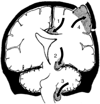
The goals of managing a patient with intracranial hypertension include preventing cerebral ischemia and preventing brain herniation (Figure 132-2).

Respiratory

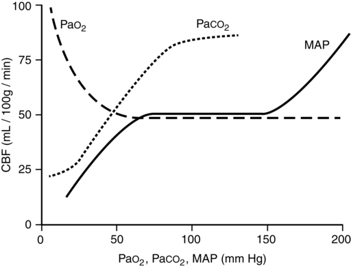
PaCO2 is the single most potent physiologic determinant of CBF (Figure 132-3) and CBV. At a PaCO2 between 20 and 80 mm Hg, CBF decreases 1 mL/100 g brain weight/min and CBV decreases 0.05 mL/100 g brain weight for each 1-mm Hg decrease in PaCO2. Decreasing PaCO2 to 25 to 28 mm Hg should provide near-maximal reductions in ICP, lasting up to 24 h, without adversely affecting acid-base or electrolyte status or decreasing cerebral O2 delivery (i.e., resulting from combined cerebral vasoconstriction and leftward shift in the oxyhemoglobin dissociation curve). Accordingly, in the setting of severe traumatic brain injury, the Brain Trauma Foundation states that aggressive hyperventilation (i.e., PaCO2 ≤ 25 mm Hg) is not recommended, as further reductions in PaCO2 may result in iatrogenic brain injury.

Hypoxia (PaO2 <50 mm Hg) will increase CBF and ICP. Application of positive end-expiratory pressure may decrease venous effluent from the cranium and exacerbate intracranial hypertension.

Coughing against a closed glottis (i.e., Valsalva maneuver) will increase ICP. Intravenously administered lidocaine, esmolol, or opioids can be used to attenuate the ICP response to direct laryngoscopy, tracheal intubation, or coughing.

Cardiovascular

Mean arterial pressure (MAP) is a determinant of cerebral perfusion pressure (CPP) (i.e., CPP = MAP – ICP). The blood-brain barrier and autoregulation may be disrupted at the site of cerebral ischemic, traumatic, hemorrhagic, or osmolar insults. In these regions, it is correct to assume that CBF is passively dependent on CPP. Before the dura is opened, it is prudent to treat all hypertensive episodes by deepening the level of anesthesia, administering antihypertensive drugs that lack the ability to dilate cerebral vessels—and thus elevate ICP—(e.g., esmolol, labetalol, metoprolol), or both deepen the level of anesthesia and administer antihypertensive drugs. With respect to CPP, the critical threshold for ischemia is approximately 50 to 60 mm Hg. However, routine use of vasopressors and intravenously administered fluids to maintain the CPP greater than 70 mm Hg is not advised. Taking this information together, one can infer that maintaining the CPP near 60 to 70 mm Hg is advisable in the setting of traumatic brain injury.

Intravenous fluid administration should not be spared at the expense of hemodynamic stability. Osmolar, not oncotic, pressure is the primary determinant of fluid shifts within the brain. Therefore, maintaining intravascular isovolemia with a near-isoosmolar solution (e.g., normal saline, lactated Ringer’s solution) is safe and beneficial to end-organ preservation. Hypoosmolar glucose-containing fluids (e.g., D5W) have the ability to (1) increase cerebral edema, (2) increase ICP, and (3) induce hyperglycemia, which exacerbates ischemic neurologic injury.

Metabolic

Cerebral metabolism decreases approximately 6% to 7% per 1 of temperature reduction. Mild hypothermia (i.e., temperature reductions of 1°C – 6°C) has been reported to improve neurologic outcome following focal or global brain ischemia in laboratory studies. Conversely, fever may worsen postischemic neurologic outcome. Proposed mechanisms for temperature modulation of postischemic neurologic outcome include alterations in cerebral metabolism, blood-brain barrier stability, membrane depolarization, ion homeostasis (e.g., calcium), neurotransmitter release (e.g., glutamate or aspartate), enzyme function (e.g., phospholipase, xanthine oxidase, or nitric oxide synthase), and free radical production or scavenging. Despite convincing laboratory data, large clinical trials have produced mixed results.