Hypofunction of the Ovaries

Published on 25/03/2015 by admin

Filed under Pediatrics

Last modified 22/04/2025

Print this page

rate 1 star rate 2 star rate 3 star rate 4 star rate 5 star
Your rating: none, Average: 0 (0 votes)

This article have been viewed 3001 times

Chapter 580 Hypofunction of the Ovaries

Hypofunction of the ovaries can be either primary or central in etiology. It may be caused by congenital failure of development, postnatal destruction (primary or hypergonadotropic hypogonadism), or lack of central stimulation by the pituitary and/or hypothalamus (secondary or tertiary hypogonadotropic hypogonadism). Primary ovarian insufficiency (hypergonadotropic hypogonadism), which is also termed premature ovarian failure, is characterized by the arrest of normal ovarian function before the age of 40 yr. Mutations of certain genes could result in primary ovarian insufficiency. Hypofunction of the ovaries due to lack of central stimulation (hypogonadotropic hypogonadism) can be associated with other processes such as multiple pituitary hormone deficiencies and some chronic diseases. Table 580-1 details the etiologic classification of ovarian hypofunction.

580.1 Hypergonadotropic Hypogonadism in the Female (Primary Hypogonadism)

Diagnosis of hypergonadotropic hypogonadism before puberty is difficult. Except in the case of Turner syndrome, most affected patients have no prepubertal clinical manifestations.

Turner Syndrome

Turner described a syndrome consisting of sexual infantilism, webbed neck, and cubitus valgus in adult females (Chapter 76). Ullrich described an 8 yr old girl with short stature and many of the same phenotypic features. The term Ullrich-Turner syndrome is frequently used in Europe but rarely used in the USA. The condition is defined as the combination of the characteristic phenotypic features accompanied by complete or partial absence of the second X chromosome with or without mosaicism.

Pathogenesis

Half the patients with Turner syndrome have a 45,X chromosomal complement. About 15% of patients are mosaics for 45,X and a normal cell line (45,X/46,XX). Other mosaics with isochromosomes, 45,X/46,X,i(Xq); with rings, 45,X/46,X,r(X); or with fragments, 45,X/46fra, occur less often. Mosaicism is detected most commonly when more than one tissue is examined. The single X is of maternal origin in nearly 80% of 45,X patients. The mechanism of chromosome loss is unknown, and the risk for the syndrome does not increase with maternal age. The genes involved in the Turner phenotype are X-linked genes that escape inactivation. A major locus involved in the control of linear growth has been mapped within the pseudoautosomal region of the X chromosome (PAR1). SHOX, a homeobox-containing gene of 170 kb of DNA within the PAR1, is thought to be important for controlling growth in children with Turner syndrome, Leri-Weill syndrome, and rarely in patients having idiopathic short stature. Genes for the control of normal ovarian function are postulated to be on Xp and perhaps two “supergenes” on Xq.

Turner syndrome occurs in about 1/1,500-2,500 live born females. The frequency of the 45,X karyotype at conception is about 3.0%, but 99% of these are spontaneously aborted, accounting for 5-10% of all abortuses. Mosaicism (45,X/46,XX) occurs in a proportion higher than that seen with any other aneuploid state, but the mosaic Turner constitution is rare among the abortuses; these findings indicate preferential survival for mosaic forms.

The normal fetal ovary contains about 7 million oocytes, but these begin to disappear rapidly after the 5th mo of gestation. At birth, there are only 2 million (1 million active follicles); by menarche, there are 400,000-500,000; and at menopause, 10,000 remain. In the absence of 1 X chromosome, this process is accelerated, and nearly all oocytes are gone by 2 yr of age. In aborted 45,X fetuses, the number of primordial germ cells in the gonadal ridge appears to be normal, suggesting that the normal process is accelerated in patients with Turner syndrome. Eventually, the ovaries are described as “streaks” and consist only of connective tissue, with only a few germ cells present.

Clinical Manifestations

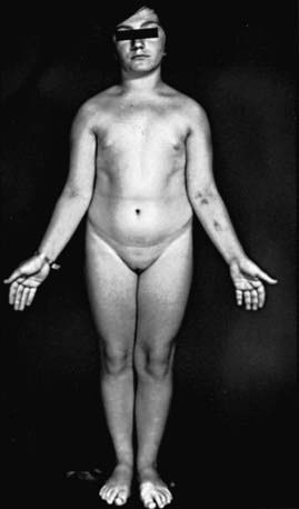
Many patients with Turner syndrome are recognizable at birth because of a characteristic edema of the dorsa of the hands and feet and loose skinfolds at the nape of the neck. Low birthweight and decreased length are common (Chapter 76). Clinical manifestations in childhood include webbing of the neck, a low posterior hairline, small mandible, prominent ears, epicanthal folds, high arched palate, a broad chest presenting the illusion of widely spaced nipples, cubitus valgus, and hyperconvex fingernails. The diagnosis is often first suspected at puberty when breast development fails to occur.

Short stature, the cardinal finding in virtually all girls with Turner syndrome, may be present with little in the way of other clinical manifestations. The linear growth deceleration begins in infancy and young childhood, gets progressively more pronounced in later childhood and adolescence, and results in significant adult short stature. Sexual maturation fails to occur at the expected age. Among untreated patients with Turner syndrome, the mean adult height is 143-144 cm in the USA and most of northern Europe, but 140 cm in Argentina and 147 cm in Scandinavia (Fig. 580-1). The height is well correlated with the midparental height (average of the parents’ heights). Specific growth curves for height have been developed for girls with Turner syndrome.

Associated cardiac defects are common. In the girls with Turner syndrome, life-threatening consequences of X-chromosome haploinsufficiency involve the cardiovascular system. There is a 4- to 5-fold increased rate of premature mortality secondary to congenital heart disease and premature coronary heart disease in adults with Turner syndrome. Clinically silent cardiac defects, mainly bicuspid aortic valve, but also ascending aortic dilation and partial anomalous pulmonary venous connections are present in patients with Turner syndrome. Regardless of the age, all patients with Turner syndrome at the time of diagnosis need comprehensive cardiovascular evaluation by a cardiologist specializing in congenital heart disease. Complete cardiologic evaluation, including echocardiography, reveals isolated nonstenotic bicuspid aortic valves in one third to one half of patients. In later life, bicuspid aortic valve disease can progress to dilatation of the aortic root. Less frequent defects include aortic coarctation (20%), aortic stenosis, mitral valve prolapse, and anomalous pulmonary venous drainage. In a study of 170/393 females with Turner syndrome in Denmark, 38% of patients with 45,X chromosomes had cardiovascular malformations compared with 11% of those with mosaic monosomy X; the most common were aortic valve abnormalities and aortic coarctation. Webbed neck in patients with or without recognized syndromes is associated with both flow-related and non–flow-related heart defects. Among patients with Turner syndrome, those with webbed neck have a much greater chance of having coarctation of the aorta than do those without webbed necks. Recent studies have suggested that in Turner syndrome there is a broader spectrum of cardiovascular abnormalities than previously recognized. Transthoracic echocardiogram in young girls is adequate if cardiac anatomy is clearly seen; otherwise magnetic resonance angiographic screening studies should be considered in asymptomatic individuals with Turner syndrome. During adolescence, and certainly before pregnancy is contemplated, repeat cardiac evaluation should be considered even in those without prior findings of cardiac abnormalities. Blood pressure should be routinely monitored even in the absence of cardiac or renal lesions and especially in those with suggestions of aortic root dilatation. Cardiac MRI is a valuable tool to detect and monitor aortic root dilation.

Renal ultrasound should be performed in all girls with Turner syndrome at diagnosis. One fourth to one third of patients have renal malformations on ultrasonographic examination (50% of those with 45,X karyotypes). The more serious defects include pelvic kidney, horseshoe kidney, double collecting system, complete absence of one kidney, and ureteropelvic junction obstruction. Some of the malformations may increase the risk of hypertension and urinary tract infection. Idiopathic hypertension is also common. Girls with Turner syndrome who had normal baseline renal ultrasound did not develop renal disease during a follow-up period averaging 6 yr.

When the ovaries were examined by ultrasonography, older studies found a significant decrease in percentage of detectable ovaries from infancy to later childhood. A subsequent report found no such age-related differences in a cross-sectional and longitudinal study conducted in Italy; 27-46% of patients had detectable ovaries at various ages; 76% of those with X mosaicism and 26% of those with 45,X karyotypes had detectable ovaries.

Sexual maturation usually fails to occur, but 10-20% of girls have spontaneous breast development, and a small percentage may have menstrual periods. Primary gonadal failure is associated with early onset of adrenarche (elevation in DHEA sulfate) but delayed pubarche (pubic hair development). Spontaneous pregnancies have been reported in menstruating patients with Turner syndrome. Premature menopause, increased risk of miscarriage, and offspring with increased risk of trisomy 21 have been reported in some of these women. A woman with a 45,X/46,X,r(X) karyotype treated with hormone replacement therapy had 3 pregnancies, resulting in a normal 46,XY male infant, a spontaneous abortion, and a healthy term female with Turner syndrome 45,X/46,Xr(X).

Antithyroid antibodies (thyroid peroxidase, and/or thyroglobulin antibodies) occur in 30-50% of patients. The prevalence increases with advancing age. Ten to 30% have autoimmune thyroid disease, with or without the presence of a goiter. Age-dependent abnormalities in carbohydrate metabolism characterized by abnormal glucose tolerance and insulin resistance and, only rarely, frank type 2 diabetes occur in patients with Turner syndrome. Impaired insulin secretion has been described in 45,X women. Cholesterol levels are elevated in adolescence, regardless of body mass index or karyotype.

Inflammatory bowel disease, both Crohn disease and ulcerative colitis; gastrointestinal bleeding due to abnormal mesenteric vasculature; and delayed gastric emptying time have all been reported. Screening for celiac disease is recommended by recent guidelines, since the risk of celiac disease is increased in Turner syndrome, with 4-6% of individuals affected. Although autoimmune diseases have been associated with Turner syndrome, the prevalence of type 1 diabetes with Turner syndrome is not very high.

Sternal malformations can be detected by lateral chest radiography. An increased carrying angle at the elbow is usually not clinically significant. Scoliosis occurs in about 10% of adolescent girls. Congenital hip dysplasia occurs more commonly than in the general population. Reported eye findings include anterior segment dysgenesis and keratoconus. Pigmented nevi become more prominent with age; melanocytic nevi are common. Essential hyperhidrosis, torus mandibularis, and alopecia areata occur rarely.

Recurrent bilateral otitis media develops in about 75% of patients. Sensorineural hearing deficits are common, and the frequency increases with age. Problems with gross and fine motor-sensory integration, failure to walk before 15 mo of age, and early language dysfunction often raise questions about developmental delay, but intelligence is normal in most patients. However, mental retardation does occur in patients with 45,X/46,X,r(X); the ring chromosome is unable to undergo inactivation and leads to 2 functional X chromosomes.

A special attention should be given to psychosocial development in girls with Turner syndrome. In general the behavior function is normal in girls with Turner syndrome, but they are at an increased risk for social isolation, immaturity, and anxiety. Other conditions such as dyslexia, nonverbal learning disability, and attention deficit disorder have been reported in girls with Turner syndrome. In adults, deficits in perceptual spatial skills are more common than they are in the general population. Some unconfirmed data suggest the existence of an imprinted X-linked locus that affects cognitive function such as verbal and higher-order executive function skills. These functions are apparently better when the X is paternal in origin.

The prevalence of mosaicism depends in large part on the techniques used for studying chromosomal patterns. The use of fluorescent in situ hybridization and reverse transcription–polymerase chain reaction (PCR) has increased the reported prevalence of mosaic patterns to as high as 60-74%.

Mosaicism involving the Y chromosome occurs in 5%. A population study of Danish women using PCR with 5 different primer sets found Y chromosome material in 12.2%. Gonadoblastoma among Y-positive patients occurred in 7-10%. Therefore, the current recommendation is that prophylactic gonadectomy should be performed even in the absence of MRI or CT evidence of tumors. The recommended timing of this procedure is at the time of diagnosis, but this may need to be re-evaluated in the future. The gonadoblastoma locus on the Y chromosome (GBY) maps close to the Y centromere. The presence of only the SRY (sex determining region on Y) locus is not sufficient to confer increased susceptibility for the development of gonadoblastoma. A careful study of 53 patients with Turner syndrome by nested PCR excluded low-level Y mosaicism in almost all cases. A 2nd round of PCR detected SRY on the distal short arm of the Y chromosome in only 2 subjects. Therefore, routine PCR for Y chromosome detection for the purpose of assigning gonadoblastoma risk does not seem indicated. High-throughput quantitative genotyping may provide an effective and inexpensive method for the identification of X chromosome abnormalities and Y chromosome material identification.

In patients with 45,X/46,XX mosaicism, the abnormalities are attenuated and fewer; short stature is as frequent as it is in the 45,X patient and may be the only manifestation of the condition other than ovarian failure (see Fig. 580-1).

Laboratory Findings

Chromosomal analysis must be considered routinely in short girls. In a systematic search, using Southern blot analysis of leukocyte DNA, Turner syndrome was detected in 4.8% of girls referred to an endocrinology service because of short stature. Patients with a marker chromosome in some or all cells should be tested for DNA sequences at or near the centromere of the Y chromosome for GBY.

Ultrasonography of the heart, kidneys, and ovaries is indicated after the diagnosis is established. The most common skeletal abnormalities are shortening of the 4th metatarsal and metacarpal bones, epiphyseal dysgenesis in the joints of the knees and elbows, Madelung deformity, scoliosis, and in older patients, inadequate osseous mineralization.

Plasma levels of gonadotropins, particularly follicle-stimulating hormone (FSH), are markedly elevated to greater than those of age-matched controls during infancy; at 2-3 yr of age, a progressive decrease in levels occurs until they reach a nadir at 6-8 yr of age, and by 10-11 yr, they rise to adult castrate levels.

Thyroid antiperoxidase antibodies should be checked periodically, and if positive, levels of thyroxine and thyroid-stimulating hormone should be obtained. Turner syndrome girls should be screened by measuring tissue transglutaminase (TGG) IgA antibodies for celiac disease. Initial testing should be done around age 4 yr and repeated every 2-5 yr. Extensive studies have failed to establish that growth hormone deficiency plays a primary role in the pathogenesis of the growth disorder. Defects in normal secretory patterns of growth hormone are seen in adolescents due to lack of gonadal steroids but not in younger girls with Turner syndrome. In vitro, monocytes and lymphocytes show decreased sensitivity to insulin-like growth factor-1 (IGF-1).

The American Academy of Pediatrics has published a comprehensive guide to the health supervision of children with Turner syndrome. A guide to the care of girls and women with Turner syndrome was published in 2007.

Treatment

Treatment with recombinant human growth hormone increases height velocity and ultimate stature in most but not all children. Many girls achieve heights of greater than 150 cm with early initiation of treatment. In a large, multicenter, placebo-controlled U.S. clinical trial, 99 patients with Turner syndrome who started receiving growth hormone at a mean age of 10.9 yr at doses between 0.27 and 0.36 mg/kg/wk achieved a mean height of 149 cm, with nearly one third reaching heights greater than 152.4 cm (60 in). In the Netherlands, higher doses of growth hormone (up to 0.63 mg/kg/wk in the 3rd yr of treatment) resulted in 85% of the subjects reaching adult heights in the normal range for the Dutch reference population. Growth hormone treatment should be initiated in early childhood and/or when there is evidence of height velocity attenuation on specific Turner syndrome growth curves. The starting dose of growth hormone is 0.375 mg/kg/wk. Growth hormone therapy does not significantly aggravate carbohydrate tolerance and does not result in marked adverse events in patients with Turner syndrome. Serum levels of IGF-1 should be monitored if the patient is receiving high doses of growth hormone.

Oxandrolone has also been used to treat the short stature associated with Turner syndrome, either alone or in combination with growth hormone. This synthetic anabolic steroid has weak androgenic effects, and patients should be monitored for signs of pubarche, as well as hepatotoxicity. The latter is rare.

Replacement therapy with estrogens is indicated, but there is little consensus about the optimal age at which to initiate treatment. The psychologic preparedness of the patient to accept therapy must be taken into account. The improved growth achieved by girls treated with growth hormone in childhood permits initiation of estrogen replacement at 12-13 yr. Delaying estrogen therapy to optimize height potential until 15 yr of age, as previously recommended, seems unwarranted. This change of starting early estrogen therapy was considered due to psychologic importance of age appropriate pubertal maturation. Also delaying estrogen therapy could be deleterious for bone health and other aspects of child’s health. Low dose estrogen replacement at 12 yr of age permits a normal pace of puberty without interfering with the positive effect of growth hormone on the final adult height. Estrogen therapy improves verbal and nonverbal memory in girls with Turner syndrome. In young women with age-appropriate pubertal development who achieve normal height, health-related quality-of-life questionnaires have yielded normal results.

Although many forms of estrogen are available, oral estrogens have been mostly used. Both transdermal and injectable depot forms of estradiol may be more alternative physiological options. A conjugated estrogen (Premarin), 0.15-0.625 mg daily, or micronized estradiol (Estrace), 0.5 mg, given daily for 3-6 mo is usually effective in inducing puberty. The estrogen then is cycled (taken on days 1-23), and a progestin (Provera) is added (taken on days 10-23) in a dose of 5-10 mg daily. In the remainder of the calendar month, during which no treatment is given, withdrawal bleeding usually occurs.

Prenatal chromosome analysis for advanced maternal age has revealed a frequency of 45,X/46,XX that is 10 times higher than when diagnosed postnatally. Most of these patients have no clinical manifestations of Turner syndrome, and levels of gonadotropins are normal. Awareness of this mild phenotype is important in counseling patients.

Psychosocial support for these girls is an integral component of treatment. A comprehensive psychologic education evaluation is recommended either at the time of Turner syndrome diagnosis depending on the patient’s age, or any of the components of behavior or cognition become obvious or immediately preceding school entry. The Turner Syndrome Society, which has local chapters in the USA, and similar groups in Canada and other countries provide a valuable support system for these patients and their families in addition to that given by the health care team.

Successful pregnancies have been carried to term using ovum donation and in vitro fertilization. Adolescents with few signs of spontaneous puberty may have ovaries with follicles. There remains a future possibility of using cryopreserved ovarian tissue with immature oocytes before the regression of the ovaries for the future pregnancies. In adult women with Turner syndrome, there seems to be a high prevalence of undiagnosed bone mineral density, lipid, and thyroid abnormalities. Glucose intolerance, diminished 1st-phase insulin response, elevated blood pressure, and lowered fat free mass are common. Glucose tolerance worsens, but fat free mass and blood pressure and general physical fitness improve with sex hormone replacement. The neurocognitive profile of adult women is unaffected by estrogen status.

XX Gonadal Dysgenesis

Some phenotypically and genetically normal females have gonadal lesions identical to those in 45,X patients but without somatic features of Turner syndrome; their condition is termed pure gonadal dysgenesis or pure ovarian dysgenesis.

The disorder is rarely recognized in children because the external genitals are normal, no other abnormalities are visible, and growth is normal. At pubertal age, sexual maturation fails to take place. Plasma gonadotropin levels are elevated. Delay of epiphyseal fusion results in a eunuchoid habitus. Pelvic ultrasonography reveals streak ovaries.

Affected siblings, parental consanguinity, and failure to uncover mosaicism all point to female-limited autosomal recessive inheritance. The disorder appears to be especially frequent in Finland (1/8,300 liveborn girls). In this population, several mutations in the FSH receptor gene (on chromosome 2p) were demonstrated as the cause of the condition. FSH receptor gene mutations were not detected in Mexican women with 46,XX gonadal dysgenesis. In some patients, XX gonadal dysgenesis has been associated with sensorineural deafness (Perrault syndrome). A patient with this condition and concomitant growth hormone deficiency and virilization has also been reported. There may be distinct genetic forms of this disorder. Müllerian agenesis, or the Mayer-Rokitansky-Küster-Hauser syndrome, which is 2nd to gonadal dysgenesis as the most common cause of primary amenorrhea, occurring in 1 : 4,000 to 1 : 5,000 females, has been reported in association with 46,XX gonadal dysgenesis in a 17 yr old adolescent with primary amenorrhea and lack of breast development. One case of dysgerminoma with syncytiotrophoblastic giant cells was reported. An 18 yr old woman with primary amenorrhea and an absence of müllerian-derived structures, unilateral renal agenesis, and clinical signs of androgen excess—a phenotype resembling the Mayer-Rokitansky-Küster-Hauser syndrome was found to have a loss-of-function mutation in the WNT4 gene. Treatment consists of estrogen replacement therapy.

45,X/46,XY Gonadal Dysgenesis

45,X/46,XY gonadal dysgenesis, also called mixed gonadal dysgenesis, has extreme phenotypic variability postnatally that may extend from a Turner-like syndrome to a male phenotype with a penile urethra; it is possible to delineate 3 major clinical phenotypes. Short stature is a major finding in all affected children. Ninety percent of prenatally diagnosed cases have a normal male phenotype.

Some patients have no evidence of virilization; they have a female phenotype and often have the somatic signs of Turner syndrome. The condition is discovered prepubertally when chromosomal studies are made in short girls, or later when chromosomal studies are made because of failure of sexual maturation. Fallopian tubes and uterus are present. The gonads consist of intra-abdominal undifferentiated streaks; chromosomal study of the streak often reveals an XY cell line. The streak gonad differs somewhat from that in girls with Turner syndrome; in addition to wavy connective tissue, there are often tubular or cordlike structures, occasional clumps of granulosa cells, and frequently, mesonephric or hilar cells.

Some children have mild virilization manifested only by prepubertal clitorimegaly. Normal müllerian structures are present, but at puberty virilization occurs. These patients usually have an intra-abdominal testis, a contralateral streak gonad, and bilateral fallopian tubes.

Many children present with frank ambiguity of the genitals in infancy. A testis and vas deferens are found on one side in the labioscrotal fold, and a streak gonad is identified on the contralateral side. Despite the presence of a testis, fallopian tubes are often present bilaterally. An infantile or rudimentary uterus is almost always present.

Other genotypes and phenotypes have been described. About 25% of 200 analyzed patients have a dicentric Y chromosome (45,X/46,X,dic Y). In some patients, the Y chromosome may be represented by only a fragment (45,X/45,X +fra); application of Y-specific probes can establish the origin of the fragment. It is not clear why the same genotype (45,X/46,XY) can result in such diverse phenotypes. Mutations in the SRY gene have been described in some patients.

Children with a female phenotype present no problem in gender of rearing. Patients who are only slightly virilized are usually assigned a female gender of rearing before a diagnosis is established. Patients with ambiguity of the genitals are readily often clinically indistinguishable from patients with various types of 46,XY disorders of sex development (46,XY DSD). In many but not all instances, these children are best reared as females; the short stature, the ease of genital reconstruction, and the predisposition of the gonad to the development of malignancy may favor this choice. In some patients followed to adulthood, the putative normal testis proves to be dysgenetic with eventual loss of Leydig and Sertoli cell function (Chapter 577). In an analysis of 22 Australian patients with mixed gonadal dysgenesis, no significant associations or correlations were found between internal and external phenotypes or endocrine function and gonadal morphologic features. The sex of rearing was determined by the appearance of the external genitals. In 11 patients, basal and human chorionic gonadotropin–stimulated testosterone levels were lower than in control subjects.

Gonadal tumors, usually gonadoblastomas, occur in about 25% of these children. As described above, a gonadoblastoma locus has been localized to a region near the centromere of the Y chromosome (GBY). These germ cell tumors are preceded by the changes of carcinoma in situ. Accordingly, both gonads should be removed in all patients reared as girls, and the undifferentiated gonad should be removed in the patients reared as boys.

There is no correlation among the proportion of 45,X/46,XY cell lines in either blood or fibroblasts and phenotype. In the past, all patients came to clinical attention because of their abnormal phenotypes. However, 45,X/46,XY mosaicism is found in about 7% of fetuses with true chromosome mosaicism encountered prenatally. Of 76 infants with 45,X/46,XY mosaicism diagnosed prenatally, 72 had a normal male phenotype, 1 had a female phenotype, and only 3 males had hypospadias. Of 12 males whose gonads were examined, only 3 were abnormal. These data must be taken into account when counseling a family in which a 45,X/46,XY infant is discovered prenatally.

XXX, XXXX, and XXXXX Females

Noonan Syndrome

Girls with Noonan syndrome show certain anomalies that also occur in girls with 45,X Turner syndrome, but they have normal 46,XX chromosomes. The most common abnormalities are the same as those described for males with Noonan syndrome (Chapter 577.1). The phenotype differs from Turner syndrome in several respects. Short stature is one of the cardinal signs of this syndrome. Mental retardation is often present, the cardiac defect is most often pulmonary valvular stenosis or an atrial septal defect rather than an aortic defect, normal sexual maturation usually occurs but is delayed by 2 yr on average, and premature ovarian failure has been reported. Growth hormone therapy is approved by the U.S. Food and Drug Administration for use in Noonan syndrome patients with short stature.

Other Ovarian Defects

Some young women with no chromosomal abnormality are found to have streak gonads that may contain only occasional or no germ cells. Gonadotropins are increased. Cytotoxic drugs, especially alkylating agents such as cyclophosphamide and busulfan, procarbazine, etoposide, and exposure of the ovaries to irradiation for the treatment of malignancy are frequent causes of ovarian failure. Young women with Hodgkin disease demonstrate that combination chemotherapy and pelvic irradiation may be more deleterious than either therapy alone. Teenagers are more likely than older women to retain or recover ovarian function after irradiation or combined chemotherapy; normal pregnancies have occurred after such treatment. Current treatment regimens may result in some ovarian damage in most girls treated for cancer. The LD50 for the human oocyte has been estimated to be about 4 Gy; doses as low as 6 Gy have produced primary amenorrhea. Ovarian transposition before abdominal and pelvic irradiation in childhood can preserve ovarian function by decreasing the ovarian exposure to less than 4-7 Gy.

Autoimmune ovarian failure occurs in 60% of children older than 13 yr of age with type I autoimmune polyendocrinopathy (Addison disease, hypoparathyroidism, candidiasis). This condition, also known as polyglandular autoimmune disease (PGAD) type 1 is rare worldwide but not in Finland, where, as a result of a founder gene effect, it occurs in 1 : 25,000 people. The gene for this disorder is located on chromosome 21 and is associated with HLA-DR5. In patients with PGAD-1 and ovarian failure, an association with HLA-A3 has been described. Affected girls may not develop sexually, or secondary amenorrhea may occur in young women. The ovaries may have lymphocytic infiltration or appear simply as streaks. Most affected patients have circulating steroid cell antibodies and autoantibodies to 21-hydroxylase. Among patients with polyglandular autoimmune syndromes, 5% were found to have hypogonadism.

The condition also occurs in young women as an isolated event or in association with other autoimmune disorders, leading to secondary amenorrhea (premature ovarian failure, POF). It occurs in 0.2-0.9% of women younger than 40 yr of age. Premature ovarian failure is a heterogeneous disorder with many causes: chromosomal, genetic, enzymatic, infectious, and iatrogenic. When associated with autoimmune adrenal disease, steroid cell autoantibodies are always present. These antibodies react with P450scc, 17α-OH, or 21-OH enzymes. When associated with an entire host of endocrine and nonendocrine autoimmune diseases and not adrenal autoimmunity, steroid cell autoantibodies are rarely found. A second autoimmune disorder, often subclinical, is found in 10-39% of adult patients with POF. One 17 yr old with idiopathic thrombocytopenic purpura and 47,XXX chromosomes had autoimmune POF. Patients with POF do not have the neurocognitive defects found in Turner syndrome patients.

Galactosemia, particularly the classical form of the disease, usually results in ovarian damage, beginning during intrauterine life. Levels of FSH and luteinizing hormone (LH) are elevated early in life. Ovarian damage may be due to deficient uridine diphosphate-galactose (Chapter 81). The Denys-Drash syndrome, caused by a WT1 mutation, can result in ovarian dysgenesis.

Ataxia-telangiectasia may be associated with ovarian hypoplasia and elevated gonadotropins; the cause is unknown. Gonadoblastomas and dysgerminomas have occurred in a few girls.

Hypergonadotropic hypogonadism has been postulated to also occur because of the resistance of the ovary to both endogenous and exogenous gonadotropins (Savage syndrome). This condition occurs also in women with POF. Antiovarian antibodies or FSH receptor abnormalities may cause this condition. Mutation of the FSH receptor gene has been reported as an autosomal recessive condition (Chapter 576). A few females with 46,XX chromosomes presenting in primary amenorrhea with elevated gonadotropin levels were found to have inactivating mutations of the LH receptor gene. This suggests that LH action is needed for normal follicular development and ovulation. Other genetic defects associated with ovarian failure include mutations in FOXL2, GNAS, CYP17, and CYP19. Some data also suggest that mutations within the gene encoding transcription factor SF-1 are associated with early ovarian failure.

Bibliography

Aittomaki K, Lucena JLD, Pakarinen P, et al. Mutation in the follicle-stimulating hormone receptor gene causes hereditary hypergonadotropic ovarian failure. Cell. 1995;82:959-968.

Berdahl LD, Wenstrom KD, Hanson JW. Web neck anomaly and its association with congenital heart disease. Am J Med Genet. 1995;56:304-307.

Betterle C, Volpato M. Adrenal and ovarian autoimmunity. Eur J Endocrinol. 1998;138:16-25.

Binder G, Kock A, Wajs E, et al. Nested polymerase chain reaction study of 53 cases with Turner syndrome: is cytogenetically undetected Y mosaicism common? J Clin Endocrinol Metab. 1995;80:3532-3536.

Bondy CA. Care of girls and women with Turner syndrome: a guideline of the Turner syndrome study group. J Clin Endocrinol Metab. 2007;92:10-25.

Calvo RM, Asuncion M, Telleria D, et al. Screening for mutations in the steroidogenic acute regulatory protein and steroidogenic factor-1 genes, and in CYP11A and dosage-sensitive sex reversal-adrenal hypoplasia gene on the X chromosome, gene-1 (DAX-1), in hyperandrogenic hirsute women. J Clin Endocrinol Metab. 2001;86:1746-1749.

Carel JC, Mathivon L, Gendrel C, et al. Near normalization of final height with adapted doses of growth hormone in Turner syndrome. J Clin Endocrinol Metab. 1998;83:1462-1466.

Chang HJ, Clark RD, Bachman H. The phenotype of 45,X/46,XY mosaicism: an analysis of 92 prenatally diagnosed cases. Am J Hum Genet. 1990;46:156-167.

De la Chesnayae E, Canto P, Ulloa-Aguirre A, et al. No evidence of mutations in the follicle-stimulating hormone receptor gene in Mexican women with 46,XX pure gonadal dysgenesis. Am J Med Genet. 2001;98:125-128.

De Vos M, Devroey P, Fauser BC. Primary ovarian insufficiency. Lancet. 2010;376:911-918.

Doherty E, Pakarinen P, Tiitinen A, et al. A novel mutation in the FSH receptor inhibiting signal transduction and causing primary ovarian failure. J Clin Endocrinol Metab. 2002;87:1151-1155.

Gicquel C, Gaston V, Cabrol S, et al. Assessment of Turner syndrome by molecular analysis of the X chromosome in growth-retarded girls. J Clin Endocrinol Metab. 1998;83:1472-1476.

Gravholt CH, Fedder J, Naeraa RW, et al. Occurrence of gonadoblastoma in females with Turner syndrome and Y chromosome material: a population study. J Clin Endocrinol Metab. 2000;85:3199-3202.

Hoek A, Schoemaker J, Drexhage HA. Premature ovarian failure and ovarian autoimmunity. Endocr Rev. 1997;18:107-134.

Holland CM. 47,XXX in an adolescent with premature ovarian failure and autoimmune disease. J Pediatr Adolesc Gynecol. 2001;14:77-80.

Iezzoni JC, Kap-Herr CV, Golden W, et al. Gonadoblastomas in 45,X/46,XY mosaicism. Am J Clin Pathol. 1997;108:197-203.

Lin AE, Lippe B, Rosenfeld RD. Further delineation of aortic dilation, dissection, and rupture in patients with Turner syndrome. Pediatrics. 1998;102:e12.

Linssen WH, van den Bent MJ, Brunner HG, et al. Deafness, sensory neuropathy, and ovarian dysgenesis: a new syndrome or a broader spectrum of Perrault syndrome? Am J Med Genet. 1994;51:81-82.

Lourenco D, Brauner R, Lin L, et al. Mutations in NRSA1 associated with ovarian insufficiency. N Engl J Med. 2009;360:1200-1210.

Mazzanti L, Cacciari E, Bergamaschi R, et al. Pelvic ultrasonography in patients with Turner syndrome: age-related findings in different karyotypes. J Pediatr. 1997;131:135-140.

Melner MH, Feltus FA. Autoimmune premature ovarian failure—endocrine aspects of a T-cell disease: review. Endocrinology. 1999;140:3401-3403.

Migeon BR, Luo S, Jani M, et al. The severe phenotype of females with tiny ring X chromosomes are associated with inability of these chromosomes to undergo X inactivation. Am J Med Genet. 1994;55:497-504.

Nelson LM. Primary ovarian insufficiency. N Engl J Med. 2009;360:606-614.

Pasquino AM, Passeri F, Pucarelli I, et al. Spontaneous pubertal development in Turner syndrome. J Clin Endocrinol Metab. 1997;82:1810-1813.

Patwardham AJ, Brown WE, Bender BG, et al. Reduced size of the amygdala in individuals with 47,XXY and 47,XXX karyotypes. Am J Med Genet. 2002;114:93-98.

Quigley CA, Crowe BJ, Anglin DG, et al. Growth hormone and low dose estrogen in Turner syndrome: results of a United States multi-center trial to near-final height. J Clin Endocrinol Metab. 2002;87:2033-2041.

Rongen-Westerlaken C, Corel K, van den Broeck J, et al. Reference values for height, height velocity and weight in Turner syndrome. Acta Paediatr. 1997;86:937-942.

Rosenfeld R, Devine N, Julius Hunold J, et al. Treatment of girls with Turner syndrome (TS) with very low doses of estrogen beginning at 12 years enhances the growth-promoting effect of GH. J Clin Endocrinol Metab. 2005;90:6424-6430.

Ross JL, Roeltgen D, Feuillan P, et al. Use of estrogen in young girls with Turner syndrome. Neurology. 2000;54:164-170.

Schorry EK, Lovell AM, Milatovich A, et al. Ullrich-Turner syndrome and neurofibromatosis-1. Am J Med Genet. 1996;66:423-425.

Seashore MR, Cho S, Desposito F, et al. American Academy of Pediatrics Committee on Genetics: health supervision for children with Turner syndrome. Pediatrics. 1995;96:1166-1173.

Sempe M, Hansson Bodallaz C, Limoni C. Growth curves in untreated Ullrich-Turner syndrome: French reference standards 1–22 years. Eur J Pediatr. 1996;155:862-869.

Sybert VP. Cardiovascular malformations and complications in Turner syndrome. Pediatrics. 1998;101:E11.

Telvi L, Lebar A, DelPino O, et al. 45,X/46,XY mosaicism: report of 27 cases. Pediatrics. 1999;104:304-308.

Thibaud E, Ramirez M, Brauner R, et al. Preservation of ovarian function by ovarian transposition performed before pelvic irradiation during childhood. J Pediatr. 1992;121:880-884.

580.2 Hypogonadotropic Hypogonadism in the Female (Secondary Hypogonadism)

Hypofunction of the ovaries can result from failure to secrete normal pulses of the gonadotropins LH (luteinizing hormone) and FSH (follicle stimulating hormone). Hypogonadotropic hypogonadism (HH) may occur if the hypothalamic-pituitary-gonadal axis is interrupted either at the hypothalamic or pituitary level. The mechanisms that result in HH include failure of the hypothalamic LHRH (luteinizing hormone–releasing hormone, also known as GnRH or gonadotropin-releasing hormone) pulse generator or inability of the pituitary to respond with secretion of LH and FSH. It is often difficult to distinguish between marked constitutional delay and hypogonadotropic hypogonadism.

Etiology