Published on 19/03/2015 by admin
Filed under Dermatology
Last modified 22/04/2025
This article have been viewed 2176 times
Herbert Hönigsmann
Evidence Levels: A Double-blind study B Clinical trial ≥ 20 subjects C Clinical trial < 20 subjects D Series ≥ 5 subjects E Anecdotal case reports
Hydroa vacciniforme (HV) is a very rare, idiopathic photodermatosis that mainly starts in childhood, frequently resolving by adolescence or young adulthood. Its prevalence is 0.1–0.5 cases per 100 000 per year. It is characterized by recurrent crops of papulovesicles or vesicles, most commonly on the face and the dorsa of the hands, but other sun-exposed areas of the skin, such as the lower lips, may also be involved. The vesicles resolve with pock-like scarring. The disease was first described by Bazin in 1862, and it is possible that before the clear definition of erythropoietic protoporphyria by Magnus et al. in 1961, some cases may have been protoporphyria rather than hydroa because of the similarity of symptoms. There are now a number of reports of an association with Epstein–Barr virus (EBV) infection mainly from Japan and China, but not all these cases are typical: they are associated with NK/T-cell lymphoproliferative disorders with a frequently fatal outcome. Recently such cases were also reported from South America. EBV DNA blood load was strongly positive in the seven French adult patients with HV. A Swedish study in four children with EBV-associated HV treated with acyclovir/valacyclovir showed a good clinical response.
Hydroa vacciniforme usually presents in childhood, sometimes with spontaneous improvement during adolescence. Parents generally seek specialist advice because their children are unable to tolerate sunshine (play outdoors or travel abroad) and because the eruption can result in considerable scarring, both of which cause significant morbidity.
Hydroa vacciniforme is almost always refractory to any treatment, but restriction of sun exposure, appropriate clothing, and regular use of broad-spectrum sunscreens with an effective UVA filter can help in mild to moderate disease. Windows in the car and home can be covered with films that filter UV wavelengths less than 380 nm.
In patients with more severe disease, however, courses of narrowband UVB phototherapy or psoralen with UVA (PUVA) administered as for polymorphic light eruption may help occasionally. Both phototherapy regimens usually consist of thrice-weekly treatments for an average of 3 to 4 weeks. It is important to administer these therapies carefully to avoid provoking disease exacerbations.
Antimicrobial therapy has also been tried, as have antimalarials and systemic immunosuppressive therapy, including intermittent oral corticosteroids, but although occasionally helpful, none of these appear to be reliably effective. β-Carotene, used in several studies, however, was mostly shown to be ineffective.
For severe and refractory HV unresponsive to other therapies, immunosuppressive agents including azathioprine and cyclosporine may be effective, but thalidomide does not seem to be. However, the use of immunosuppressive drugs for an admittedly unpleasant, but otherwise benign, disease should be carefully considered.
In two reports, dietary fish oil rich in omega-3 polyunsaturated fatty acids was associated with clinical improvement in three of four patients. The mechanism may be through inhibition of prostanoid production and by their proposed buffering effect against free radical-induced damage.
The rare nature of this condition means that there are no large or randomized trials. Evidence for treatment is based on case series or single reports.
Erythrocyte and plasma protoporphyrin levels, red cell photohemolysis, and stool analysis
Photoprovocation testing with UVA
Serology for antinuclear antibody and extractable nuclear antigens
Screening for EBV infection and detection of EBV-infected cells by T-cell receptor-γ gene rearrangement with polymerase chain reaction
A porphyrin screen will exclude erythropoietic protoporphyria
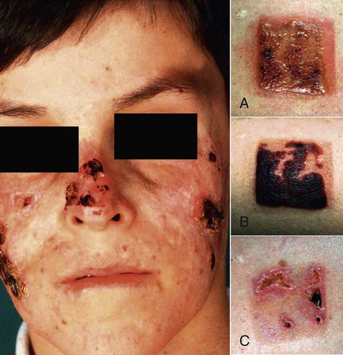
Photoprovocation testing induces typical blisters. Light tests are abnormal in the UVA range. Photographs to the right of the figure show the result of photoprovocation with UVA (three times 30 J/cm2 on 3 consecutive days): (A) after 24 hours; (B) after 48 hours; and (C) after 2 weeks.
Serology for antinuclear antibody and extractable nuclear antigens (anti-Ro, La, and Sm) will exclude bullous lupus erythematosus, which quite commonly can be ruled out by its clinical symptoms.
Rare cases have been associated with metabolic disorders, such as Hartnup disease, and so aminoaciduria should be ruled out.
Screening for EBV should be done in view of the increasing number of reports of the possible association.
Jaschke E, Hönigsmann H. Hautarzt 1981; 32: 350–3.
Successful photoprovocation with UVA in one case.
Sonnex TS, Hawk JLM. Br J Dermatol 1988; 118: 101–8.
Successful photoprovocation with UVA in several cases.
Gupta G, Man I, Kemmett D. J Am Acad Dermatol 2000; 42: 208–13.
Eight of 14 patients were sensitive in the UVA spectrum. UVA provocation tests showed a papulovesicular response in six of 14 patients.
There is now strong evidence that UVA radiation is the causal factor. In addition to reduced UVA minimal erythema dose values, repetitive broad-spectrum UVA has been shown to reproduce lesions that are clinically and histologically identical to those produced by natural sunlight and which heal with scarring. All cases seen so far by this author (H.H.) had their action spectrum in the UVA range.
Iwatsuki K, Satoh M, Yamamoto T, Oono T, Morizane S, Ohtsuka M, et al. Arch Dermatol 2006; 142: 587–95.
T cells positive for EBV-encoded small nuclear RNA (EBER) were detected, to various degrees, in cutaneous infiltrates in 28 (97%) of 29 patients, including all six patients with definite HV having a positive phototest reaction.
Hirai Y, Yamamoto T Kimura H, Ito Y, Tsuji K, Miyake T, et al. J Invest Dermatol 2012; 132: 1401–8.
The observations indicate that cutaneous lesions of both typical and severe HV are induced by EBER + T cells, associated with a larger number of EBER − cytotoxic T lymphocytes, without apparent involvement of NK cell infiltration.
Verneuil L, Gouarin S, Comoz F, Agbalika F, Creveuil C, Varna M, et al. Br J Dermatol 2010; 163: 174–82.
EBV was involved in HV pathogenesis and persisted in adult patients with HV. A positive EBV DNA load, specific to HV in the spectrum of photosensitive disorders, might be a useful biomarker in HV.
Disease in nine of 15 patients was controlled satisfactorily with high-factor broad-spectrum sunscreens and sunlight avoidance.
Disease severity was reduced in eight of 10 patients using either Coppertone® Supershade 15 or RoC® factor 10.
Most sunscreens offer good UVB but not UVA protection. The new Dundee sunscreen Reflectant Sun Screen, which is available from Tayside Pharmaceuticals, Ninewells Hospital and Medical School, Dundee, UK, offers better protection in the UVA and visible spectra.
Dawe R, Russell S, Ferguson J. Br J Dermatol 1996; 135: 1016–17.
The Museum 200 Film (manufactured by Sun Guard, Florida, USA) prevents transmission of all wavelengths less than 380 nm.
This is a clear, lightweight film that can be stuck on to any glass surface without causing visual impairment. It may be a useful adjunct in the treatment of most photodermatoses, but in some patients with HV, particularly those who are sensitive in the 380–400 nm wavelengths, it may not be beneficial.
Collins P, Ferguson J. Br J Dermatol 1995; 132: 956–63.
This was an open clinical trial in which four patients were treated on average 10 times on a daily basis. Two of these patients reported an increase in tolerance to sunshine from 1 hour to 3 to 6 hours.
Five of 15 patients who had not responded to conservative measures were treated with narrowband UVB phototherapy. In three patients there was good or moderate disease control. In the other two, narrowband UVB phototherapy was not helpful.
Jury CS, McHenry P, Burden AD, Lever R, Bilsland D. Clin Exp Dermatol 2006; 31: 196–9.
Narrowband UVB phototherapy is a useful and well tolerated treatment for children with severe or intractable inflammatory skin disease.
Two of 10 patients were treated with UVB and had improvement of their disease. There was a flare of in the one patient treated with PUVA.
It is likely that broadband UVB was used in this report, but the methodology is unclear.
One patient received PUVA therapy and had good control of his disease.
Millard TP, Hawk JL. Am J Clin Dermatol 2002; 3: 239–46.
A review of the management of various photodermatoses, with reference to the use of UVB and PUVA.
Lysell J, Wiegleb Edström D, Linde A, Carlsson G, Malmros-Svennilson J, Westermark A, et al. Acta Derm Venereol 2009; 89: 393–7.
Successful treatment in four children. Acyclovir/valacyclovir therapy is a safe treatment and should be tried. However, further studies are required to confirm these results.
Four of 10 patients were treated with either hydroxychloroquine (two patients) or chloroquine (two patients). Hydroxychloroquine 100 mg daily was ineffective, but the two patients on chloroquine (100–125 mg daily) had a reduction in the severity of their disease.
Leenutaphong V. J Am Acad Dermatol 1991; 25: 892–5.
One patient treated with chloroquine phosphate 500 mg daily did not find it beneficial.
Ketterer R, Morier P, Frenk E. Dermatology 1994; 189: 428–9.
One patient treated with chloroquine 100 mg daily and broad-spectrum sunscreens showed good disease control.
It is unclear whether the response was due to chloroquine or the sunscreen.
Bickers DR, Demar LK, DeLeo V, Poh-Fitzpatrick MB, Aronberg JM, Harber LC. Arch Dermatol 1978; 114: 1193–6.
Two patients reported an improvement of their disease with β-carotene 180 mg daily.
Halasz CLG, Leach EE, Walther RR, Poh-Fitzpatrick MB. J Am Acad Dermatol 1983; 8: 171–6.
The one patient treated with β-carotene 180 mg daily reported some subjective improvement.
Goldgeier MH, Nordlund JJ, Lucky AW, Sibrack LA, McCarthy MJ, McGuire J. Arch Dermatol 1982: 118: 588–91.
β-Carotene 120 mg daily for 2 months in the one patient was ineffective.
Blackwell V, McGregor JM, Hawk JLM. Clin Exp Dermatol 1998; 23: 73–6.
There was good control of disease with cyclosporine 3 mg/kg daily over a 2-month period.
The report does not provide details of follow-up.
Modeste AB, Cordel N, Balguerie X, Leroy D, Lauret P, Joly P. Ann Derm Venereol 2001; 128: 247–9.
One patient was successfully treated with dietary fish oil after unsuccessful treatment with antimalarials.
Rhodes LE, White SI. Br J Dermatol 1998; 138: 173–8.
Three patients were treated with dietary fish oil, five capsules daily for 3 months. A mild to good improvement was noted in two patients, but no improvement in the third. The latter patient responded to azathioprine.
Cruces MJ, de la Torre C. Photodermatology 1986; 3: 109–10.
In the one patient treated with thalidomide there was initial improvement.
In the one patient, thalidomide 100 mg daily was ineffective. Cyclosporine proved helpful at 3 mg/kg/day. Azathioprine 2.5–3.5 mg/kg daily was ineffective in the one patient studied.
Treatment of Skin Disease Comprehensive Therapeutic Strategies 4e
WhatsApp us


 High-factor broad-spectrum sunscreens and behavioral sunlight avoidance
 Narrowband UVB phototherapy (TL-01)
 PUVA
PUVA Acyclovir/valacyclovir
 Antimalarials
 β-Carotene
 Azathioprine
 Cyclosporine
 Dietary fish oil
 Thalidomide