Histoplasmosis

Published on 19/03/2015 by admin

Filed under Dermatology

Last modified 19/03/2015

Print this page

rate 1 star rate 2 star rate 3 star rate 4 star rate 5 star
Your rating: none, Average: 0 (0 votes)

This article have been viewed 1183 times

Histoplasmosis

Mahreen Ameen and Wanda Sonia Robles

Evidence Levels:  A Double-blind study  B Clinical trial ≥ 20 subjects  C Clinical trial < 20 subjects  D Series ≥ 5 subjects  E Anecdotal case reports

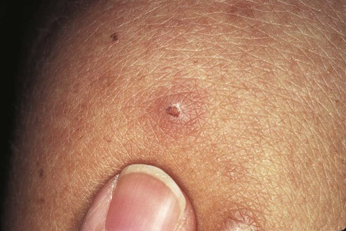
image

(From Lebwohl, M.G., 2003. The Skin and Systemic Disease: A Color Atlas, 2nd edn. Churchill Livingstone, with permission of Elsevier.)

Histoplasmosis is an endemic mycosis caused by a dimorphic fungus, Histoplasma capsulatum, of which there are two varieties that are pathogenic to humans. The most common variety worldwide is H. capsulatum var. capsulatum, which is highly endemic in the US, particularly in the Mississippi and the Ohio valleys. H. capsulatum var. duboisii is endemic only in Central and West Africa (where it coexists with H. capsulatum var. capsulatum), and it is therefore sometimes referred to as African histoplasmosis The infection is usually contracted through inhalation of spores in dry soil or bird or bat droppings. Disease progression and severity depend on the intensity of exposure to H. capsulatum and host immunity. There is a wide clinical spectrum of disease. Acute pulmonary disease is the most common presentation, and most cases are either asymptomatic or mild and self-limiting. Chronic pulmonary histoplasmosis, usually occurring in patients with underlying lung disease, produces cavities, progressing to pulmonary fibrosis. Hematogenous dissemination occurs within the first few weeks but resolves with the development of cell-mediated immunity to H. capsulatum. Progressive disseminated histoplasmosis occurs in the immunocompromised. Patient groups at risk include those with AIDS, hematological malignancies, on immunosuppressive therapy (including corticosteroids and tumor necrosis factor antagonists), transplant recipients, and infants. Disseminated histoplasmosis is an AIDS-defining illness. It is usually a reactivation of prior infection and a sign of advanced immunosuppression, generally occurring in those with CD4 counts <150 cells/mm3.

Disseminated histoplasmosis can involve any organ, most commonly presenting with hepatosplenomegaly and mucocutaneous lesions. Molluscum-like papules, nodules or ulcerative lesions affect the skin, and are usually localized to the face, upper chest, and arms. Oropharyngeal ulcers affect the buccal mucosa, tongue, gingiva, lips, pharynx, and larynx. Histoplasmosis may also present to the dermatologist as a cause of erythema multiforme or erythema nodosum. Rarely, primary cutaneous lesions may arise from direct inoculation.

Management strategy

Pulmonary histoplasmosis in the acute phase usually resolves spontaneously and only requires treatment if symptoms persist for more than 1 month. Oral itraconazole is the treatment of choice for mild to moderate histoplasmosis. Severe pulmonary infection, disseminated histoplasmosis, and infection in the immunocompromised, particularly in association with AIDS, should be treated with amphotericin B (AmB) formulations (AmB deoxycholate, liposomal AmB or AmB lipid complex). The lipid formulations carry a lower risk of nephrotoxicity but are more expensive. Treatment is commenced with amphotericin until there is clinical improvement, and then stepped down to oral itraconazole. Fluconazole and ketoconazole are second-line alternatives to itraconazole. Ketoconazole carries higher risks of adverse effects than the other azoles. The new triazoles, voriconazole and posaconazole demonstrate in vitro activity against H. capsulatum, and have been successfully used in individual cases for different forms of histoplasmosis infection.

Patients with AIDS-related histoplasmosis who undergo antiretroviral treatment have better outcomes than those who are not treated with antiretrovirals. Some clinicians defer commencement of antiretroviral therapy until there is a reduction in fungal burden to avoid precipitating immune reconstitution inflammatory syndrome (IRIS). However, IRIS is a rare complication of histoplasmosis and is not usually severe. Therefore, others advocate early antiretroviral therapy to improve cellular immunity, a key defence against H. capsulatum.

Specific investigations

The gold standard method for diagnosing histoplasmosis is the isolation of H. capsulatum from culture. The use of several specimens (sputum, bronchoalveolar lavage, skin lesions, blood, bone marrow or liver) for culture will increase the yield. Culture is highly specific but is limited by slow growth, and plates must be kept for as long as 12 weeks. Blood culture using the lysis-centrifugation system is more rapid and increases sensitivity. Histopathology is also more rapid but its sensitivity is <50% in patients with disseminated disease, and even lower in pulmonary histoplasmosis. Biopsy specimens may demonstrate the distinctive 2–4 µm oval, narrow-based budding yeasts of H. capsulatum. Serological tests are rapid but may be falsely negative in immunosuppressed patients, and during the first 2 months following exposure while antibodies are still developing. In addition, elevated antibody titers persist for several years following initial infection. Antigen detection is a rapid means of diagnosis in patients with disseminated disease. Sensitivity is greater with urine than with other body fluids (plasma, bronchoalveolar lavage fluid or cerebrospinal fluid). Antigen quantitation enables treatment response to be monitored.

In disseminated disease, general laboratory tests reveal pancytopenia, hyperbilirubinemia, elevated liver enzymes, and serum lactate dehydrogenase levels.

First-line therapies

Buy Membership for Dermatology Category to continue reading. Learn more here
imageAmphotericin B B