Published on 19/03/2015 by admin
Filed under Dermatology
Last modified 22/04/2025
This article have been viewed 2181 times
Robin A.C. Graham-Brown and Susan M. Burge
Evidence Levels: A Double-blind study B Clinical trial ≥ 20 subjects C Clinical trial < 20 subjects D Series ≥ 5 subjects E Anecdotal case reports
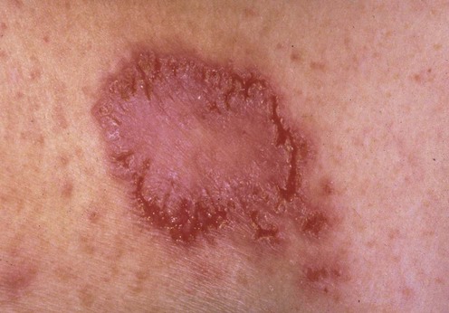
Hailey–Hailey disease (benign familial pemphigus) is a rare blistering disorder first described by two medical brothers in 1939 and characterized by recurrent vesicles and erosions, particularly involving flexural areas. Signs usually appear for the first time from the late teens to the third or fourth decades. The disease is generally of relatively limited extent, although widespread and severe involvement can occur. The most commonly affected sites are the axillae, groins, and other intertriginous areas such as the inframammary folds. The neck may be affected where the collar rubs. Lesions may also occur on the trunk. The disease may koebnerize into other inflammatory dermatoses, such as seborrheic dermatitis.
Hailey–Hailey disease is a dominantly inherited condition. The defective gene, ATP2C1, encodes an ATP-powered calcium channel pump on the Golgi membrane. There are clinical and histopathologic similarities with Darier disease and Grover disease (see relevant chapters).
The lesions of Hailey–Hailey disease are frequently precipitated by friction. Infections with various bacteria, yeasts, and viruses also appear to be aggravating factors in some patients. Thus, avoidance of precipitating trauma and skin infections can help to reduce the frequency and severity of outbreaks.
Simple anti-infective agents, topical or systemic, reduce the severity of exacerbations and remain the mainstay of treatment. Topical tetracyclines, fusidic acid, and imidazoles have all been recommended. Tetracyclines and semi-synthetic penicillins are probably the best systemic agents. If secondary infection with herpes simplex is suspected, appropriate oral antiviral therapy should be instituted.
Combining anti-infective therapy with topical corticosteroids seems to be particularly helpful, but corticosteroids alone may reduce the severity of lesions. Generally, moderate to potent agents are required, though some patients gain benefit from milder preparations. Caution should be exercised with long-term use because the skin of the axillae and groins is prone to atrophy. Topical calcineurin inhibitors tacrolimus and pimecrolimus, either alone or in combination with topical corticosteroids, have been reported to be effective and are certainly worth trying, although some authors dispute this. Some success has also been recorded with calcitriol. Analgesia is important.
Patients with Hailey–Hailey disease are at high risk of developing contact allergic dermatitis, and patch testing should be performed if there is a poor response to therapy.
Electron beam therapy has been used occasionally for recalcitrant disease.
Patients with major exacerbations may benefit from a short course of systemic corticosteroids, but control seldom lasts, and there may be a rebound of the disease on withdrawal.
Systemic alternatives that have been tried in severe disease include dapsone, cyclosporine, methotrexate, and retinoids, but there is little evidence for their effectiveness beyond anecdotal case reports.
There may be a place for surgical approaches to disease of limited extent, including excision and grafting, dermabrasion, the use of CO2 and other lasers. Injections of botulinum toxin have been advocated to reduce sweating in axillary Hailey-Hailey disease.
Unsurprisingly, perhaps, reports of treatment with biologic agents are beginning to appear in mainstream journals. It is much too soon to recommend their use routinely, even in severe disease.
Biopsy
Microbiologic cultures for bacteria, yeast, and herpes virus
Consider patch testing to topical medicaments
Burge SM. Br J Dermatol 1992; 126: 275–82.
In this series 86% of patients found combinations of topical corticosteroids and anti-infective agents helpful, especially if they were started as soon as the patient noticed the onset of discomfort.
This helpful article remains a key review of clinical and therapeutic aspects of Hailey–Hailey disease.
Sand C, Thomsen HK. Arch Dermatol 2003; 139: 1401–2.
Laffitte E, Panizzon RG. Arch Dermatol 2004; 140: 1282.
Umar SA, Bhattacharjee P, Brodell RT. J Drugs Dermatol 2004; 3: 200–3.
These authors recommend alternating clobetasol and tacrolimus.
Tchernev G, Cardoso JC. Rev Med Chil 2011; 139: 633–7.
A report of success with pimecrolimus
Bianchi L, Chimenti M, Giunta A. J Am Acad Dermatol 2004; 51: 475–6.
Marsch WC, Stüttgen G. Br J Dermatol 1978; 99: 553.
The use of systemic corticosteroids was successful in controlling particularly extensive Hailey–Hailey disease, but cessation of therapy resulted in significant rebound of the disease.
Sire DJ, Johnson BL. Arch Dermatol 1971; 103: 262.
Jitsukawa K, Ring J, Weyer U, Kimmig W, Radloff H. J Am Acad Dermatol 1992; 27: 625–6.
Ormerod AD, Duncan J, Stankler L. Br J Dermatol 1991; 124: 299–300.
Berth-Jones J, Smith SG, Graham-Brown RA. Clin Exp Dermatol 1995; 20: 70–2.
Konrad H, Karamfilov T, Wollina U. J Cosmet Laser Ther 2001; 3: 181–4.
Lapiere JC, Hirsh A, Gordon KB, Cook B, Montalvo A. Dermatol Surg 2000; 26: 371–4.
Koeyers WJ, Van Der Geer S, Krekels G. J Dermatol Treat 2008: 19: 251–4.
These papers all report small numbers or individual case reports of apparent success. Tacrolimus, pimecrolimus and calcitriol are at least safe. Dapsone is generally safe and may be worth a try. Cyclosporine has a long, daunting list of side effects, but can be used safely if doses do not exceed 5 mg/kg and patients are properly monitored. There are now a number of reports suggesting that Botulinum toxin may be a useful adjuvant to other therapeutic modalities.
Shelley WB, Randall P. Arch Dermatol 1969; 100: 275.
Zachariae H. J Am Acad Dermatol 1992; 27: 136.
LeBlanc KG Jr, Wharton JB, Sheehan DJ. Skinmed 2011; 9: 263–4.
Kartamaa M, Reitamo S. Arch Dermatol 1992; 128: 646.
Awadalla F, Rosenbach A. J Cosmet Laser Ther 2011; 13: 191–2.
Surgery must remain a last resort in this condition, especially as many authors report some recurrences, either around the edges of the treated areas or on further friction or trauma. Treatment is painful and the trauma of dressings may exacerbate the disease.
Narbutt J, Chrusciel A, Rychter A, Fijuth J, Lesiak A, Sysa-Jedrzejowska A. Acta Derm Venereol 2010; 90: 179–82.
Vilarinho C, Ventura F, Brito C. J Eur Acad Dermatol Venereol 2010; 24: 106.
Hunt MJ, Salisbury EL, Painter DM, Lee S. Australas J Dermatol 1996; 37: 196–8.
Berger EM, Galadari HI, Gottlieb AB. J Drugs Dermatol 2007; 6: 734–6.
Dammak A, Camus M, Anyfantakis V, Guillet G. Br J Dermatol 2009; 161: 967–8.
Norman R, Greenberg RG, Jackson JM. J Am Acad Dermatol 2006; 54(3 Suppl 2): S139–42.
Hurd DS, Johnston C, Bevins A. Br J Dermatol 2008; 158: 399–401.
Fernandez Guarino M, Ryan AM, Perez-Garcia B, Arrazola JM, Jaen P. J Dermatol Treat 2008; 19: 288.
An interesting idea but the treatment appeared to be extremely painful and the outcomes variable.
Treatment of Skin Disease Comprehensive Therapeutic Strategies 4e
WhatsApp us


 Anti-infectives and antibiotics
 Topical corticosteroids
 Topical calcineurin inhibitors
 Calcitriol
 Systemic corticosteroids
 Dapsone
 Cyclosporine
 Botulinum toxin
 Surgical excision (with healing by secondary intention or by grafting)
 Dermabrasion
 Laser therapy
 Superficial irradiation (Grenz rays or electron beam)
 Methotrexate
 Oral retinoids
 5-Fluorouracil
 Biologics
 Photodynamic therapy