Published on 19/03/2015 by admin
Filed under Dermatology
Last modified 22/04/2025
This article have been viewed 1961 times
Nevianna Tomson and Susan E. Handfield-Jones
Evidence Levels: A Double-blind study B Clinical trial ≥ 20 subjects C Clinical trial < 20 subjects D Series ≥ 5 subjects E Anecdotal case reports
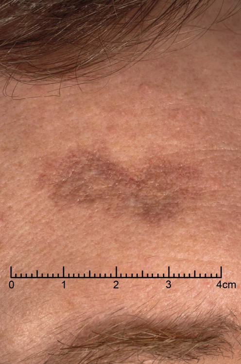
(© Addenbrooke’s Hospital.)
Granuloma faciale is a rare, benign, chronic inflammatory dermatosis caused by a localized form of cutaneous vasculitis. It presents primarily in middle-aged Caucasian males, usually as a single lesion on the face. Multiple lesions occur in up to a third of patients and there are isolated reports of similar conditions affecting the eye and upper airways.
Lesions are red-brown, violaceous or flesh-colored plaques or nodules with accentuation of follicular openings. Clinical diagnosis is difficult. Differential diagnosis includes sarcoid, lupus, lymphocytoma cutis, persistent insect bite reactions, and lymphoma. The histological differential diagnosis includes erythema elevatum diutinum, and angiolymphoid hyperplasia with eosinophilia.
Granuloma faciale is a chronic condition; spontaneous remission is unusual. Lesions are usually asymptomatic, but treatment is needed to reduce disfigurement. It is notoriously resistant to treatment and, because of the rarity of the condition, there are no formal trials of therapy. Treatment modalities can be divided into destructive techniques and anti-inflammatory approaches. The optimal treatment depends on the size, site, and thickness of the lesions. For isolated or small numbers of lesions intralesional steroid or destructive treatments such as cryotherapy, laser or surgical excision can be used. For multiple or widespread lesions systemic treatment, such as dapsone or clofazimine, can be considered.
Cosmetic camouflage can be helpful for some patients with flatter lesions.
Skin biopsy
Hematology (complete blood count)
Histological findings include a dense eosinophilic and neutrophilic infiltrate, often perivascular, affecting the upper and sometimes deep dermis. The epidermis is spared and there is a Grenz zone. Telangiectasia is common. Vasculitis with leukocytoclasis is reported. Dermal fibrosis is often seen.
Ortonne N, Wechsler J, Bagot M, Grosshans E, Cribier B. J Am Acad Dermatol 2005; 53: 1002–9.
Peripheral blood eosinophilia is sometimes found.
Arundell FD, Burdick KH. Arch Dermatol 1960; 82: 437–8.
This paper reports response to dexamethasone, but triamcinolone acetonide and triamcinolone hexacetonide have also been used. Patients should be warned of the risk of skin atrophy and pigment change.
Panagiotopoulos A, Anyfantakis V, Rallis E, Chasapi V, Stavropoulos P, Boubouka C, et al. Br J Dermatol 2006; 154: 357–60.
Nine patients were treated with either spray or closed probe cryotherapy. The open-spray technique was given as one or two freeze–thaw cycles of 20–30 seconds. The cryoprobe was given as one to three freeze–thaw cycles of 15–20 seconds. Patients were re-treated after 1 and 3 months. All were clear of disease activity at 6 months. One patient developed severe inflammatory reaction with blistering. Two had hypopigmentation, but this resolved in 4 months. There was no recurrence within 2 to 4 years’ follow-up.
Dowlati B, Firooz A, Dowlati Y. Int J Dermatol 1997; 36: 548–51.
Cryotherapy for 20–30 seconds was followed by triamcinolone acetonide 5 mg/mL intralesionally.
Cheung S-T, Lanigan SW. Clin Exp Dermatol 2005; 30: 373–5.
Four patients who had all failed with cryotherapy were treated with Candela Vbeam PDL at 595 nm. In two patients the lesions resolved. Nasal lesions, especially flatter ones, seem to respond better to treatment.
Ludwig E, Allam J-P, Bieber T, Novak N. Br J Dermatol 2003; 149: 634–7.
A report of two cases of sizeable facial lesions responding rapidly to laserscope potassium-titanyl-phosphate 532 nm laser.
Apfelberg DB, Druker D, Maser MR, Lash H, Spence B, Deneau D. Arch Dermatol 1983; 119: 573–6.
A report of three cases responding to argon laser with no recurrence from five to 23 months. A ‘white, collagenous scar’ resulted.
Wheeland RG, Ashley JR, Smith DA, Ellis DL, Wheeland DN. J Dermatol Surg Oncol 1984; 10: 730–3.
A single treatment resulted in healing with no discernible scar. There was no recurrence at 1 year.
Phillips DK, Hymes SR. Arch Dermatol 1994; 130: 1436–7.
Although surgery is mentioned in many papers, recurrence can occur even after full-thickness excision and grafting.
Dinehart SM, Gross DJ, Davis CM, Herzberg AJ. Arch Otolaryngol Head Neck Surg 1990; 116: 849–51.
Combined electrosurgery and dermabrasion was compared with CO2 laser treatment in a patient with two similar lesions. Skin texture at 6 weeks was better on the laser-treated side, but laser treatment was more time-consuming.
Van de Kerkhof PCM. Acta Derm Venereol 1994; 74: 61–2.
A 4 cm plaque showed ‘impressive improvement’ with dapsone 200 mg daily. Dapsone needs careful monitoring, and many patients would not tolerate 200 mg daily.
Many authors mention dapsone as a treatment that has been tried but failed.
Eetam I, Ertekin B, Unal I, Alper S. J Dermatol Treat 2006; 17: 238–40.
A case report of a 1 cm × 5 cm lesion on the central face showing ‘dramatic recovery’ following pimecrolimus cream 1% twice daily for 2 months.
Marcoval J, Moreno A, Bordas X, Peyri J. J Am Acad Dermatol 2006; 55: S110–11.
This and other papers describe response to topical tacrolimus, sometimes within a few months. In some patients lesions had previously failed to respond to other therapies. Remission for up to 2 years is described.
The possible risks of topical calcineurin inhibitors in sun-exposed sites must be considered.
Gomez-de la Fuente E, del Rio R, Guerra A, Rodriguez-Peralto JL, Iglesias L. Acta Derm Venereol 2000; 80: 144.
A patient with a 10-year history of histologically proven disease on the nose was treated with 300 mg clofazimine once daily for 5 months, with ‘remarkable improvement.’ Two similar reports are cited.
Hudson LD. J Am Acad Dermatol 1983; 8: 559.
A 62-year-old man with a 1-month history of biopsy-proven granuloma faciale affecting the nasal alae responded to 24 J UVA given over 10 weeks, showing a very marked improvement with no evidence of residual lesion at 6 months.
There are old reports of treatment with intralesional gold and bismuth, radiotherapy, oral colchicine, isoniazid, potassium arsenite, testosterone, and antimalarials, but within the last 25 years there have been no reports of successful response to these agents.
Treatment of Skin Disease Comprehensive Therapeutic Strategies 4e
WhatsApp us