Published on 19/03/2015 by admin
Filed under Dermatology
Last modified 22/04/2025
This article have been viewed 2728 times
Carlo Gelmetti
Evidence Levels: A Double-blind study B Clinical trial ≥ 20 subjects C Clinical trial < 20 subjects D Series ≥ 5 subjects E Anecdotal case reports
Gianotti–Crosti syndrome was described in 1955 by Ferdinando Gianotti as an exanthematic papular rash symmetrically distributed on the face, buttocks, and extremities. The disease had mild constitutional symptoms, was not preceded by fever or severe prodromes and had a benign course. The term ‘Gianotti–Crosti syndrome’ (GCS) is now used to include all eruptive acrolocated dermatoses characterized by papular or papulovesicular lesions caused by microorganisms or vaccines. The first association was with hepatitis B (HBV). In addition to viruses, GCS has been reported to occur after immunization with live, killed, and recombinant vaccines. As the number of immunizations increases and more combinations will be available, an increasing number of these post-immunization cases are expected.
Synonyms include papular acrodermatitis of childhood, infantile papular acrodermatitis, papulovesicular acrolocated syndrome, Crosti–Gianotti disease, Gianotti disease.
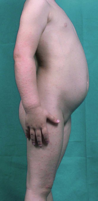
The diagnosis of GCS is clinical. Like other viral exanthems, it mainly affects children of preschool age, though adult cases have occurred. The typical eruption consists of monomorphic, lentil-sized lesions symmetrically distributed on the face, buttocks, and limbs. The lesions are papular or papulovesicular, sometimes edematous, and rarely purpuric. The trunk, antecubital, and popliteal surfaces are usually spared, even though a transient rash may be noted on the trunk in the early stage. Mucous membranes are not affected. The eruption develops within a week, typically beginning on the thighs and buttocks, then involving the extensor aspects of the arms, and finally the face. Individual lesions are a few millimeters in diameter (bigger lesions are uncommon), and vary in color from rose to red-brown. The pruritus is usually mild, and excoriation is rare. The lesions fade in 3 to 4 weeks with mild desquamation, and relapse is rare. A longer course of up to 6 to 8 weeks is occasionally seen. Inguinal and axillary lymphadenopathy is common.
In cases associated with HBV, the hepatitis begins at the same time as, or 1 to 2 weeks after, the onset of the GCS. The liver is usually enlarged but not tender; jaundice is exceptional. There are high levels of liver enzymes, and the viral markers become detectable in the serum depending on the duration of infection. Complications are rare, although some patients developed chronic periportal hepatitis. Abnormalities in the peripheral blood are inconsistent: there may be a leukopenia or a slight leukocytosis with 2–15% of monocytes; the erythrocyte sedimentation rate is not raised.
In HBV-negative cases, hepatomegaly and liver function abnormalities, if present, are mild, probably because some viruses are considered minor hepatitic viruses (e.g., Epstein–Barr virus, EBV).
There is no specific recommended treatment for GCS. However, when itching is disturbing, oral antihistamines or topical antipruritics can be used symptomatically. Although topical and systemic corticosteroids are sometimes used, the benefit is not established, since corticosteroids could prolong or delay the recovery of the disease.
In cases associated with HBV, the hepatitis can be treated with Interferon Alpha-2b or pegylated interferon with or without nucleoside analogs such as adefovir, entecavir, telbivudine, tenofovir, and lamivudine, the last approved also for children. It is important to investigate and treat other family members who may be carriers of the HBV or may benefit from vaccination. Vaccination against HBV, which will potentially eradicate all cases of GCS due to this virus, seems also effective in reducing the incidence of hepatocellular carcinoma. Other infective causes may also require treatment if identified.
Liver enzymes
Viral serology for Epstein–Barr virus, hepatitis B, hepatitis A, cytomegalovirus
Total IgE (PRIST) and specific IgE (RAST)
Other possible causes include coxsackie virus, adenovirus, enterovirus, human herpes virus 6, reovirus, varicella, roseola, rotavirus, respiratory syncytial virus, Lyme borreliosis, Mycoplasma pneumoniae, meningococcal infection, etc., and immunization.
Pise GA, Vetrichevvel TP, Agarwal KK, Thappa DM. Indian J Dermatol Venereol Leprol 2007; 73: 123–4.
A 9-year-old boy, treated with multiple transfusions for his disease, presented with GCS. Liver function tests showed indirect hyperbilirubinemia and mildly elevated liver enzymes. HBsAg was detected. The patient was managed with antihistamines and skin lesions cleared in 2 weeks. He had no recurrences in the subsequent 6 months of follow-up.
Karakas M, Durdu M, Tuncer I, Cevlik F. J Dermatol 2007; 34: 117–20.
A 5-year-old boy presented with a 1-month history of GCS. The lesions had appeared 3 weeks after a first dose of HBV recombinant vaccine. Antibody to HBsAg was positive; other viral serologic tests, including CMV, EBC, HAV, HCV, and EBV, were negative. The patient was treated with an emollient and the eruption subsided within 4 weeks.
Retrouvey M, Koch LH, Williams JV. Pediatr Dermatol 2012; 29: 666–8.
A 19-month-old boy developed GCS 2 days after immunization with measles, mumps and rubella vaccine through the right thigh, and the varicella virus live vaccine and diphtheria, tetanus, and pertussis vaccines through the left thigh. It is suggested that combined vaccinations carry a higher risk for precipitating GCS or that the risk of developing GCS is greater when an active viral infection is present at the time of vaccination.
Kwon NH, Kim JE, Cho BK, Park HJ. Ann Dermatol 2011; 23: 554–5.
A 7-year-old boy presented with a 2-day history of GCS appeared 5 days after a single dose of an H1N1 vaccine. The patient was otherwise healthy. Routine tests were within normal ranges and serological tests for CMV, EBV, HAV, HBV, enterovirus pool, parvovirus B19, and M. pneumoniae were negative. Screening for the H1N1 was negative. Additionally, the result of polymerase chain reaction of nasopharyngeal secretion was negative for H1N1 virus.
Brandt O, Abeck D, Gianotti R, Burgdorf W. J Am Acad Dermatol 2006; 54: 136–45.
EBV is now the most common cause of GCS. Other viruses have been connected, including HVA, CMV, HHV6, coxsackie A16, B4, and B5; rotavirus; parvo B19; respiratory syncytial virus; mumps virus and parainfluenza virus types 1 and 2. Apparently HIV can also be associated. Bacteria (Bartonella henselae, M. pneumoniae, β-hemolytic streptococci and Borrelia burgdorferi) also appear capable of triggering GCS. Despite the proven connection between HBV and GCS, immunization against HBV only rarely causes GCS.
Cocciolone R, Morey A, Panasiuk P, Whitfeld MJ. Australas J Dermatol 2011; 52: 32–6.
The authors describe an atypical GCS in two unrelated patients with coexistent hepatitis B and HIV infection. The protracted duration of the exanthem, between 4 and 25 months, may have been related to their underlying HIV infection. Immunoperoxidase studies suggest the presence of hepatitis B surface antigens within the vessels of both lesional and perilesional skin, providing further support for the proposed, immune-mediated pathogenesis of paraviral eruptions.
Ricci G, Patrizi A, Neri I, Specchia F, Tosti G, Masi M. Acta Derm Venereol 2003; 83: 202–5.
In 29 children affected by GCS and 59 age- and sex-matched controls, the presence of atopic dermatitis (24.1%) was significantly higher (p < 0.005) than in the control group (6.8%). In addition, the percentage of patients with total IgE >2 SD for age was higher than in controls (27.6% vs 13.7%), as was the percentage with specific IgE present (31% vs 17.2%).
Linke M, Géraud C, Schneider SW, Goerdt S, Utikal J. Acta Derm Venereol 2011; 91: 491–4.
A 7-year-old girl presented with moderately pruritic lesions consistent with GCS. In addition, tense blisters were found on the hands and feet. The face and mucous membranes were not affected. Oral corticosteroid and an oral antihistamine were administered, and an antiseptic cream (triclosan 2%) was applied topically. The skin lesions subsided within a week without relapse.
Metelitsa AI, Fiorillo L. J Am Acad Dermatol 2011; 65: 876–7.
A previously healthy 2-year-old boy presented with GCS. Two weeks after onset of the eruption, he was immunized with influenza vaccination and new lesions appeared at the immunization site. Laboratory revealed a positive urine culture for CMV, and positive IgM for EBV. He was treated with 2.5% hydrocortisone cream, and the eruption resolved. Twelve months later, he presented with a similar eruption at the site of a recent intramuscular influenza vaccination. As GCS is a mild and self-limiting disease, further vaccinations are not contraindicated.
Baldari U, Cattonar P, Nobile C, Celli B, Righini MG, Trevisan G. Acta Derm Venereol 1996; 76: 242–3.
One patient was treated with ceftriaxone (500 mg IM daily for 30 days) and the second with josamicin (250 mg/day for 14 days).
Zawar V, Chuh A. J Dermatol Case Rep 2008; 2: 63–6.
A 6-year-old girl presented with a history of a generalized intensely pruritic rash consistent with GCS, following a febrile illness with common cold symptoms. Remission was not achieved despite of several medications: topical emollients, topical and systemic corticosteroids, topical and systemic antibiotics, and oral histamines. A course of oral ribavirin, a drug said to be efficacious against influenza virus, parainfluenza viruses, respiratory syncytial virus, coronavirus, and hepatitis C virus at a dose of 300 mg daily for 5 days lead to dramatic remission 5 days later.
Treatment of Skin Disease Comprehensive Therapeutic Strategies 4e
WhatsApp us


 Systemic antihistamines
 Systemic corticosteroids
 Topical corticosteroids
 Topical antiseptics
 Topical antipruritics
 Emollients
 Vitamin C
 Systemic antibiotics
 Ribavirin
 Interferon-α
 Nucleoside analogs
 Hepatitis B vaccine