[level-membership-for-dermatology-category]
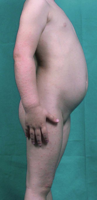
Gianotti–Crosti syndrome
Specific investigations


First-line therapies
 Systemic antihistamines
 Systemic corticosteroids
 Topical corticosteroids
 Topical antiseptics
 Topical antipruritics
 Emollients
 Vitamin C
 Systemic antibiotics
 Ribavirin
 Interferon-α
 Nucleoside analogs
 Hepatitis B vaccine
[/level-membership-for-dermatology-category][not-level-membership-for-dermatology-category]
Gianotti–Crosti syndrome
[level-membership-for-dermatology-category]
[/level-membership-for-dermatology-category][not-level-membership-for-dermatology-category]
