Gaucher Disease

Published on 21/04/2017 by admin

Filed under Pathology

Last modified 22/04/2025

Print this page

rate 1 star rate 2 star rate 3 star rate 4 star rate 5 star
Your rating: none, Average: 3 (26 votes)

This article have been viewed 4575 times

 Results in hepatosplenomegaly and pancytopenia

• Variable disease progression
• Enzyme replacement therapy represents mainstay therapy

image Early diagnosis is crucial to improving outcome

Microscopic

• Accumulation of glucocerebroside in Kupffer cells and portal tract macrophages results in uniquely linear amphophilic cytoplasm

• Affected cells show characteristic linear, tissue paper-like, fibrillary, or corrugated amphophilic cytoplasm
image Cells are positive for PAS-D
• Hepatocytes and other hepatic structures are spared

Ancillary Tests

• Electron microscopy demonstrates intralysosomal compact long tubular structures

Top Differential Diagnoses

• Niemann-Pick disease

image Enlarged Kupffer cells but foamy cytoplasm with small round vacuoles
• Wolman disease

image Frozen section-stained slide stained with oil red O reveals abundant lipid, and polarized light highlights needle-shaped cholesterol crystals
• Pseudo-Gaucher cells in bone marrow biopsy
image
Kupffer Cell Clusters in Hepatic Lobule
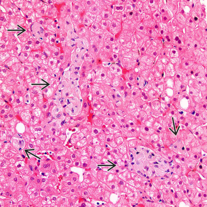
Clusters of enlarged foamy Kupffer cells image with fibrillary, amphophilic cytoplasm are seen in the hepatic lobules. The clusters vary in size and shape.

image
Gaucher Cells With Linear Cytoplasmic Striations
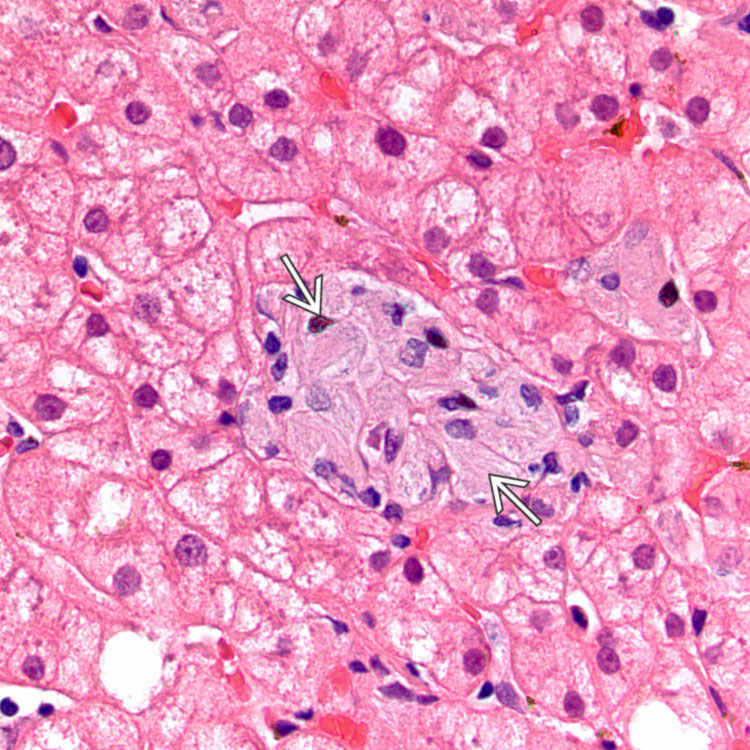
These Gaucher cells demonstrate the characteristic fibrillary or striated cytoplasm reminiscent of wrinkled tissue paper image .
image
Gaucher Cells Compared to Hepatocytes
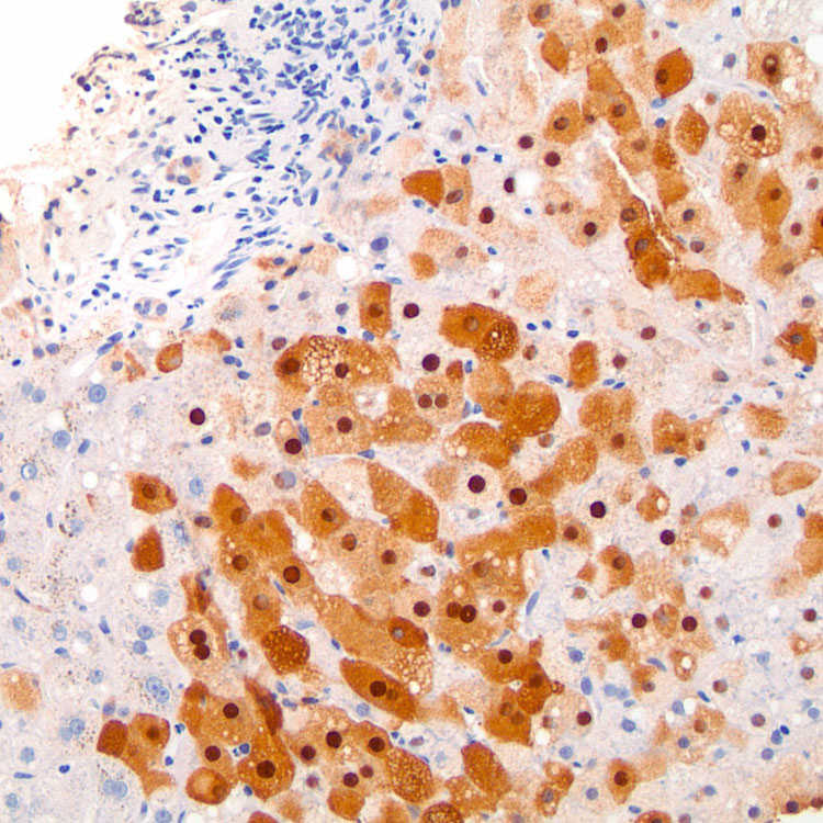
In Gaucher disease, the characteristic Kupffer cell glucocerebroside inclusions exhibit a fibrillary or striated appearance image
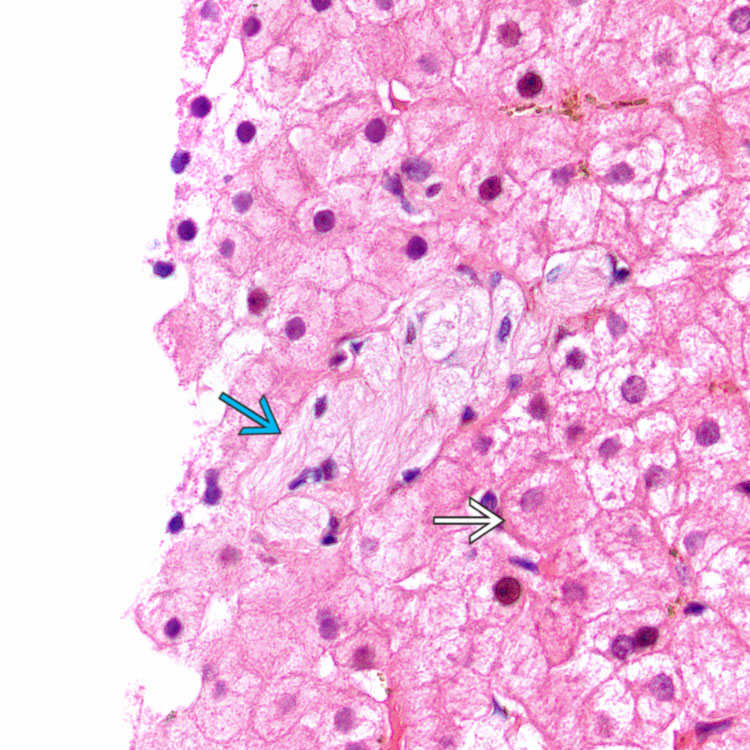
. These cells have small, eccentric, and often wrinkled nuclei. In contrast, hepatocytes have more eosinophilic, granular cytoplasm and rounded nuclei image .
image
Electron Micrograph of Gaucher Cell
Electron microscopy of a Gaucher cell demonstrates lysosomes containing numerous elongated tubular structures image 
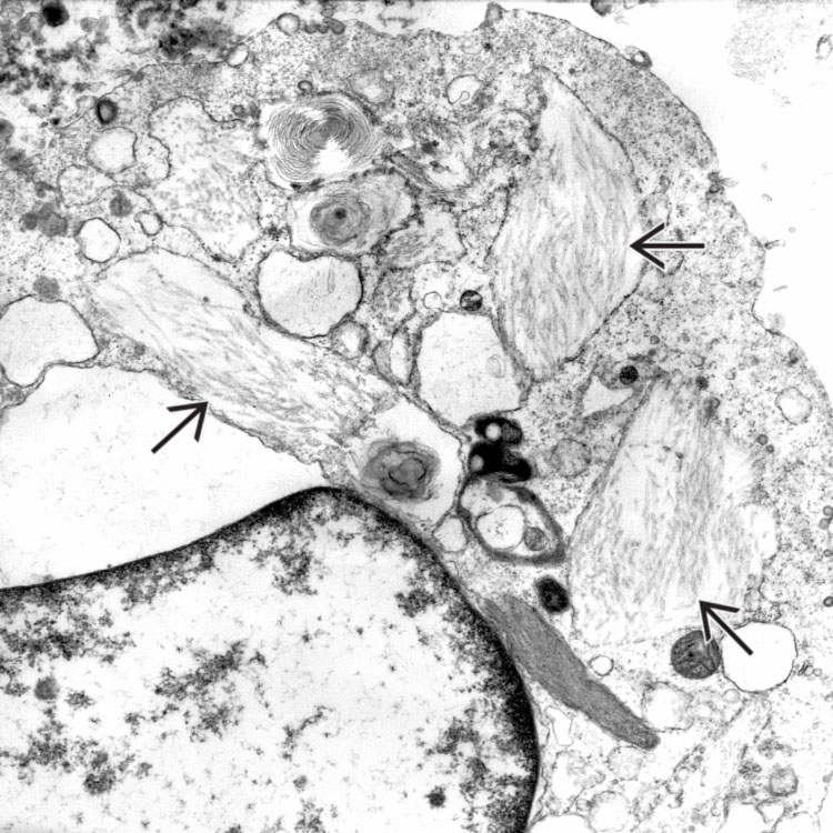
that are arranged in compact bundles. (Courtesy Z. Laszik, MD, PhD.)

TERMINOLOGY

Synonyms

• Glucocerebrosidase deficiency

Definitions

• Inherited deficiency of lysosomal enzyme glucocerebrosidase

ETIOLOGY/PATHOGENESIS

Inborn Error of Metabolism

• Most common lysosomal glycolipid storage disorder

image Acid β-glucosidase (glucocerebrosidase) enzyme deficiency
image Accumulation of glucocerebroside (also called glucosylceramide) in phagocytic cells

Autosomal Recessive Trait

• Mutation in GBA, encoding acid β-glucosidase, on 1q21
• Homozygotes are affected

CLINICAL ISSUES

Site

• Visceral organs such as liver, spleen, and lung
• Bone marrow and bone
• Variably present central nervous system involvement

Presentation

• Hepatosplenomegaly

• Pancytopenia
image Anemia
image Thrombocytopenia
• 3 classic clinical variants

image Type 1 (nonneuronopathic)

– Most common form
– 55-60% diagnosed before 20 years of age
– 30% diagnosed before 10 years of age
image Types 2 and 3 (neuronopathic)

Treatment

• Enzyme replacement therapy

Prognosis

• Early diagnosis is crucial to improving outcome
• Variable disease progression

MICROSCOPIC

Histologic Features

• Accumulation and storage of glucocerebroside in Kupffer cells and macrophages

image Spares hepatocytes
• Characteristic linear, tissue paper-like, fibrillary, or corrugated amphophilic cytoplasm

image Cells are positive for PAS-D
• Effects secondary to sinusoidal Kupffer cell involvement are rare

image May see atrophic hepatocytes and eventual sinusoidal fibrosis
image Micronodular cirrhosis and hepatocellular carcinoma have been reported

ANCILLARY TESTS

Electron Microscopy

• Enlarged Kupffer cells and portal tract macrophages

image Cytoplasm expanded by enlarged, irregular, single membrane-bound lysosomes
image Lysosomes filled with stored glucocerebroside substance
– Compact long tubular structures or finely reticular to flocculent material

DIFFERENTIAL DIAGNOSIS

Niemann-Pick Disease

• Enlarged Kupffer cells but foamy cytoplasm with small round vacuoles

image Enlarged pale hepatocytes may be indistinguishable from Kupffer cells
• Distinguished by electron microscopy

image Concentric lamellar lipid inclusions in lysosomes of hepatocytes and Kupffer cells in Niemann-Pick disease

Wolman Disease

• Histologically similar to Niemann-Pick disease

image Frozen section-stained slide stained with oil red O reveals abundant lipid, and polarized light highlights needle-shaped cholesterol crystals
• Distinguished by electron microscopy

image Membrane-bound lipid droplets in hepatocytes and Kupffer cells
image Cholesterol crystals in hepatocytes and Kupffer cells

Pseudo-Gaucher Cells in Bone Marrow Biopsy

• Resulting from high rate of cell turnover
• Can be found in chronic myelogenous leukemia

DIAGNOSTIC CHECKLIST

Pathologic Interpretation Pearls

• Accumulation of glucocerebroside in Kupffer cells and macrophages but not hepatocytes
• Electron microscopy demonstrates intralysosomal rod-shaped inclusions

SELECTED REFERENCES

1.Chen, M, et al. Gaucher disease: review of the literature. Arch Pathol Lab Med. 2008; 132(5):851–853.

2.Niederau, C, et al. Gaucher’s disease: a review for the internist and hepatologist. Hepatogastroenterology. 2000; 47(34):984–997.

3.Pastores, GM. Gaucher’s Disease. Pathological features. Baillieres Clin Haematol. 1997; 10(4):739–749.

4.Lee, RE. The pathology of Gaucher disease. Prog Clin Biol Res. 1982; 95:177–217.

5.James, SP, et al. Liver abnormalities in patients with Gaucher’s disease. Gastroenterology. 1981; 80(1):126–133.

6.Lee, RE, et al. Gaucher’s disease. I. Modern enzymatic and anatomic methods of diagnosis. Arch Pathol Lab Med. 1981; 105(2):102–104.

7.Lee, RE, et al. Gaucher’s disease: clinical, morphologic, and pathogenetic considerations. Pathol Annu. 1977; 12(Pt 2):309–339.

8.Hibbs, RG, et al. A histochemical and electron microscopic study of Gaucher cells. Arch Pathol. 1970; 89(2):137–153.