Gait Disorders

Published on 12/04/2015 by admin

Filed under Neurology

Last modified 22/04/2025

Print this page

rate 1 star rate 2 star rate 3 star rate 4 star rate 5 star
Your rating: none, Average: 0 (0 votes)

This article have been viewed 7339 times

Chapter 22 Gait Disorders

The maintenance of an upright posture and the act of walking are among the first, and ultimately the most complex, motor skills humans acquire. From an early age, walking skills are modified and refined. In later years, the interplay between voluntary and automatic control of posture and gait provides a rich and complex repertoire of motion that ranges from walking to running, hopping, dancing, and so on. The pattern of walking may be so distinctive that individuals can be recognized by the characteristics of their gait. Many diseases of the nervous system are identified by the disturbances of gait and posture they produce.

Physiological and Biomechanical Aspects of Gait

Humans assume a stable upright posture before beginning to walk. Mechanical stability when standing is based on musculoskeletal linkages between the trunk and legs. Dynamic equilibrium during walking is maintained by coordinated synergies of axial and proximal limb muscle contraction and a hierarchy of postural reflexes and responses. The latter include automatic righting reflexes keeping the head upright on the trunk, supporting reactions controlling antigravity muscle tone, anticipatory (feed-forward) postural reflexes occurring before limb movement, and reactive (feedback) postural adjustments counteracting body perturbations during movement. Postural responses are also modified by voluntary control according to the circumstances, such as rescue reactions to preserve the upright posture (a step or windmill arm movements), and protective reactions to prevent injury (an outstretched arm to break a fall). Postural reflexes and responses are generated by the integration of visual, vestibular, and proprioceptive inputs in the context of voluntary intent and ongoing changes in the environment in which the subject is moving. Once the trunk is upright and stable, locomotion can begin. The initiation of gait is heralded by a series of shifts in the center of pressure beneath the feet during the course of an anticipatory postural adjustment—first posteriorly, then laterally toward the stepping foot, and finally toward the stance foot to allow the stepping foot to swing forward. This sequence is then followed by the stereotyped stance, swing, and step phases of the gait cycle.

Anatomical Aspects of Gait

The neuroanatomical structures responsible for equilibrium and locomotion in humans are inferred from studies in lower species that suggest two basic systems. First, brainstem locomotor centers (Takakusaki, 2008) project through descending reticulospinal pathways into the ventromedial spinal cord. Stimulation of brainstem locomotor centers results in an increase in axial and limb muscle tone to assume an upright posture before stepping begins. Second, assemblies of spinal interneurons (central pattern generators or spinal locomotor centers) activate motoneurons of limb and trunk muscles in a patterned and repetitive manner to drive stepping movements and stimulate propriospinal networks that link the trunk and limbs to facilitate the synergistic coordinated limb and trunk movements of locomotion. In quadrupedal animals, spinal locomotor centers are capable of maintaining and coordinating rhythmic stepping movements after spinal transection. The cerebral cortex and corticospinal tract are not necessary for experimentally induced locomotion in quadrupeds but are required for precision stepping. The isolated spinal cord in humans can produce spontaneous movements, but it cannot generate rhythmic stepping or maintain truncal balance, indicating that brainstem and higher cortical connections are necessary for bipedal walking in humans. In monkeys, spinal stepping requires preservation of the descending ventromedial brainstem and ventrolateral spinal motor pathways. Lesions of the medial brainstem in monkeys interrupt descending reticulospinal, vestibulospinal, and tectospinal systems, resulting in dysequilibrium.

The control of posture and locomotion in humans may be mediated by similar networks, with an additional level of supraspinal control needed to maintain bipedal stance and the complex repertoire of gait. Frontal cortex, via corticospinal tract and the basal ganglia, provide the signals to brainstem locomotor centers such as the pedunculopontine and adjacent nuclei of the midbrain to increase postural tone and commence rhythmic stepping. The corticospinal tract also projects to spinal cord motoneurons, enabling precision foot movements for stepping or dancing. The parietal cortex integrates sensory inputs indicating where the individual is in space, the relationship to gravitational forces, the speed and direction of movement, and the characteristics of the terrain and environment. The cerebellum modulates the rate, rhythm, amplitude, and force of stepping and also contributes to the medial brainstem efferent system controlling equilibrium and truncal posture through projections from the flocculonodular and anterior lobes. Much of the automatic control of truncal posture and walking in humans must be derived from integration of these various functions at the highest levels of motor organization, but the precise details remain unknown.

History and Common Symptoms of Gait Disturbance

A detailed account of the patient’s walking difficulty and its evolution provide the first clues to the underlying diagnosis. When evaluating the history, it is helpful to note the particular circumstances in which the walking difficulty occurs, the leg movements most affected, and any associated symptoms. Because disorders at many levels of the peripheral and central nervous systems give rise to difficulty walking, it is necessary to consider whether the problem is caused by muscle weakness, a defect of higher motor control, or imbalance due to cerebellar disease or proprioceptive sensory loss. Walking over uneven ground exacerbates most walking difficulties, leading to tripping, stumbling, and falls. A ligamentous ankle strain or even a bony fracture may result from tripping, and falling may be the presenting symptom of a gait disorder. Fear of falling may lead to a variety of voluntary protective measures to minimize the risk of injury. In some patients, particularly the elderly, compensatory strategies and a fear of falling lead to a “cautious” gait that dominates the clinical picture. Often an individual is unaware of their gait abnormality, and family or friends note altered cadence, shuffling, veering, or slowness.

Slowness and Stiffness

Slowness of walking is encountered in the elderly and in most gait disorders. Recent pooled analysis from nine selected cohorts has provided evidence that the speed of gait may correlate with longer survival in older adults (Studenski et al., 2011). Walking slowly is a normal reaction to unstable or slippery surfaces that cause postural insecurity and threaten balance. Similarly, those who feel their balance is less secure because of any musculoskeletal or neurological disorders walk slower. In Parkinson disease (PD) and other basal ganglia diseases, slowness of walking is due to shuffling with short, shallow steps. Difficulty initiating stepping when starting to walk (start hesitation) and when encountering an obstacle or turning (freezing) are common in more advanced stages of parkinsonian syndromes.

Difficulty rising from a chair or turning in bed and a general decline in agility may be clues to loss of truncal mobility in diffuse cerebrovascular disease, hydrocephalus, and extrapyramidal diseases. Axial muscle weakness due to peripheral neuromuscular diseases may also interfere with truncal mobility. Fatigue during walking accompanies muscular weakness of any cause and is a frequent symptom of the extra effort required to walk in upper motor neuron syndromes and basal ganglia disease.

The circumstances in which leg stiffness occurs when walking may be revealing. It is important to recognize that leg muscle tone in some upper motor neuron syndromes and dystonia may be normal when the patient is examined in the supine position but is increased during walking. An action dystonia of the foot is a common initial symptom of primary torsion dystonia in childhood. Stiffness, inversion, and plantar flexion of the foot and a tendency to walk on the toes may only become evident after walking some distance or running. Patients with dopa-responsive dystonia and prominent diurnal fluctuation typically develop symptoms in the afternoon. Exercise-induced dystonia of the foot when running may be the presenting symptom of PD.

Examination of Posture and Walking

A scheme for the examination of posture and walking is summarized in Box 22.1. A convenient starting point is to observe the overall pattern of limb and body movement during walking. Normal walking progresses in a smooth and effortless manner. The truncal posture is upright, and the legs swing in a fluid motion with a regular stride length. Synergistic head, trunk, and upper-limb movement flow with each step. Observation of the pattern of body and limb movement during walking also helps the examiner decide whether the gait problem is caused by a focal abnormality (e.g., shortening, hip disease, muscle weakness) or a generalized disorder of movement, and whether the problem is unilateral or bilateral. After observing the overall walking pattern, the specific aspects of posture and gait should be examined (see Box 22.1).

Stance

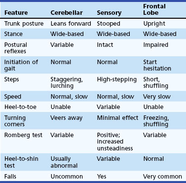
The width of the stance base (the distance between the feet) during quiet arising from sitting, standing, and walking gives some indication of balance. Wide-based gaits are typical of cerebellar or sensory ataxia but also may be seen in diffuse cerebrovascular disease and frontal lobe lesions (Table 22.1). Widening the stance base is an efficient method of reducing body sway in the lateral and anteroposterior planes. Persons whose balance is insecure for any reason tend to adopt a wider stance and a posture of mild generalized flexion and to take shorter steps. Those who have attempted to walk on ice or other slippery surfaces will recognize this phenomenon. Eversion of the feet is another manner in which to increase stability and is particularly common in patients with diffuse cerebrovascular disease. Spontaneous sway, drift of the body in any direction, postural tremor, or ability to stay upright without touching furniture or assistance of another person are important clues.

Trunk Posture

The trunk is normally upright during standing and walking. Flexion of the trunk and a stooped posture are prominent features in PD. Slight flexion at the hips to lower the trunk and shift the center of gravity forward to minimize posterior body sway and reduce the risk of falling backward is common in many unsteady cautious gait syndromes. Neck and trunk extension is characteristic of progressive supranuclear palsy. Neck flexion occurs with weakness of the neck extensors in motor neuron disease and myasthenia and as a dystonic manifestation in multiple system atrophy and parkinsonism. An exaggerated lumbar lordosis, caused by hip-girdle weakness, is typical of proximal myopathies. Paraspinal muscle spasm and rigidity also produces an exaggerated lumbar lordosis in the stiff person syndrome. Tilt of the trunk to one side in dystonia is accompanied by axial muscle spasms, the most common being an exaggerated flexion movement of the trunk and hip with each step. Truncal tilt away from the affected side is observed in some acute vascular lesions of the thalamus and basal ganglia. Misperception of truncal posture and position results in inappropriate movements to correct the perceived tilt in the pusher syndrome, associated with posterolateral thalamic hemorrhages (Karnath et al., 2005). Acute vestibular imbalance in the lateral medullary syndrome leads to sway or tilt toward the side of the lesion (lateropulsion). Abnormal truncal postures occur in paraspinal myopathies that produce weakness of trunk extension and a posture of truncal flexion (camptocormia). Dystonia and parkinsonism also may alter truncal posture and lead to camptocormia or lateral truncal flexion (Pisa syndrome). Abnormal thoracolumbar postures also result from spinal ankylosis and spondylitis. A restricted range of spinal movement and persistence of the abnormal spinal posture when supine or during sleep are useful pointers toward a bony spinal deformity as the cause of an abnormal truncal posture. Truncal postures, particularly in the lumbar region, can be compensatory for shortening of one lower limb, lumbar or leg pain, or disease of the hip, knee, or ankle.

Postural Responses

Reactive postural responses are examined by sharply pulling the upper trunk backward or forward while the patient is standing. The pull should be sufficient to require the patient to step to regain their balance. This maneuver is referred to as the pull test (Hunt and Setni, 2006). The examiner must be prepared, generally by having a wall behind them, to prevent the patient from falling. A few short, shuffling steps backward (retropulsion) or a backwards fall after backward displacements, or forward (propulsion) after forward displacements, suggest impairment of postural righting (reactive postural) reactions. Falls after postural changes such as arising from a chair or turning while walking suggest impaired anticipatory postural responses. Falls without rescue arm movements or stepping movements to break the fall indicate loss of protective postural responses. Injuries sustained during falls provide a clue to the loss of these postural responses. A tendency to fall backward spontaneously is a sign of impaired postural reflexes in progressive supranuclear palsy and gait disorders associated with diseases of the frontal lobes.

Walking

Stepping

Once walking is underway, the length and trajectory of each step and the rhythm of stepping should be noted. Short, regular, shallow steps or shuffling and a slow gait are characteristic of the akinetic-rigid syndromes. Shuffling is most evident when starting to walk, stopping, or turning corners. Specifically examining these maneuvers may reveal a subtle tendency to shuffle. Once the person is underway, freezing may interrupt walking, with further shuffling and start hesitation. Festination (increasingly rapid, small steps) is common in PD but rare in other akinetic-rigid syndromes, which frequently are associated with poor balance and falls rather than festination. Unilateral loss of synergistic arm swing while walking is a valuable sign of early PD. A reduction in associated trunk movement and arm swing also may be evident in unilateral upper motor neuron and acute cerebellar syndromes. A slow gait also is seen in ataxic and spastic syndromes. Jerky steps of irregular rhythm and variable length and direction suggest ataxic or choreic syndromes. Subtle degrees of cerebellar ataxia may be unmasked by asking the patient to walk in a straight line heel-to-toe (tandem gait), to stand on one leg, or to walk and turn quickly. When vision is important in helping maintain balance, as in sensory ataxia caused by proprioceptive loss, the removal of vision greatly exaggerates the ataxia. This is the basis of the Romberg test, in which eye closure leads to a dramatic increase in unsteadiness and even falls in the patient with sensory ataxia. When performing the Romberg test, it is important that the patient be standing comfortably before eye closure and to remember that a modest increase in body sway is a normal response to eye closure. Distinctive leg postures and foot trajectories occur during stepping in sensory ataxia, foot drop, spasticity, and dystonia. It may be necessary to examine the patient running to identify an action dystonia of the legs in the early stages of primary torsion dystonia.

Motor and Sensory Examination

After the patient has been observed walking, motor and sensory function in the limbs is examined with the patient sitting or supine to confirm the suspicions raised by the gait features. The size and length of the limbs should be measured in any child presenting with a limp. Asymmetry in leg size suggests a congenital malformation of the spinal cord or brain, or (rarely) local overgrowth of tissue. The spinal column should be inspected for scoliosis, and the lumbar region for skin defects or hairy patches indicative of spinal dysraphism.

Changes in muscle tone such as spasticity, lead-pipe or cogwheel rigidity, or paratonic rigidity (gegenhalten) point toward diseases of the upper motor neuron, basal ganglia, and frontal lobes, respectively. In the patient who complains of symptoms in only one leg, a detailed examination of the other leg is important. If signs of an upper motor neuron syndrome are present in both legs, a disorder of the spinal cord or parasagittal region is likely. Muscle bulk and strength are examined for evidence of muscle wasting and the presence and distribution of muscle weakness. Examination reveals whether the abnormal posture of the leg in a patient with foot drop (Box 22.2) is caused by spasticity or weakness of ankle dorsiflexors, which in turn may be due to anterior horn cell disease, a peripheral neuropathy, a peroneal compression neuropathy, or an L5 root lesion. Joint position sense should be examined for defects of proprioception in the ataxic patient or awkward posturing of the foot. Other signs such as a supranuclear gaze palsy, ataxia, and frontal lobe release signs should be sought where relevant.

Classification of Gait Patterns

The goal of classification is to arrange gait patterns into a scheme or order that reflects the physiological basis of human gait and helps the clinician recognize what levels of the nervous system are deranged. A scheme based on Hughlings Jackson’s three orders of neurological centers—lower (simplest), middle, and higher (complex and integrative)—enables a classification according to function. Each center contributes sensory and motor function, but that of higher centers is more complex and dispersed within the nervous system.

Lower-Level Gait Disorders

Lower-level disorders have physical signs of neurological dysfunction such as weakness or sensory loss. Lower-level motor function and muscle contraction produce the force required for postural responses and locomotion. Lower-level motor dysfunction arises from muscle and peripheral nerve disease. Gaits that typify this category include foot drop or steppage gaits associated with distal weakness, usually due to neuropathy, and waddling gaits associated with proximal (usually myopathic) weakness. Lower-level sensory dysfunction arises from disorders of the three basic senses thought most important for gait and balance: proprioception, vision, and vestibular sensation. Lower-level sensory disorders arise from diseases affecting the peripheral transmission of these sensory modalities. Sensory ataxia results from deafferentation due to peripheral nerve, dorsal root ganglion, or posterior column lesions and is associated with loss of proprioception or joint position sense. Imbalance in acute peripheral vestibulopathies or visual impairment are other examples of lower-level sensory disorders.

Middle-Level Gait Disorders

Middle-level gait disorders are primarily motor dysfunctions and appear to be related to impaired modulation of force generated by the lower-level motor system. The middle-level motor disorders include: (1) spasticity from disruption of the corticospinal tracts, (2) ataxia arising from disturbances of the cerebellum and its connections, (3) hypokinetic gaits associated with parkinsonism, and (4) hyperkinetic gaits associated with chorea, dystonia, and other movement disorders.

Spastic Gait

Spasticity of the arm and leg on one side produces the characteristic clinical picture of a spastic hemiparesis. The arm is held adducted, internally rotated at the shoulder, and flexed at the elbow, with pronation of the forearm and flexion of the wrist and fingers. The leg is slightly flexed at the hip and extended at the knee, with plantar flexion and inversion of the foot. The swing phase of each step is accomplished by slight lateral flexion of the trunk toward the unaffected side and hyperextension of the hip on that side to allow slow circumduction of the stiffly extended paretic leg as it is swung forward from the hip, dragging the toe or catching it on the ground beneath. A minimum of associated arm swing occurs on the affected side. The stance may be slightly widened, and the speed of walking is slow. Balance may be poor because the hemiparesis interferes with corrective postural adjustments on the affected side. Muscle tone in the affected limbs is increased, clonus may be present, and tendon reflexes are abnormally brisk, with an extensor plantar response. Examination of the sole of the shoe may reveal wear of the toe and outer borders of the shoe, suggesting that the spastic gait is of long standing. After identifying a spastic hemiparesis, determining the site at which the corticospinal tract is involved, determining the motor level, and confirmatory imaging with magnetic resonance imaging (MRI) of the brain (and where indicated, the spinal cord) should be the next steps in the diagnostic process.

Spasticity of both legs gives rise to a spastic paraparesis. The legs are stiffly extended at the knees, plantar flexed at the ankles, and slightly flexed at the hips. Both legs circumduct, and the toes of the plantar flexed feet catch on the floor with each step. The gait is slow and labored as the legs are dragged forward with each step. There is a tendency to adduct the legs, particularly when the disorder begins in childhood, an appearance described as scissors gait. The causes of a spastic paraparesis include hereditary spastic paraplegia, in which the arms and sphincters are unaffected and there may be little or no leg weakness, and other myelopathies. An indication of the extent and level of the spinal cord lesion can be obtained from the presence or absence of weakness or sensory loss in the arms, a spinothalamic sensory level or posterior column sensory loss, and alterations in sphincter function. Patients with paraparesis of recent onset should be investigated with MRI of the spinal cord to exclude potentially treatable causes such as spinal cord compression.

Occasionally, bilateral leg dystonia (dystonic paraparesis) mimics a spastic paraparesis. This typically occurs in dopa-responsive dystonia in childhood and may be misdiagnosed as hereditary spastic paraplegia or cerebral diplegia. Clinical differentiation between these conditions can be difficult. Brisk tendon reflexes occur in both, and spontaneous extension of a great toe in patients with striatal disorders may be interpreted as a Babinski response. Fanning of the toes and knee flexion suggest spastic paraplegia. Other distinguishing features include changes in muscle tone, such as spasticity in hereditary spastic paraparesis and rigidity in dystonic paraparesis. In young children, the distinction is important because a proportion of such patients can be treated successfully with levodopa (discussed in the following sections).

Cerebellar Ataxia

Disease of the midline cerebellar structures, the vermis, and anterior lobe produces loss of truncal balance, increased body sway, dysequilibrium, and gait ataxia. When standing, the patient adopts a wide-based stance; the legs are stiffly extended and the hips slightly flexed to crouch forward and minimize truncal sway. The truncal gait ataxia of midline cerebellar pathology has a lurching and staggering quality that is more pronounced when walking on a narrow base or during heel-to-toe walking. A pure truncal ataxia may be the sole feature of a midline (vermis) cerebellar syndrome and escape notice if the patient is not examined when standing, because leg coordination during the heel-to-shin test may be relatively normal when examined supine. Midline cerebellar pathologies also include masses, hemorrhage, paraneoplastic syndromes, and malnutrition in alcoholism. Patients with anterior lobe atrophy develop a 3-Hz anteroposterior sway of the trunk and a rhythmic truncal and head tremor (titubation) that is superimposed on the gait ataxia. This combination of truncal gait ataxia and truncal tremor is characteristic of some late-onset cerebellar degenerations affecting the anterior lobe.

Lesions of the cerebellar flocculonodular lobe (the vestibulocerebellum) exhibit multidirectional body sway, dysequilibrium, and severe impairment of body and truncal motion. Standing and even sitting can be impossible, although when lying down, results of the heel-to-shin test may appear normal, and upper-limb function may be relatively preserved.

Limb ataxia due to involvement of the cerebellar hemispheres is characterized by a decomposition of normal leg movement. Steps are irregular and variable in timing (dyssynergia), length, and direction (dysmetria). Steps are taken slowly and carefully to reduce the tendency to lurch and stagger. These defects are accentuated when attempting to walk heel to toe in a straight line. With lesions confined to one cerebellar hemisphere, ataxia is limited to the ipsilateral limbs, and there is little postural instability or truncal imbalance if the vermis is not involved. Vascular disease and mass lesions are generally responsible for hemisphere lesions.

Cerebellar gait ataxia is exacerbated by the rapid postural adjustments needed to change direction, turn a corner or avoid obstacles, and when stopping or starting to walk. Minor support, such as holding the patient’s arm during walking, and visual compensation help the patient with a cerebellar ataxia reduce body sway. Eye closure may heighten anxiety about falling and increase body sway, but not to the extent observed in a sensory ataxia.

Episodic ataxias produce periods of impaired gait that typically last seconds to hours. Alcohol and drug use must be considered in the differential of episodic ataxias.

Hypokinetic (Parkinsonian) Gait

The most common hypokinetic-bradykinetic gait disturbance is that encountered in PD. In early PD, an asymmetrical reduction of arm swing and slight slowing in gait, particularly with turns, is characteristic. In more advanced PD, the posture is stooped, with flexion of the shoulders, neck, trunk, and knees. During walking, there is little associated or synergistic body movement. Arm swing is reduced or absent, and the arms are held immobile at the sides or slightly forward of the trunk. Tremor of the upper limbs frequently increases when walking, but parkinsonian tremor of the legs rarely affects walking. A characteristic feature of a parkinsonian gait is the tendency to begin walking with a few rapid, short, shuffling steps (start hesitation) before breaking into a more normal stepping pattern with small, shallow steps on a narrow base. Once underway, walking may be interrupted by shuffling or even cessation of movement (freezing) if an obstacle is encountered, when walking through doorways, or when attempting to undertake multiple tasks at once. These signs may be alleviated by levodopa treatment. In the long-term, levodopa therapy may induce dyskinesias, resulting in choreic and dystonic gaits as described later.

The posture of generalized flexion of the patient with PD exaggerates the normal tendency to lean forward when walking. To maintain balance when walking and avoid falling forward, the patient may advance with a series of rapid, small steps (festination). Retropulsion and propulsion are similar manifestations of a flurry of small, shuffling steps made in an effort to preserve equilibrium. Instead of a single large step, a series of small steps are taken to maintain balance. Freezing becomes increasingly troublesome in the later stages of PD, at which time sensory cues may be more useful in triggering a step than medication. The shuffling gait of PD that is responsive to levodopa characterizes the midlevel gait pattern. As the disease progresses, dysequilibrium and falls emerge that are features of a higher-level gait disorder (discussed later).

Dystonic Gait

Of all gait disturbances, dystonic syndromes produce some of the more bizarre and difficult diagnostic problems. The classic presentation of childhood-onset primary torsion dystonia is an action dystonia of a leg, with a sustained abnormal posture of the foot (typically plantar flexion and inversion) on attempting to run. In contrast, walking forward or backward or even running backward may be entirely normal at an early stage. An easily overlooked sign in the early stages is tonic extension of the great toe (the striatal toe) when walking. This may be a subtle finding but occasionally is so pronounced that a hole is worn in the toe of the shoe. With the passage of time, dystonia may progress to involve the whole leg and then become generalized.

More difficult to recognize are those dystonic syndromes that present with bizarre, seemingly inexplicable postures of the legs and trunk when walking. A characteristic feature common to dystonic gaits is excessive flexion of the hip when walking. Patients may hop or walk sideways in a crablike fashion, with hyperflexion of the hips producing an attitude of general body flexion in a simian posture, or a birdlike (peacock) gait with excessive flexion of the hip and knee and plantar flexion of the foot during the swing phase of each step. Many of these patients have been thought to be hysterical because of the unusual gait disturbance and because formal neurological examination is often normal when the patient is examined lying supine. Each of these gait patterns has been described in association with primary and secondary dystonic syndromes. Tardive dystonia following neuroleptic drug exposure may produce similar bizarre abnormalities of gait.

It is important to look for asymmetry in the assessment of childhood-onset dystonia. Symptomatic causes should be excluded in hemidystonic presentations, isolated leg dystonia in an adult, and early loss of postural responses and righting reflexes in association with a dystonic gait.

Dopa-responsive dystonia characteristically presents in childhood with walking difficulties and diurnal fluctuations in severity of dystonia. Typically the child walks normally in the early morning but develops increasing rigidity and dystonic posturing of the legs as the day progresses or after exercise; these difficulties may be relieved by a nap. Examination reveals a dystonic plantar flexion and inversion of the foot, with the additional feature of brisk tendon reflexes. Some of these patients respond dramatically to levodopa, so early recognition is important. Indeed, all children presenting with a dystonic foot or leg should have a therapeutic trial of levodopa before other therapies such as anticholinergic drugs are commenced.

Paroxysmal dyskinesias also may present with difficulty walking. Paroxysmal kinesigenic choreoathetosis may present with the sudden onset of difficulty walking as the result of dystonic postures and involuntary movements of the legs, often appearing after standing from a seated position. The attacks with this disorder are typically very brief, lasting a matter of seconds.

Higher-Level Gait Disorders

Higher-level gait disorders are characterized by varying combinations of dysequilibrium, falls due to inappropriate or absent postural responses, and shuffling steps or freezing. In contrast to lower- and middle-level gait patterns, formal neurological examination fails to reveal signs that adequately explain the gait disturbance, though brisk reflexes and extensor plantar responses or depressed reflexes and minor distal sensory loss may be encountered. Slowness of sequential leg movement and poor control of truncal movement are often present. The stepping patterns are influenced by environmental cues that may facilitate stepping but may also may induce freezing of gait if present. The performance of multiple simultaneous tasks may precipitate freezing or falls.

There are many descriptions of similar gait patterns in the literature, often focusing on one element of the gait disturbance. Because of uncertainty about the pathophysiology of these clinical manifestations, there has been no agreement on the terminology used to describe them.

Hypokinetic Higher-Level Gait Patterns

With progression of PD, freezing of gait, dysequilibrium, and falls become increasingly troublesome and do not respond to increasing doses of levodopa. There is some evidence from clinical, imaging, and pathological studies to suggest that dysequilibrium is mediated via mechanisms other than dopaminergic deficiency, and subcortical cholinergic projections from the pedunculopontine nucleus have been implicated (Bohnen et al., 2009). Falls occur late in the course of PD, and a number of causes must be considered. These include festinating steps that are too small to restore balance, tripping or stumbling over rough surfaces because shuffling steps fail to clear small obstacles, and failure to step, with start hesitation and freezing. In each of these examples, falling stems from locomotor hypokinesia and a lack of normal-sized, rapid, compensatory voluntary movements. These falls are forward onto knees and outstretched arms (indicating preservation of rescue reactions). Other falls, in any direction, occur when changing posture or turning in small spaces and result from loss of postural and righting responses, either spontaneously when multitasking or after minor perturbations. It is also important to consider collapsing falls related to orthostatic hypotension, a common finding in PD.

Unlike the hypokinetic steps and flexed truncal posture, the loss of postural and righting responses generally does not respond to levodopa. Deep brain stimulation (DBS) of the subthalamic nucleus (STN) or the globus pallidus interna (GPi) may improve gait but also may worsen gait and may be associated with increased falls (Ferraye et al., 2008; Weaver et al., 2009). DBS in the region of the pedunculopontine nucleus is under investigation for disequilibrium and freezing of gait, with mixed results (Ferraye et al., 2010).

Similar slowness of leg movement and shuffling occurs in a variety of other akinetic-rigid syndromes (Box 22.4), the most common of which are multiple system atrophy, corticobasal degeneration, and progressive supranuclear palsy. A number of clinical signs help distinguish among these conditions (Table 22.2). In progressive supranuclear palsy, the typical neck posture is one of extension, with axial and nuchal rigidity rather than neck and trunk flexion as in PD. A stooped posture with exaggerated neck flexion is sometimes a feature of multiple system atrophy. A distinguishing feature of progressive supranuclear palsy and multiple system atrophy is the early appearance of falls due to loss of postural and righting responses, in comparison to the preservation of these reactions in PD until later stages of the illness. There also may be an element of ataxia in these akinetic-rigid syndromes that is not evident in PD. Falls occur in 80% of patients with progressive supranuclear palsy and can be dramatic, leading to injury. The disturbance of postural control in progressive supranuclear palsy is coupled with impulsivity due to frontal executive dysfunction that leads to reckless lurching movements during postural changes, such as sitting or arising, and toppling falls. Accordingly, the patient who presents with falls and an akinetic-rigid syndrome is more likely to have one of these variants of parkinsonism rather than PD. Finally, the dramatic response to levodopa that is typical of PD does not occur in these variants of parkinsonism, although some cases of multiple system atrophy respond partially for a short period.

Table 22.2 Summary of Clinical Features Differentiating Parkinson Disease from Symptomatic Parkinsonism in Patients with an Akinetic-Rigid Gait Syndrome

Feature Parkinson Disease Symptomatic Parkinsonism
Posture Stooped (trunk flexion) Stooped or upright (trunk flexion/extension)
Stance Narrow Often wide-based
Initiation of walking Start hesitation Start hesitation, magnetic feet
Steps Small, shuffling Small, shuffling
Stride length Short Short
Freezing Common Common
Leg movement Stiff, rigid Stiff, rigid
Speed Slow Slow
Festination Common Rare
Arm swing Minimal or absent Reduced or excessive
Heel-to-toe walking Normal Poor (truncal ataxia)
Postural reflexes Preserved in early stages Absent at early stage
Falls Late (forward, tripping) Early and severe (backward, tripping, or without apparent reason)

In addition to the advanced hypokinetic disorders discussed previously, frontal lobe tumors (glioma or meningioma), anterior cerebral artery infarction, obstructive or communicating hydrocephalus (especially normal-pressure hydrocephalus), and diffuse cerebrovascular disease (multiple lacunar infarcts and Binswanger disease) also produce disturbance of gait and balance. These pathologies interrupt connections between the frontal lobes and other cortical and subcortical structures. The clinical appearance of the gait of patients with such lesions varies from a predominantly wide-based ataxic gait to an akinetic-rigid gait with slow, short steps and a tendency to shuffle. It is common for a patient to present with a combination of these features. In the early stages, the stance base is wide, with an upright posture of the trunk and shuffling when starting to walk or turning corners. There may be episodes of freezing. Arm swing is normal or even exaggerated, giving the appearance of a “military two-step” gait. The normal fluidity of trunk and limb motion is lost. Examination reveals normal voluntary upper-limb and hand movements and a lively facial expression. This “lower-half parkinsonism” is commonly seen in diffuse cerebrovascular disease. The marche à petits pas of Dejerine and Critchley’s atherosclerotic parkinsonism refer to a similar clinical picture. Patients with this clinical syndrome commonly are misdiagnosed as having PD. The normal motor function of the upper limbs, retained arm swing during walking, upright truncal posture, wide-based stance, upper motor neuron signs including pseudobulbar palsy, and the absence of a resting tremor distinguish this syndrome from PD. In addition, the lower-half parkinsonism of diffuse cerebrovascular disease does not respond to levodopa treatment (see Box 22.4). Walking speed in subcortical arteriosclerotic encephalopathy is slower than in cerebellar gait ataxia or PD (Ebersbach et al., 1999). Slowness of movement and the lack of heel-to-shin ataxia distinguish the wide-based stance of this syndrome from that of cerebellar gait ataxia (see Table 22.1).

As the underlying condition progresses, the unsteadiness and slowness of movement become more pronounced. There may be great difficulty initiating a step, as if the feet were glued to the floor (magnetic feet). Attempts to take a step require assistance, with the patient seeking the support of nearby objects or persons. There may be excessive upper-body movement as the patient tries to free the feet to initiate walking. Once walking is underway, steps may be better, but small, shuffling, ineffective steps (freezing) reemerge when the patient tries to turn. Such patients rarely exhibit the festination of PD, but a few steps of propulsion or retropulsion may be taken. Postural and righting reactions are impaired and eventually lost. Falls are common and follow the slightest perturbation. In contrast, these patients are often able to move the legs with greater facility when seated or lying supine than when standing. Stepping, walking, or bicycling leg movements are possible when lying or seated but not when standing. This apparent discrepancy may reflect an inability to stand secondary to dysequilibrium, which in turn reflects poor truncal control. Impaired truncal mobility with truncal imbalance commonly appears in the advanced stages of a frontal gait disorder, so patients are unable to stand or turn over when lying in bed. Walking then becomes impossible, and even simple leg movements are slow and clumsy when lying down. Paratonic rigidity (gegenhalten) of the arms and legs is common. Tendon reflexes may be brisk, with extensor plantar responses. Grasp reflexes in the hands and feet may be elicited. Urinary incontinence and dementia frequently occur. Investigation of the brain with MRI reveals the majority of conditions causing this syndrome, such as diffuse cerebrovascular disease, cortical atrophy, or hydrocephalus.

Some patients display fragments of this clinical picture. Those with the syndrome of gait initiation failure exhibit profound start hesitation and freezing, but step size and rhythm are normal once walking is underway. Sensory cues may facilitate stepping. Balance while standing or walking is normal. These findings are similar to those seen with walking in PD, but speech and upper-limb function are normal, and there is no response to levodopa. Brain imaging results are normal. The cause of this syndrome is unknown, but the slowly progressive evolution of symptoms suggests a degenerative condition. Occasionally, isolated episodic festination with truncal flexion is encountered. Others complain of a loss of the normal fluency of stepping when walking; a conscious effort is required to maintain a normal stepping rhythm and step size. These symptoms may be associated with subtle dysequilibrium, manifesting as a few brief staggering steps to one side or a few steps of retropulsion after standing up, turning quickly, or making other rapid changes in body position. Finally, some elderly patients experience severe walking difficulties that resemble those described in frontal lobe disease. The history in these syndromes is one of gradual onset, without stroke-like episodes or identifiable structural or vascular lesions of the frontal lobes or cerebral white matter on imaging. The cause of these syndromes is unknown. The criteria for normal pressure hydrocephalus are not fulfilled, there are no signs of parkinsonism, levodopa is ineffective, and there is no evidence of more generalized cerebral dysfunction, as occurs in Alzheimer disease. Indeed, it is rare for patients with Alzheimer disease to develop difficulty walking until the later stages of the disease.

Elderly Gait Patterns, Cautious Gaits, and Fear of Falling

Healthy, neurologically normal elderly people tend to walk at slower speeds than their younger counterparts. The slower speed of walking is related to shorter and shallower steps with reduced excursion at lower limb joints. In addition, stance width may be slightly wider than normal, and synergistic arm and trunk movements are less vigorous. The rhythmicity of stepping is preserved. These changes give the normal elderly gait a cautious or guarded appearance. Factors contributing to a general decline in mobility of the elderly include degenerative joint disease, reducing range of limb movement, and decreased cardiovascular fitness, limiting exercise capacity. The changes in gait pattern of the elderly also provide a more secure base to compensate for a subtle age-related deterioration in balance.

In unselected elderly populations, a more pronounced deterioration in gait and postural control is evident. Steps are shorter, stride length is reduced, and the stance phase of walking is increased, leading to a reduction in walking speed. These changes are most marked in those who fall.

Elderly patients with an insecure gait characterized by slow short steps, en bloc turns, and falls often have signs of multiple neurological deficits such as peripheral motor (mild proximal) weakness, subtle sensory loss (mild distal light touch and proprioceptive loss, blunted vestibular or visual function), and even mild spastic paraparesis due to cervical myelopathy, without any one lesion being severe enough to explain the walking difficulty. The cumulative effect of these multiple deficits may interfere with perceived stability and equilibrium. Along with musculoskeletal disorders, postural hypotension, and loss of confidence (especially after falls), these factors contribute to a cautious gait pattern. In this situation, brain imaging is valuable to look for subcortical white-matter ischemia (especially in the frontal lobes and periventricular regions) that correlates with imbalance, increased body sway, falls, and cognitive decline (Baezner et al., 2008).

A cautious gait is a normal response related to the perception of impaired or threatened balance, as exemplified by walking on a slippery icy surface, and accordingly should be interpreted as compensatory and not specific for any level of the gait classification. Due account must be taken of the fear of falling that frequently accompanies gait difficulties. This may lead to a marked loss of confidence when walking and a cautious or protected gait. Such patients may be unable to walk without support. They hold onto furniture, lean on walls, and avoid crowded or open spaces because of a fear of falling. The gait may improve dramatically when support is provided. A formal program of gait retraining may help restore confidence and improve the ability to walk.

Perceptions of Instability and Illusions of Movement

A number of syndromes have been described in which middle-aged individuals complain of unsteadiness and imbalance associated with “dizziness,” sensations or illusions of semicontinuous body motion, sudden brief body displacements, or body tilt. In some, the symptoms develop in open spaces where there are no visible supports (space phobia). Others develop sensations in particular situations such as on bridges, stairs, and escalators or in crowded rooms, leading to the development of phobic avoidance behavior and the syndrome of phobic postural vertigo (Brandt, 1996). Prolonged illusory swaying and unsteadiness after sea or air travel is referred to as the mal de débarquement syndrome. Past episodes of a vestibulopathy may suggest the possibility of a subtle semicircular canal or otolith disturbance, but a disorder of vestibular function is rarely confirmed in these syndromes. A fear of falling, anxiety, and related complaints are common accompaniments. These symptoms must be distinguished from the physiological “vertigo” and unsteadiness accompanying visual vestibular mismatch or conflict when observing moving objects, focusing on distant objects in large panoramas, or looking upward at moving objects.

Hysterical and Psychogenic Gait Disorders

The wide range of abnormalities of gait seen in lesions of different parts of the nervous system make hysterical and psychogenic gaits among the most difficult to diagnose. The hallmark of hysterical gait paralysis is the inability to use one or both legs when walking, but normal synergistic movements of the affected leg(s) when the patient is examined while lying down or observed when changing position. This discrepancy is further illustrated by the Hoover sign in the patient with an apparently paralyzed leg when examined supine. As the patient lifts the normal leg, the examiner places a hand under the “paralyzed” leg and feels the presence (and strength) of synergistic hip extension. The apparent severe weakness of hysterical paresis often presents little disability or inconvenience in some patients; others with hysterical paraplegia are confined to bed and may even develop contractures from lack of leg movements.

A gait disorder is one of the more common manifestations of a psychogenic or hysterical movement disorder. The typical gait patterns encountered include transient fluctuations in posture while walking; knee buckling without falls; excessive slowness and hesitancy; a crouched, stooped, or other abnormal posture of the trunk; complex postural adjustments with each step; exaggerated body sway or excessive body motion especially brought out by tandem walking; and trembling, weak legs (Hayes et al., 1999). The more acrobatic hysterical disorders of gait indicate the extent to which the nervous system is functioning normally and is capable of high-level coordinated motor skills to perform various complex maneuvers. Suggestibility, variability, improvement with distraction, and a history of sudden onset or a rapid, dramatic, and complete recovery are other features of psychogenic gait (and movement) disorders. One must be cautious in accepting a diagnosis of hysteria, however, because a bizarre gait may be a presenting feature of primary torsion dystonia, and unusual truncal and leg postures may be encountered in truncal and leg tremors. Finally, higher-level gait disorders often have a disconnect between the standard neurological exam and the gait pattern.

Musculoskeletal Disorders and Antalgic Gait

References

Baezner H., Blahak C., Poggesi A., et al. Association of gait and balance disorders with age-related white matter changes: the LADIS study. Neurology. 2008;70:935-942.

Bohnen N.I., Muller M.L., Koeppe R.A., et al. History of falls in Parkinson’s disease is associated with reduced cholinergic activity. Neurology. 2009;73:1670-1676.

Brandt T. Phobic postural vertigo. Neurology. 1996;46:1515-1519.

Ebersbach G., Sojer M., Valldeoriola F., et al. Comparative analysis of gait in Parkinson’s disease, cerebellar ataxia and subcortical arteriosclerotic encephalopathy. Brain. 1999;122:1349-1355.

Ferraye M.U., Debu B., Fraix V., et al. Effects of subthalamic nucleus stimulation and levodopa on freezing of gait in Parkinson’s disease. Neurology. 2008;70:1431-1437.

Ferraye M.U., Debu B., Fraix V., et al. Effects of pedunculopontine nucleus area stimulation on gait disorders in Parkinson’s disease. Brain. 2010;133:205-214.

Hayes M.W., Graham S., Heldorf P., et al. A video review of the diagnosis of psychogenic gait: appendix and commentary. Mov Disord. 1999;14:914-921.

Hunt A.L., Sethi K.D. The pull test: a history. Mov Disord. 2006;21:894-899.

Karnath H.O., Johannsen L., Broets D., et al. Posterior thalamic haemorrhage induces “pusher syndrome. Neurology. 2005;64:1014-1019.

Studenski S., Perera S., Patel K., et al. Gait speed and survival in older adults. JAMA. 2011;305:50-58.

Takakusaki K. Forebrain control of locomotor behaviours. Brain Res Rev. 2008;57:192-198.

Weaver F.M., Follet K., Stern M., et al. Bilateral deep brain stimulation vs best medical therapy for patients with advanced Parkinson’s disease: a randomized controlled trial. JAMA. 2009;301:63-73.