Functional neurosurgery

Published on 07/02/2015 by admin

Filed under Anesthesiology

Last modified 22/04/2025

Print this page

rate 1 star rate 2 star rate 3 star rate 4 star rate 5 star
Your rating: none, Average: 0 (0 votes)

This article have been viewed 1242 times

Functional neurosurgery

Jeffrey J. Pasternak, MS, MD

Functional neurosurgery is a broad term applied to a variety of neurosurgical procedures performed to treat conditions in which the function of the brain is abnormal, typically in the context of normal gross structure and anatomy. These conditions include Parkinson disease, essential tremor, dystonia, obsessive compulsive disorder, and, possibly, Tourette syndrome, depression, refractory obesity, and epilepsy. The major challenge during functional neurosurgical procedures is to accurately and safely identify the abnormal regions of brain tissue, which is usually accomplished via neurologic assessment in an awake or partially sedated patient. Alternatively, radiographically guided or electrophysiologically guided techniques may be employed in patients having general anesthesia.

Implantation of deep brain stimulators

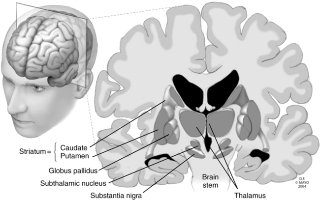
Deep brain stimulation (DBS) involves the implantation of electrodes into select regions of the brain, allowing for electrical stimulation of the area to modulate brain activity, resulting in attenuation, if not elimination, of the symptoms and signs of a number of disease states (Table 133-1). The specific site of electrode implantation depends on the disorder for which the patient requires treatment (Figure 133-1). Despite the use of DBS for many decades, the exact mechanism or mechanisms accounting for its clinical efficacy are not well understood, but the electric current is believed to somehow modulate abnormal neuronal function, either by acting directly on neuronal action potentials or altering neurotransmitter release. Use of DBS has generally replaced ablative procedures, such as pallidotomy or thalamotomy (i.e., thermal, mechanical, or electrical destruction of a region of the globus pallidus or thalamus, respectively), for the treatment of Parkinson disease. Unlike these earlier procedures, DBS is reversible.

Table 133-1

Disease States and Potential Anatomic Targets for Deep Brain Stimulation

Disease Potential Deep Brain Stimulation Target(s)
Parkinson disease and essential tremor Subthalamic nucleus
Globus pallidus
Dystonia Globus pallidus
Cerebellar tremor from multiple sclerosis Thalamic ventral intermediate nucleus
Pantothenate kinase–associated neurodegeneration Globus pallidus
Medically refractory depression Subgenual cingulate region
Tourette syndrome Anterior limb of the internal capsule
Thalamic centromedian-parafascicular complex
Obsessive-compulsive disorder Nucleus accumbens
Anterior limb of the internal capsule
Central pain syndromes Motor cortex
Periaqueductal gray matter
Periventricular gray matter
Thalamus
Medically refractory epilepsy Anterior nucleus of the thalamus
Centromedian nucleus of the thalamus
Subthalamic nucleus
Cluster headaches Posterior hypothalamus
Obesity Lateral hypothalamus
Ventromedial hypothalamus
Nucleus accumbens

Modified, with permission, from Siddiqui MS, Ellis TL, Tatter SB, Okun MS. Deep brain stimulation: Treating neurological and psychiatric disorders by modulating brain activity. NeuroRehabilitation. 2008;23:105-113.

DBS implantation is typically conducted via frame-based stereotactic techniques. In essence, a stereotactic head frame is applied, usually under monitored anesthesia care, and the neurosurgeon injects a local anesthetic agent at the sites where the pins of the head frame will be inserted to immobilize the skull within the head frame, after which the patient undergoes imaging (i.e., computed tomography or magnetic resonance imaging) to localize the deep brain target relative to the stereotactic head frame. An electrode is advanced via a burr hole to the approximate location of the target. Implantation of the electrode into the exact target nucleus is usually facilitated by single-neuron recordings and then confirmed, if possible, by the resolution of symptoms upon stimulation. The nature of various disorders, such as obesity, epilepsy, and obsessive-compulsive disorder, may not allow for immediate confirmation of symptom resolution. Following electrode implantation, the wires are tunneled under the skin to reach a generator, which is typically implanted in the pectoral region.

Generally, electrode implantation is performed with the patient in a semiseated position with monitored anesthesia care with sedatives administered to keep the patient “comfortable” but not so sedated that the surgeon cannot intraoperatively assess and optimize the efficacy of electrode placement. Providing an adequate but not excessive level of sedation can prove to be very challenging because the procedures tend to be of long duration and access to the airway is limited due to the presence of the head frame, which is rigidly fixed to the operating table. A means to rapidly secure the airway (i.e., laryngeal mask airway, fiberoptic bronchoscope) should be readily available. Some sedative drugs (i.e., propofol, benzodiazepines) can inhibit neuronal activity, thus influencing the ability to utilize single-neuron recordings to accurately identify the deep brain target. Short-acting opioids (e.g., fentanyl, remifentanil) and dexmedetomidine have been used successfully to provide sedation for these procedures.

Alternatively, in patients not able to tolerate the procedure with sedation (e.g., children or adults with impaired cognitive or intellectual abilities) implantation of a depth electrode can be conducted with general anesthesia. Drugs used to maintain general anesthesia may significantly impact the ability to identify and monitor neuronal electrical recordings. In these situations, proper placement of the depth electrode is usually dependent on imaging data referenced to the stereotactic head frame; thus, the likelihood of improper or ineffective electrode position may be greater when using general anesthesia.

Clinically consequential venous air embolism has been reported with the implantation of electrodes for deep brain stimulation, and the use of precordial Doppler sonography monitoring should be considered. Of note, electrical impedance from precordial Doppler sonography may impair neuronal electrical recording and may need to be suspended during recording of neuronal activity. Tunneling of electrode leads and implantation of the pulse generator are usually conducted during general anesthesia following removal of the stereotactic head frame.

Cervical denervation for dystonia

Dystonias are a group of disorders in which inappropriate and sustained muscle contractions lead to twisting movements and abnormal postures. The causes are many and types include congenital, idiopathic, trauma-induced, and drug-induced dystonias. Conservative treatment options include antiparkinsonian drugs (such as trihexyphenidyl or the combination of carbidopa and levodopa), antiepileptics, benzodiazepines, and β-adrenergic receptor blocking agents. If these medications do not produce satisfactory results, injection of botulinum toxin into the affected muscles may also provide some improvement and is the most commonly performed invasive procedure. DBS has been approved by the Food and Drug Administration in the United States for treatment of cervical dystonia and is currently under investigation, assessing efficacy compared to other treatment options, for other dystonias.

Refractory cervical dystonia may also be treated with selective peripheral muscular denervation, which involves identifying and transecting the nerves supplying the affected muscles. This procedure is usually conducted under general anesthesia with the patient in either the prone or sitting position. In either case, the surgeon will directly stimulate nerves with an electric current to identify specific muscule innervation; hence, the use of neuromuscular blocking agents is contraindicated during this segment of the procedure. In patients undergoing cervical denervation in the sitting position, techniques used to monitor for (i.e., transesophageal echocardiography, precordial Doppler sonography) and to treat (i.e., central venous catheter) air entrained into the venous system should be considered.

Epilepsy surgery

Epilepsy, or recurrent seizure disorder, affects 50 million people worldwide and occurs in all age groups. Initial management of epilepsy is usually with the use of one or more antiepileptic drugs. Despite this, many patients either continue to experience frequent seizures despite the use of multiple antiepileptic agents or are unable to tolerate the side effects of these drugs, which include somnolence, ataxia, hepatitis, cutaneous reactions, or aplastic anemia. Active epilepsy has a major negative impact on quality of life. For example, patients are unable to drive automobiles, may have work limitations, and also suffer the embarrassment of having seizures in public. In these patients, surgical management should be considered as a treatment option. Although epilepsy surgery was long considered as a last resort, the loss of developmental milestones in children and young adults who continue to experience seizures or have unacceptable side effects with the use of antiepileptic medications has increased the number and decreased the age of those having surgery. There are two major types of epilepsy surgery: (1) resective and (2) nonresective or functional procedures.

Resective procedures

The goal of resective procedures is to remove an abnormal region of brain that is thought to give rise to seizures (i.e., an epileptogenic focus). Preoperative identification of the epileptogenic focus is usually determined by history and physical examination, brain imaging, and video-electroencephalography. In the latter procedure, patients are admitted to a video telemetry unit, and simultaneous videotaping of the patient’s activity and electroencephalography are conducted to correlate the electrical characteristics of the seizure with motor or behavioral findings. In some patients, depth electrodes may be implanted prior to video-electroencephalography to allow for characterization of epileptogenic foci in deeper regions of the brain. Because many epileptogenic foci amenable to surgery are found to exist in the anterior temporal lobe, resection of this region is common, accounting for 75% of resective procedures. In many cases, intraoperative electrocorticography may be employed to allow for accurate identification of the epileptogenic focus. Electrode-containing grids are placed directly on the brain surface and abnormal epileptiform activity (i.e., abnormal background electroencephalographic activity) generated by the epileptogenic focus is recorded, allowing for a more precise resection. In cases in which electrocorticography is employed, anesthetic drugs that suppress epileptiform activity should be avoided or minimized during mapping. These drugs include inhalation anesthetic agents, sedative and anesthetic doses of barbiturates and propofol, and benzodiazepines. Nitrous oxide, opioids, diphenhydramine, droperidol, and possibly dexmedetomidine may be used to maintain sedation or general anesthesia during this period. Additionally, low-dose methohexital (0.3-1 mg/kg), etomidate (0.1-0.3 mg/kg), or alfentanil (50 μg/kg) may be administered as a bolus to enhance epileptiform activity generated by the seizure focus. Patients requiring intraoperative electrocorticography should be counseled preoperatively on the increased risk of intraoperative awareness, given the limited choices and doses of drugs that are available. In patients undergoing resection near the language center located in the temporal lobe, the procedure may be carried out with local anesthesia and sedation (i.e., awake craniotomy) allowing for intraoperative language assessment. Given the possibility of an intraprocedural seizure, the clinician should be prepared with airway equipment to secure an airway in a situation with limited airway access. Options may include temporary mask ventilation, an intubating laryngeal mask airway, and fiberoptically guided intubation. Additionally, termination of a seizure should be accomplished with agents that cause minimal respiratory depression in the setting of an unsecured airway. Additionally, the surgeon may irrigate the brain surface with cold saline solution in an effort to terminate a seizure.

Nonresective or functional procedures

In patients who continue to have frequent seizures despite resective treatment or are deemed not to be candidates for resective options (e.g., those with lesions near eloquent cortex, those in whom a primary epileptogenic focus cannot be identified, and those with multiple medical comorbid conditions that increase anesthetic risk), functional surgical procedures may be considered. Functional procedures are generally palliative and are considered a means to achieve a reduction in seizure frequency, as opposed to achieving a cure of epilepsy. Functional procedures include electrical stimulation techniques (i.e., vagal nerve, cortical, DBS), multiple subpial transection, and corpus callosotomy.

Vagal nerve stimulation involves placement of an electrode in the left vagal nerve sheath in the neck and a pulse generator in the pectoral region; it is performed with general anesthesia. The left vagus nerve is the preferred target, because parasympathetic innervation of the heart is predominantly derived from the right vagus nerve. A 50% or greater reduction in seizure frequency occurs in about 30% of patients with this technique. The exact mechanism by which vagal nerve stimulation results in a reduction in seizure frequency is not currently understood. The most common side effects are cough and hoarseness. Other stimulation techniques currently under investigation for seizure control include cortical stimulation and DBS. The major advantage of stimulation-based techniques for epilepsy control is reversibility, such that, if patients are unable to tolerate side effects or experience no benefit, the pulse generator and electrode can be removed with minimal injury to brain tissue.

The treatment goal of the remaining functional procedures is to limit seizure spread to the adjacent cortex. These techniques include multiple subpial transection and corpus callosotomy, both typically performed with general anesthesia. In the former technique, multiple incisions of about 4 mm in depth are made in cerebral cortex in an effort to transect fibers that may be involved in seizure propagation. The goal is to limit seizure spread while maintaining the function of eloquent cortex. Corpus callosotomy, performed more frequently in children than in adults, involves complete or staged transection of the corpus callosum to prevent seizure propagation to the adjacent cerebral hemisphere.