Published on 19/03/2015 by admin
Filed under Dermatology
Last modified 22/04/2025
This article have been viewed 2942 times
Justine Kluk and Malcolm H.A. Rustin
Evidence Levels: A Double-blind study B Clinical trial ≥ 20 subjects C Clinical trial < 20 subjects D Series ≥ 5 subjects E Anecdotal case reports
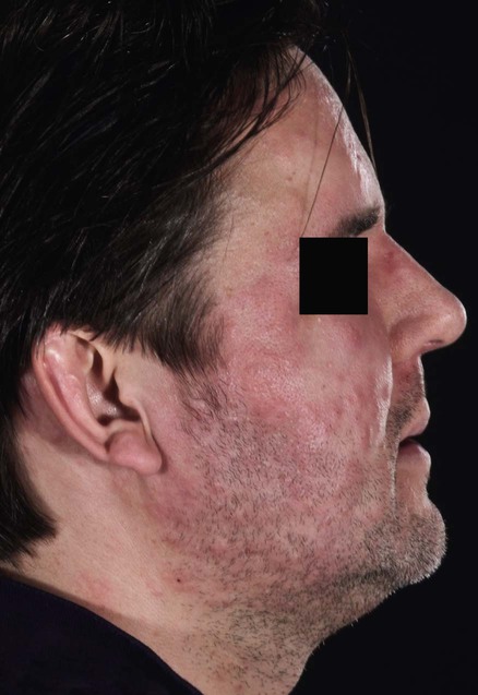
Follicular mucinosis is characterized histologically by mucinous degeneration of the follicular outer root sheaths and sebaceous glands with an inflammatory infiltrate composed of lymphocytes, histiocytes, and eosinophils. Lesions consist of erythematous, scaly, and infiltrated plaques with follicular papules or prominent follicular orifices, and may demonstrate alopecia (alopecia mucinosa). Benign follicular mucinosis tends to affect younger patients (under 40 years), with fewer lesions, usually situated on the head and neck. Although lesions may resolve spontaneously within 2 years, a more generalized form, with lesions on the trunk and extremities, may run a chronic relapsing course over many years. Follicular mucinosis is associated with lymphoma, particularly mycosis fungoides, in 15–30% of cases. It is still unclear whether follicular mucinosis is a transitional state evolving into mycosis fungoides in these cases. No single clinical or histological feature predicts which patients will have a benign course although those found to have mycosis fungoides rarely had initial lesions on the head and neck. Associated lymphoma tends (although not invariably) to be associated with age over 30 years, a wider distribution of lesions, and possibly systemic features such as night sweats, weight loss, or lymphadenopathy.
There is no standard therapy for follicular mucinosis. As spontaneous resolution occurs in the benign forms, observation alone is certainly justified, particularly in the younger patient with limited disease. However, the need for follow-up and evaluation to exclude lymphoma must be emphasized. Follicular mucinosis associated with mycosis fungoides or other neoplastic or inflammatory disorders is managed by treating the underlying associated condition.
Skin biopsy
Immunohistochemistry and T-cell gene receptor analysis may be helpful adjuncts
Consider investigations to rule out lymphoma or other underlying disorders, depending on the presenting clinical features (general examination, plain radiology, and CT scans)
Truhan AP, Roenigk HH. J Am Acad Dermatol 1986; 14: 1–18.
Rongioletti F, De Lucchi D, Meyes D, Mora M, Rebora A, Zupo S, et al. J Cutan Pathol 2010; 37: 15–19.
Two excellent reviews of the follicular mucinosis literature, including histopathology and investigation.
Emmerson RW. Br J Dermatol 1969; 81: 395–413.
Topical or intralesional corticosteroids improved surface eczematous change in eight of 22 patients with benign disease whose lesions resolved spontaneously within 2 years. Deeper follicular and dermal changes were not affected. Resolution was considered to have occurred independently of treatment. Six of 10 patients with benign chronic disease of more than 2 years’ duration showed slight improvement with topical or intralesional corticosteroids.
Dalle S, Marrou K, Balme B, Thomas L. Br J Dermatol 2007; 157: 609–10.
A solitary pink plaque on the occipital scalp of a 21-day-old newborn responded to mild potency topical corticosteroid with resolution within 2 months.
Al Harthi F, Kudwah A, Ajlan A, Nuaim A, Shehri F. Acta Derm Venereol 2003; 83: 389–90.
Itchy urticaria-like papules on the face, chest, and back of a 25-year-old man for 2 years responded to dapsone 100 mg daily long term after previously failing to respond to oral prednisolone. Attempts to reduce the dosage resulted in recurrence.
Sonnex TS, Ryan T, Dawber RPR. Br J Dermatol 1981; 105: 83–4.
Facial lesions in a 39-year-old man responded to mepacrine 100 mg twice daily. Lesions redeveloped on cessation of therapy.
Yotsumoto S, Uchimiya H, Kanzaki T. Br J Dermatol 2000; 142: 841–2.
A 36-year-old man presented with itchy papular lesions on his head, neck, and chest. After histological confirmation of the diagnosis, indometacin was tried (no time specified), but was not effective. Minocycline 100 mg daily for 6 weeks induced complete remission.
Wittenberg GP, Gibson LE, Pittelkow MR, el-Azhary RA. J Am Acad Dermatol 1998; 38: 849–51.
Two women under 40 years of age had acneiform facial lesions. One had reduced numbers and size of lesions following tretinoin gel 0.01% daily and oral pentoxifylline 400 mg three times daily, followed 2 years later by isotretinoin 40 mg daily. The second significantly improved following isotretinoin 40 mg daily and intermittent clobetasol cream.
Kenicer KJA, Lakshmipathi T. Br J Dermatol 1982; 107: 48–9.
A 79-year-old woman with facial, truncal, and limb papules with no evidence of systemic disease failed to respond to topical corticosteroids and localized radiotherapy (100 Gy over 5 days). After a total of 98 treatments of PUVA over 5 months, with total exposure dose 45.4 J/cm2, she remained disease free.
Von Kobyletzki G, Kreuter JA, Nordmeier R, Stücker M, Altmeyer P. Dermatology 2000; 201: 76–7.
A 26-year-old Caucasian woman with itchy follicular papules on the trunk for 7 months was diagnosed histologically and started on potent corticosteroids with no success. A UVA1 cold light source (340–530 nm) was used five times a week for 3 weeks and induced remission that had been sustained at 3 months.
Kodama H, Umemura S, Nohara N. J Dermatol 1988; 15: 72–5.
Plaques and papules on the face and back of a 48-year-old man with no signs of cutaneous lymphoma were unresponsive to topical corticosteroids, UVA, or dapsone. Indometacin 1% in white petrolatum was applied topically until the lesions disappeared. Oral indometacin 75 mg daily reduced untreated lesions, but was not tolerated. The patient was lesion free at 5-year follow-up.
Kim KR, Lee JY, Kim MK, Yoon TY. Ann Dermatol 2009; 21: 285–7.
A 52-year-old woman with plaques on both cheeks failed to respond to minocycline, dapsone, topical steroids and methotrexate. A remarkable improvement was achieved with indometacin 25 mg twice daily and intralesional corticosteroid administration after 3 months. Intralesional steroids were discontinued and half dose indometacin maintained remission for a further 4 months until lack of availability led to its discontinuation. A rebound flare was controlled with intralesional Interferon Alpha-2a 3 × 10⁶ bi-weekly for 5 weeks with subsequent increase in injection interval to 4 weekly. Complete remission was achieved after 6 months with no recurrence 4 months later.
Passaro EMC, Silveira MT, Valente NYS. Clin Exp Dermatol 2004; 29: 396–8.
A 36-year-old man presented with a 1-year history of acneiform follicular mucinosis and was commenced on 40 mg prednisolone for 20 days. His symptoms improved quickly and the prednisolone was weaned off by day 48. He had been clear for 7 months at the time of writing.
Coskey RJ, Mehregan AH. Arch Dermatol 1970; 102: 193–4.
Patients with one or two facial lesions were given superficial X-ray therapy in a weekly dose of 7.5 Gy for 4 weeks (three cases) or a combination of X-ray therapy and topical corticosteroid cream (six cases). In all cases lesions resolved.
Fernandez-Guarino M, Harto Castano A, Cariilo R. J Eur Acad Dermatol Venereol 2008; 22: 393–404.
A 74-year-old woman with a 4-year history of recalcitrant facial plaques cleared with one session of photodynamic therapy (topical methylaminolevulinic acid, red light source, 630 nm, 37 J/cm2, 7.5 minutes). She had previously been recalcitrant to topical corticosteroids, narrowband UVB, and sulfone. She remained clinically clear 9 months post treatment.
Schneider SW, Metze D, Bonsmann G. Br J Dermatol 2010; 163: 420–3.
Six patients were treated with hydroxychloroquine 200 mg three times daily for 10 days with subsequent adjustment for weight, usually to 200 mg twice daily. All patients demonstrated clinical improvement within 6 weeks and complete remission within 2 to 5 months with full hair regrowth. Individual patients were observed for three to 23 years and no relapses were seen.
De Cerqueira AMM, Oliveira CCC, Souza CFD, Sasajima CC. J Am Acad Dermatol 2010; 62 (Suppl 1): AB37.
A 12-year-old female was treated with topical 0.1% tacrolimus for lesions affecting the left side of her face with complete regression within 15 days. Relapse was not seen within 9 months.
Gorpelioglu C, Sarifakioglu E, Bayrak R. Clin Exp Dermatol 2008; 34: 86–7.
A 24-year-old man applied topical 1% pimecrolimus twice daily to a plaque on his chin after lack of response to local corticosteroids. Complete remission was achieved within 1 month and treatment was discontinued. He remained clear at 7-month follow-up.
Treatment of Skin Disease Comprehensive Therapeutic Strategies 4e
WhatsApp us


 Topical and intralesional corticosteroids
 Dapsone
 Mepacrine
 Tetracycline
 Isotretinoin
 Psoralen and UVA (PUVA)
 UVA1
UVA1 Indometacin
 Interferon
 Systemic corticosteroids
 Superficial radiotherapy
 Photodynamic therapy
 Hydroxychloroquine
 Topical tacrolimus
 Topical pimecrolimus