Evoked potential monitoring

Published on 07/02/2015 by admin

Filed under Anesthesiology

Last modified 22/04/2025

Print this page

rate 1 star rate 2 star rate 3 star rate 4 star rate 5 star
Your rating: none, Average: 1 (1 votes)

This article have been viewed 2957 times

Evoked potential monitoring

Jeffrey J. Pasternak, MS, MD

Recording of evoked potentials (EPs) is used to assess the integrity of select neuronal pathways within the central and peripheral nervous systems. EP monitoring is especially useful intraoperatively when general anesthesia otherwise limits or prevents performance of a clinical neurologic examination. Evaluation of four major neuronal systems can be accomplished via four EP measurements: somatosensory (SSEP), brainstem auditory evoked responses (BAER), visual (VEP), and motor (MEP). BAERs are the most resistant to the effects of anesthetic agents, and VEPs are the most sensitive; SSEP and MEP responses are intermediate in sensitivity to the effects of anesthetic agents.

The evoked potential waveform

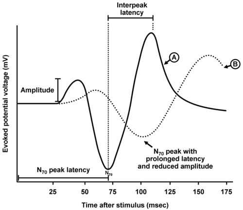
All four EP techniques involve the application of a stimulus that generates a neuronal response with measurement of that response. Typical recordings are expressed as a graph of time (in milliseconds) on the abscissa (i.e., x-axis) and voltage (mV) as the ordinate (i.e., y-axis) (Figure 20-1). The responses are very low voltage and require signal averaging to enhance their quality, that is, recorded waveforms are a composite of 50 to 100 or more measurements following multiple stimulation measurement cycles that serve to “subtract out” higher-voltage interference (e.g., electrocardiogram, electroencephalogram, and electrical noise within the operative suite). Peak voltages in the measured waveform refer to positive or negative deflections, designated by a P or N, respectively.

Two major characteristics of the measured waveform are usually described: amplitude and latency. Amplitude refers to the voltage difference between either a successive peak or a designated reference voltage. Latency refers to the length of time following stimulation for a specific peak to appear and is usually designated as a subscript of the positively or negatively deflected peak (e.g., N20 is a negatively deflected peak occurring 20 ms after stimulation). Interpeak latency refers to the time difference (in ms) between two different peaks.

Many factors influence the recorded waveform. Monitoring variables may include displacement of monitoring leads or electrical impedance, and improper patient positioning may compress a nerve, which can then interfere with conduction, even if the surgical site is remote (e.g., ulnar nerve compression in the prone position during spine operations).

Anesthetic agents have variable effects, depending on the EP modality and the anesthetic drug. Surgical factors (e.g., injury to a neural pathway from compression, reduced perfusion, or transection) are the reasons to intraoperatively monitor EPs. Physiologic variables include decreased O2 delivery to the neural pathway being monitored, which can occur with hypotension, anemia, and hypoxia. Hypothermia can also reduce the rate of neural conduction and impact recordings.

Brainstem auditory evoked responses

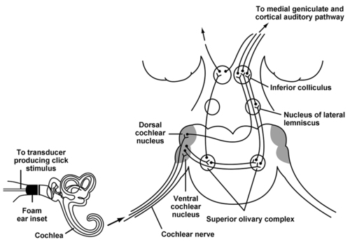
BAERs allow monitoring of the integrity of the auditory pathway both peripherally and centrally. Stimuli are loud, repetitive clicks produced by a device placed over or in the auditory canal or canals. Measurement of the response is from electrodes placed on the scalp or external ears to record contralateral and ipsilateral signals that have and have not decussated, respectively. BAER monitoring allows assessment of the acoustic transduction system of the middle and inner ear, the cochlear nerve (i.e., cranial nerve VIII), and the entire central auditory pathway rostrally to the primary auditory cortex located in the temporal lobe of the brain (Figure 20-2). Some anesthetic agents may cause minor changes in amplitude or latency of recorded waveforms; however, these changes are usually very small, even with large changes in anesthetic dose. Therefore, significant intraoperative BAER changes are usually indicative of a surgical trespass.

Potential indications for intraoperative BAERs include monitoring for microvascular decompression of cranial nerve V or VII, resection of tumors in the cerebellopontine angle, or resection of brainstem lesions, and in the intensive care unit BAERs may help in the declaration of brain death.

Placement of the cerebellar retractor during microvascular decompression of cranial nerve V or VII can stretch cranial nerve VIII and increase the risk of subsequent postoperative hearing impairment. Brainstem compression, direct trauma to cranial nerve VIII or its blood supply, or cerebellar retraction during resection of tumors in the cerebellopontine angle, such as acoustic neuromas, may result in injury to the auditory pathway.

Somatosensory evoked potentials

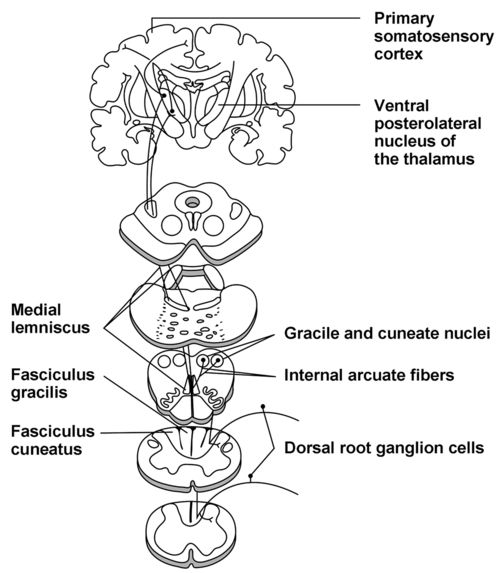
Monitoring of SSEPs permit assessment of major sensory pathways (within the dorsal column medial lemniscus) responsible for transmission of touch, vibration, and proprioception (Figure 20-3). These sensory pathways consist of first-order neurons originating at peripheral sensory receptors and entering the spinal cord to travel rostrally within the ipsilateral posterior column to synapse on the second-order neurons located within the gracile and cuneate nuclei at the cervicomedullary junction. These neurons then decussate and proceed rostrally through the brainstem as the medial lemniscus to reach the ventral posterolateral nucleus of the thalamus. The second-order neurons synapse here with third-order neurons that then travel rostrally to the primary somatosensory cortex located on the postcentral gyrus.

Stimulation and monitoring of SSEPs from the median, posterior tibial, or peroneal nerve are commonly performed. Measurement of the response can be accomplished by recording more proximally along the same nerve, over the spine, or from the contralateral scalp. Maintenance of adequate blood supply to the central portions of the pathway within the spinal cord is of critical importance when interpreting the results. Specifically, the dorsal columns within the spinal cord are supplied by the posterior spinal arteries. Accordingly, SSEP measurements are generally not reliable for the detection of ischemia in regions of the cord supplied by the anterior spinal artery (e.g., motor pathways).

SSEP monitoring can be used for procedures involving the spine (e.g., scoliosis surgery, spinal cord tumor resection, laminectomy with fusion, vertebral fractures with instability), posterior fossa operations (e.g., tumor resection), and vascular operations (e.g., carotid endarterectomy, aneurysm clipping). Anesthetic agents have variable effects on recordings of SSEPs. Stimulation and measurement of potentials from peripheral nerves and subcortical regions are often minimally affected by anesthetic agents. However, anesthetic agents can have a significant modulatory effect on the waveforms recorded from the cortex. In general, agents that cause increased latency and decreased amplitude include inhalation anesthetic agents (e.g., isoflurane, sevoflurane, desflurane), N2O, propofol, benzodiazepines, and opioids. The magnitude of the effect on either latency or amplitude varies among these agents. For example, inhalation anesthetic agents and benzodiazepines have a much greater suppressant effect of EPs than do opioids. Therefore, moderate to high doses of inhalation anesthetic agents or benzodiazepines may obliterate SSEP waveforms. On the other hand, reliable signal recording can often be accomplished even with high-dose opioid technique. Increased latency or decreased amplitude can also occur with ischemia or injury to the sensory pathway. Conversely, ketamine and etomidate increase the amplitude of SSEP waveforms. As such, these agents can actually be used to enhance signals due to their effect on amplitude. Neuromuscular blocking agents (NMBAs) have no significant effect on SSEPs.

Motor evoked potentials

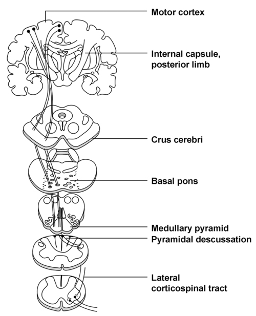
Unlike SSEP techniques that assess the integrity of afferent neural pathways, MEPs assess the efferent motor pathway: the corticospinal tract (Figure 20-4). Cell bodies of first-order motor neurons exist in the precentral gyrus of the frontal lobe. Axons extend via the internal capsule into the crux cerebri of the midbrain, basal pons, and pyramidal tracts of the medulla. In the caudal region of the medulla, these axons decussate and travel in the lateral region of the spinal cord (i.e., the lateral corticospinal tract) to synapse on secondary motor neurons. These secondary motor neurons then leave the ventral spinal cord, combine to form nerves and plexuses, and proceed to innervate muscles. Blood to the primary motor pathway comes from branches of the middle and anterior cerebral arteries in the cerebrum, the vertebrobasilar system in the brainstem, and the anterior spinal artery in the spinal cord.

Stimulation of MEPs can be accomplished from the cerebral cortex or spinal cord by applying either an electric or magnetic stimulus. Recording can be accomplished anywhere caudal to the site stimulated; however, recording within the muscle is most commonly used.

Intraoperative recording of MEPs can be used during spine operations (e.g., scoliosis correction, spinal tumor resection) or operations involving peripheral nerves (e.g., brachial plexus or peripheral nerve reconstruction/transposition). Intraoperative MEP recording may also provide valuable information during repair of thoracoabdominal aortic aneurysms. The artery of Adamkiewicz, a branch of the aorta, often has a variable location and supplies the lower two thirds of the anterior spinal cord; occlusion of the artery of Adamkiewicz either directly or via occlusion of the aorta at a site proximal to the artery’s origin places a large portion of the spinal cord at risk. MEP recording may also be used during cerebral aneurysm clipping, especially for aneurysms located within the middle cerebral, anterior cerebral, or vertebrobasilar systems because these vascular territories supply different parts of the motor pathway, and also for resection of tumors in the posterior fossa.

As with sensory EPs, MEPs are also subject to interference by anesthetic agents and physiologic variables. Electrically induced MEPs are less sensitive to anesthetic effects than are magnetically induced MEP signals. Utilization of multipulse (versus single-pulse) electrical stimulation will further reduce the sensitivity of MEPs to the effects of anesthetic agents and will improve signal quality because of summation and recruitment of a greater number of axons to transmit the stimulus. MEPs that do not involve cerebral cortical stimulation (i.e., stimulation at the level of the spinal cord or peripheral nerve) are less sensitive to the effects of anesthetic agents than are those involving cortical stimulation.

The use of NMBAs is not absolutely contraindicated when MEPS are used. In fact, low-dose infusions of NMBAs can be helpful to reduce noise within the MEP signal; however, complete neuromuscular blockade will result in loss of myogenically recorded MEPs. If NMBAs are used, compound motor action potentials can be recorded from a peripheral nerve with care taken to retain 5% to 10% of the original compound motor action potential. Inhalation anesthetic agents, N2O, propofol, barbiturates, and benzodiazepines produce decreased signal amplitude and increased signal latency, especially when given in high doses. The use of a continuous propofol infusion or low-dose inhalation anesthesia agents is typically compatible with successful MEP monitoring. Ketamine, etomidate, dexmedetomidine, and opioids have minimal effects on MEPs such that these agents can be used at moderate doses during MEP monitoring.

Intraoperative changes in evoked potential signals

If EPs are being recorded intraoperatively, the clinician should have a very systematic approach to assess the reason for changes in signals. Ischemia, injury, or transection of a neural pathway will generally result in either a decrease in signal amplitude with an increase in signal latency or a complete loss of signal (i.e., isoelectricity). The following should be considered to rule out anesthetic and monitoring-based causes:

Once anesthetic, physiologic, and monitoring-based causes have been ruled out or corrected and the signal amplitude and latency still do not improve, surgical causes for neural compromise should be sought and corrected, if possible.