Esophagectomy

Published on 11/04/2015 by admin

Filed under Surgery

Last modified 22/04/2025

Print this page

rate 1 star rate 2 star rate 3 star rate 4 star rate 5 star
Your rating: none, Average: 3 (1 votes)

This article have been viewed 1544 times

CHAPTER 6 Esophagectomy

BACKGROUND

The esophagus extends from the hypopharynx to the stomach. The cervical esophagus begins at the cricopharyngeus muscle and is approximately 5 cm in length. The thoracic esophagus, measured from the level of the first thoracic vertebra, is typically 20 to 25 cm in length. The blood supply to the esophagus is segmental and arises from the inferior thyroid arteries proximally and the left gastric artery distally. The aortic esophageal and bronchial arteries supply the mid-esophagus. The lymphatic drainage of the esophagus is extensive. Mucosal and submucosal lymphatics communicate along the entire length of the muscular esophagus; because of this, tumors of the esophagus have a tendency to spread longitudinally. Moreover, esophageal lymphatics drain to multiple regional beds and drainage may proceed in either a proximal or a distal direction. Lesions of the upper and middle thirds of the esophagus most often drain to the hilar, periesophageal, and supraclavicular nodes, whereas lesions of the distal third drain to the lesser curvature, left gastric, and celiac nodes. Notwithstanding, positive celiac nodes are found in up to 10% of metastatic tumors of the upper esophagus and distal esophageal tumors may drain to the hilar and supraclavicular nodes.

INDICATIONS FOR RESECTION

I. Esophageal Malignancies

II. Other Indications

image

Figure 6-1 Evaluation and treatment of esophageal cancer.

(Adapted from Townsend CM, Beauchamp RD, Evers BM, Mattox KL [eds]: Sabiston Textbook of Surgery: The Biological Basis of Modern Surgical Practice, 17th ed. Philadelphia, Saunders, 2004.)

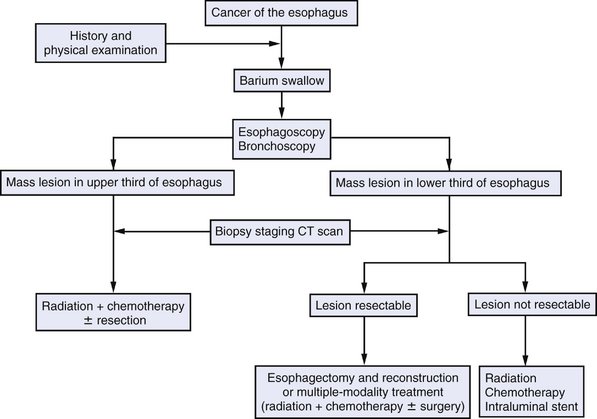
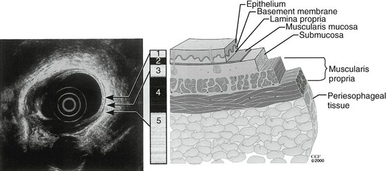
PREOPERATIVE EVALUATION

Although mortality rates have declined, particularly at high-volume centers, esophageal resection continues to be associated with significant morbidity, particularly in older patients and those with comorbidities. The preoperative evaluation should identify significant comorbidities that may increase perioperative risk. Pulmonary function studies, an electrocardiogram, and an echocardiogram should be performed. Cardiac catheterization and revascularization should be performed when appropriate. Smoking cessation and avoidance of alcohol should be encouraged. Patients who lost significant weight before surgery should receive preoperative nutritional support and reversal of malnutrition should be documented. On occasion, this may require enteral feedings via a feeding tube or intravenous parenteral nutrition. Oral hygiene should be optimized before surgery to lessen the risk of severe infection in the event of a leak from the cervical anastomosis.

image

Figure 6-2 Endoscopic ultrasonographic image demonstrating the layers of the esophageal wall.

(From Pearson FG, Cooper JD, Deslauriers J, et al [eds]: Esophageal Surgery. Philadelphia, Churchill Livingstone, 2002.)

COMPONENTS OF THE PROCEDURE AND APPLIED ANATOMY

Approaches to Esophagectomy

All approaches to esophagectomy include resection of the diseased segment of esophagus and restoration of gastrointestinal continuity. Generally, resections for cancer require disease-free margins of at least 5 cm. A variety of approaches may be used, including Ivor-Lewis, performed through a right thoracotomy and an abdominal incision; en bloc, performed through a left thoracoabdominal incision; and transhiatal, performed through abdominal and left cervical incisions and discussed later. Clinical outcomes after esophagectomy are more clearly related to the stage of the disease than to the choice of operation.

Exposure and Creation of the Gastric Conduit

image

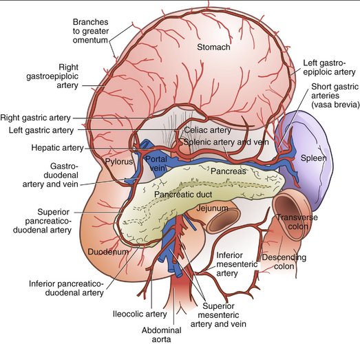
Figure 6-3 Blood supply to the stomach.

(From Zuidema G: Shackelford’s Surgery of the Alimentary Tract, 4th ed. Philadelphia, Saunders, 1995.)

image

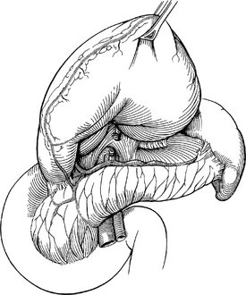
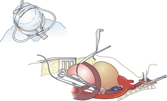
Figure 6-4 Mobilization of the stomach and ligation of the left gastric artery.

(From Cameron JL [ed]: Current Surgical Therapy, 8th ed. Philadelphia, Mosby, 2004.)

image

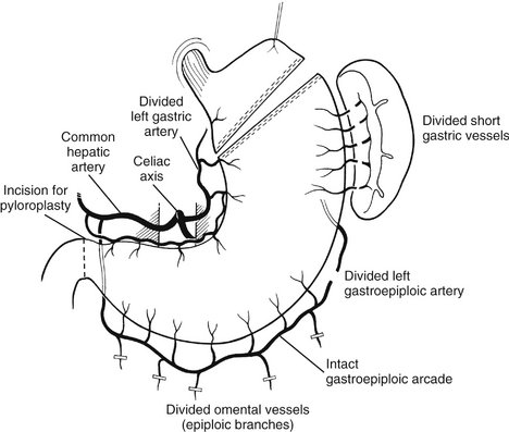
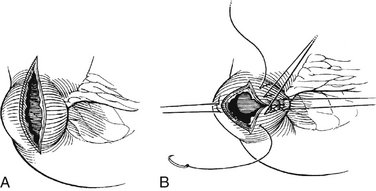
Figure 6-5 Preparation of the gastric conduit.

(From Khatri VP, Asensio JA: Operative Surgery Manual. Philadelphia, Saunders, 2003.)

image

Figure 6-6 Pyloroplasty. A, A longitudinal incision is made through the pylorus. B, The incision is closed transversely.

(From Cameron JL [ed]: Current Surgical Therapy, 8th ed. Philadelphia, Mosby, 2004.)

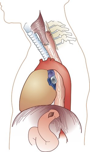
Mobilization of the Esophagus and Anastomosis

image

Figure 6-7 Mobilization of the esophagus.

(From Townsend CM, Beauchamp RD, Evers BM, Mattox KL [eds]: Sabiston Textbook of Surgery: The Biological Basis of Modern Surgical Practice, 17th ed. Philadelphia, Saunders, 2004.)

image

Figure 6-8 Transhiatal esophagectomy.

(Adapted from Orringer MB, Sloan H: Esophagectomy without thoracotomy. J Thorac Cardiovasc Surg 76:643–654, 1978.)

COMPLICATIONS