Erythroderma

Published on 19/03/2015 by admin

Filed under Dermatology

Last modified 19/03/2015

Print this page

rate 1 star rate 2 star rate 3 star rate 4 star rate 5 star
Your rating: none, Average: 0 (0 votes)

This article have been viewed 2641 times

Erythroderma

Tang Ngee Shim and John Berth-Jones

Evidence Levels:  A Double-blind study  B Clinical trial ≥ 20 subjects  C Clinical trial < 20 subjects  D Series ≥ 5 subjects  E Anecdotal case reports

image

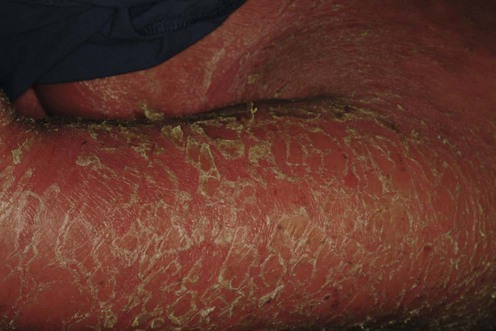
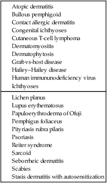
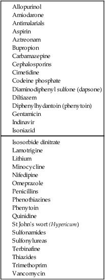
Erythroderma (exfoliative dermatitis) is defined as inflammation of at least 90% of the body surface, characterized by generalized erythema and a variable degree of scaling or desquamation. Erythroderma is commonly the result of generalization of a pre-existing chronic dermatosis or a systemic disease (see Table 75.1). These include genodermatoses and congenital disorders such as severe ichthyoses and ichthyosiform erythrodermas; severe cases of dermatoses such as psoriasis, atopic or seborrheic or contact allergic dermatitis; cutaneous T-cell lymphoma; allergic reactions to drugs (see Table 75.2); and internal malignancies (especially lymphoma and other lymphoreticular malignancies). Some cases develop without any apparent trigger and erythroderma remains ‘idiopathic’ in up to 25% of the cases.

Management strategy

Erythroderma, especially when fulminant, is a life-threatening state of skin failure that demonstrates vividly that the skin is as vital to life as any internal organ. The dangers arise from the loss of an effective barrier to entry of bacteria, from the loss of thermoregulation, from increased fluid loss through evaporation or exudation, from loss of protein due to the increased proliferative and metabolic activity that accompanies uncontrolled desquamation, and from the risk of high-output cardiac failure. All these hazards are greatest in the very young and the elderly. Many patients who are otherwise healthy can tolerate a chronic or permanently erythrodermic state. By contrast, very young and elderly patients with fulminant disease may develop septicemia and die within a matter of hours.

Many aspects of the management of a patient with erythroderma are similar regardless of the etiology, and it is often necessary to treat a case without knowing the cause. However, for optimal longer-term management it is vital to establish a more precise diagnosis whenever possible. In many cases there has been pre-existing skin disease and the diagnosis may be quite clear from the history, but when erythroderma arises de novo establishing the cause can be difficult or impossible. A comprehensive drug history should include all over-the-counter and herbal remedies such as St John’s wort. Severe pruritus may suggest atopic eczema or lymphoma. Although physical examination may be entirely non-specific, there may be clues such as bullae, indicating the presence of bullous pemphigoid or pemphigus foliaceus. Severe scaling is suggestive of psoriasis. Pityriasis rubra pilaris shows islands of uninvolved skin within erythrodermic regions with ‘orange’ palmoplantar keratoderma and follicular keratotic plugs on the knees, elbows and dorsal aspects of the hands and toes. Sparing of the flexures (‘deck-chair sign’) may suggest papuloerythroderma of Ofuji. Lymphadenopathy is often present, but is more often reactive than malignant.

Histology of the skin is often non-specific and rarely pathognomonic. Occasionally, however, the presence of atypical large lymphocytes will point to a diagnosis of cutaneous T-cell lymphoma, or there may be features suggestive of psoriasis, a lichenoid reaction, or pityriasis rubra pilaris. Repeated or multiple biopsies are sometimes helpful. Immunofluorescence for immunoglobulin deposition should be performed if an immunobullous disease is suspected.

Patients with acute onset of erythroderma are usually best managed in hospital because frequent observations and intensive supportive care are required, and bed rest may be highly therapeutic. All unnecessary medications should be discontinued. Good skin care is essential. Frequent applications of abundant quantities of bland emollients such as petrolatum are required to soothe the skin, and these help to partially restore the barrier. Oatmeal baths and wet dressings to weeping or crusted sites should be followed by application of bland emollients and low-potency topical corticosteroids. Careful attention must be paid to hydration and nutrition.

The use of more active pharmaceutical intervention requires careful consideration. Some patients with erythroderma have multiple drug allergies. Immunosuppressive drugs may be considered to be contraindicated if malignancy is suspected (particularly cutaneous lymphoma). Topical treatments may be far more irritating than expected, and systemic absorption will be greater than usual. Prophylactic antibiotics such as erythromycin are often given orally. Corticosteroids are often applied topically. Antihistamines are often prescribed, but act largely as sedatives.

If a diagnosis can be established, withdrawal of a causal drug or specific treatment for an underlying dermatosis, combined with the supportive measures described above, will usually produce a rapid improvement in the erythroderma. When a firm diagnosis cannot be made, treatment may have to be directed at the most likely cause, based on the clinical and histologic features.

Specific investigations