Published on 19/03/2015 by admin
Filed under Dermatology
Last modified 22/04/2025
This article have been viewed 2175 times
Jean Revuz
Evidence Levels: A Double-blind study B Clinical trial ≥ 20 subjects C Clinical trial < 20 subjects D Series ≥ 5 subjects E Anecdotal case reports
Erythema multiforme (EM) is a distinct cutaneous reaction pattern to a variety of stimuli, predominantly herpes simplex virus (HSV) infection. It usually runs a self-limiting course but has a tendency to recur. It is defined by the presence of ‘typical’ three-zone target lesions, with a predominantly acral distribution. The presence of mucosal involvement at more than one site distinguishes EM major from EM minor. A specific variant has mucosal lesions only, without skin involvement. EM major can be distinguished from Stevens–Johnson syndrome which lacks typical target-like lesions and acral location but, instead, irregular macule or atypical targets and a truncal location. EM is frequently misdiagnosed in cases of urticaria and more rarely in cases of cutaneous lupus, vasculitis, erythema annulare, and drug eruption.
In 30–50% of cases the etiology of EM is unknown. The most commonly recognized precipitant is HSV infection, both types I and II. HSV-specific DNA has been isolated from lesional tissue in 60–70% of cases. HSV particles are found in the circulating precursors of epidermal Langerhans cells. A variety of other viral infections (orf, VZV, EBV, CMV, HIV), bacterial infections (mainly Mycoplasma pneumoniae), and fungal infections (mainly histoplasmosis), have been implicated. An extensive list of drugs have been reported to trigger EM but most cases if not all are the result of a confusion with Stevens-Johnson syndrome. Rare cases are attributed to contact allergy.
Acute episodes of EM need only symptomatic treatment in most cases. Recurrent EM which may severely affect quality of life has a well-recognized preventive treatment in case of HSV infection.
There are no double-blind or open trials of treatments for acute episodes of EM. Most cases, particularly EM minor, run a self-limiting course. Symptomatic measures include oral antihistamines and mild- to moderate-potency topical corticosteroids to reduce pruritus. Underlying conditions, mainly Mycoplasma pneumoniae infection, should be treated. Recurrent EM (>6 attacks per year) may respond to long-term acyclovir. In acyclovir-resistant cases a variety of other therapies can be helpful (see below).
Mucosal manifestations of EM are a source of morbidity and occur in up to 70% of cases. The commonest sites affected are the buccal mucosa and lips. Symptomatic measures include mouthwashes, a soft diet, topical anesthetics (lidocaine gel, benzocaine lozenges or 0.15% benzydamine hydrochloride), and topical corticosteroids (e.g., 0.1% triamcinolone acetonide paste). Budesonide or beclomethasone inhalers (one puff three to four times daily) provide an alternative method of delivering local corticosteroid to the inflamed mucosal surfaces. Short courses of high-dose oral prednisolone may be needed for severe oral disease. Strict eye care to reduce secondary infection and scarring includes saline washes for removal of crusts, local antibiotics, and frequent debridement of tarsal and bulbar conjunctival adherences.
Histology/immunofluorescence
EM is a clinical diagnosis. Histology, with direct immunofluorescence, can be useful in atypical cases to exclude other bullous diseases that present with oral manifestations, such as pemphigus vulgaris or cicatricial pemphigoid.
Investigations directed at determining the underlying trigger factors include culture or serological testing for HSV or other infections, especially Mycoplasma pneumoniae, as indicated by clinical findings.
Schofield JK, Tatnall FM, Leigh IM. Br J Dermatol 1993; 128: 542–5.
A review of 65 patients with recurrent EM: 71% had episodes triggered by HSV infection. In one patient, EM was related to the menstrual cycle and could be precipitated by intramuscular progesterone injection. Treatment with standard doses of acyclovir for HSV was relatively disappointing; continuous acyclovir 400 mg twice daily for six months was more effective, with remission in some responders. Some patients responded to dapsone, antimalarials, azathioprine, and human immunoglobulin.
Tatnall FM, Schofield JK, Leigh IM. Br J Dermatol 1995; 132: 267–70.
Acyclovir 400 mg twice daily for 6 months suppressed EM in seven of 11 patients (including one with apparently idiopathic EM). Two patients went into complete remission.
A therapeutic trial of acyclovir is justified even when clinical evidence of HSV is lacking. Acyclovir 400 mg twice daily can be administered for 6 months to 2 years because it has a good long-term safety profile. It is ineffective in an acute episode once the herpetic lesion or EM eruption has developed.
Kerob D, Assier-Bonnet H, Esnault-Gelly P, Blanc F, Saiag P. Arch Dermatol 1998; 134: 876–7.
A reduced response to acyclovir may be due to the low oral bioavailability of the drug, and one of the second-generation antivirals, such as valacyclovir (500 mg daily) or famciclovir (250 mg twice daily) may need to be substituted.
Wetter DA, Davis MD. J Am Acad Dermatol 2010; 62: 45–53.
Of 48 patients HSV was responsible in 11 (23%); the cause remained unknown in 28 (58%). Systemic corticosteroids were used in most patients. Sixteen of 33 patients receiving continuous antiviral treatment had either partial or complete disease suppression. Mycophenolate mofetil provided partial or complete response in six of eight patients.
Vitiello M, Echeverria B, Elgart G, Kerdel F. J Cutan Med Surg 2011; 15: 115–17.
Ganciclovir was used successfully.
Mahendran R, Grant JW, Norris PG. Dermatology 2000; 200: 281–2.
Dapsone 100 mg daily was effective in controlling EM in a patient with ovarian malignancy.
Farthing PM, Maragou P, Coates M, Tatnall F, Leigh IM, Williams DM. J Oral Pathol Med 1995; 24: 9–13.
In this series of 82 patients with typical cutaneous EM, 70% had oral mucosal involvement. Five patients with resistant disease were controlled with azathioprine 100–150 mg daily.
Jones RR. Br J Dermatol 1981; 105: 465–7.
Azathioprine 100–150 mg daily was effective in two patients and permitted reduction of the corticosteroid dosage.
Moisson YF, Janier M, Civatte J. Br J Dermatol 1992; 126: 92–3.
Recurrent EM responded to thalidomide 100–200 mg daily in two patients.
Cherouati K, Claudy A, Souteyrand P, Cambazard F, Vaillant L, Moulin G, et al. Ann Dermatol Venereol 1996; 123: 375–7 (In French).
Thalidomide reduces the duration of episodes of recurrent EM by 11 days on average; dramatically effective in the exceptional continuous variant. Remission can be maintained with low-dose (25–50 mg daily) thalidomide.
Horio T, Danno K, Okamoto H, Miyachi Y, Imamura S. J Am Acad Dermatol 1983; 9: 77–81.
Fourteen of 16 subjects with EM (six related to HSV infection) responded within 1 week to 300 mg potassium iodide three times daily. Gastrointestinal and cutaneous side effects can occur with this treatment.
Ting HC, Adam BA. Dermatologica 1984; 169: 175–8.
Thirteen patients with EM minor treated with systemic corticosteroids were compared with 12 treated without. Apart from a shorter duration of fever, the corticosteroid-treated group did not respond better than the non-corticosteroid-treated group.
There is no reason to treat EM minor with steroids. This drug may still be useful in EM major.
Lozada-Nur F, Huang MZ, Zhou GA. Oral Surg Oral Med Oral Pathol 1991; 71: 283–7.
Clobetasol propionate 0.05% ointment mixed 1 : 1 with Orabase paste two to three times daily was helpful in four patients with chronic oral EM.
Lozada F, Silverman S Jr. Arch Dermatol 1980; 116: 898–901.
Topical application of 0.05% fluocinonide in an adhesive base was used in 16 patients with oral EM. All responded to therapy, and remission was induced in some cases.
Sanchis JM, Bagán JV, Gavaldá C, Murillo J, Diaz JM. J Oral Pathol Med 2010; 39: 747–52.
Systemic corticosteroids are effective in controlling the outbreaks; their use as maintenance therapy is not clearly indicated.
Lozada F. Oral Surg Oral Med Oral Pathol 1981; 52: 257–63.
In this open trial, two patients with oral EM required lower doses of prednisolone (15–20 mg on alternate days) when treated simultaneously with azathioprine (50 mg daily).
Bean SF, Quezada RK. JAMA 1983; 249: 2810–12.
In this retrospective study, patients with severe recurrent oral EM involvement were treated with prednisolone 40–60 mg daily, subsequently tapered over 2 to 3 weeks. This reduced the time taken for oral erosions to heal, but did not influence recurrences.
Some authorities, however, believe that use of corticosteroids in EM increases the frequency and chronicity of attacks.
The use of normal human immunoglobulins and antimalarials was reported in a few patients in this study. Intramuscular human immunoglobulin 750 g once a month caused suppression of EM in 11 of 13 patients. One responder remained in remission after therapy was discontinued. There was a 50% success rate with antimalarials (both hydroxychloroquine and mepacrine) used in four patients.
Dumas V, Thieulent N, Souillet AL, Jullien D, Faure M, Claudy A. Br J Dermatol 2000; 142: 1248–9.
One patient with recurrent EM and hepatitis C virus infection responded to two courses of IFN-α (9 MU weekly for 6 and 8 months, respectively).
Geraminejad P, Walling HW, Voigt MD, Stone MS. J Am Acad Dermatol 2006; 54: S18–21.
A patient with recurrent EM associated with hepatitis C had been in complete remission for 6 years after treatment with IFN-α. A new recurrence was successfully treated with IFN-α in the absence of recurring virus C infection.
Interferon may be a first-line treatment for patients with hepatitis C.
Wojnarowska F, Greaves MW, Peachey RD, Drury PL, Besser GM. J Roy Soc Med 1985; 78: 407–8.
EM linked to the luteal phase of the menstrual cycle was controlled with tamoxifen.
Brody I. Br J Dermatol 1981; 104: 191–4.
Treatment of the skin at the site of the herpetic infection with zinc sulfate solution prevented relapse of post-herpetic EM over a 2-year period of observation in one patient. For the skin, 0.025–0.05%, and for the oral mucous membrane 0.01–0.025%, zinc sulfate solution was used.
Kurkcuoglu N, Alli N. J Am Acad Dermatol 1989; 21: 814–15.
EM resistant to acyclovir responded to cimetidine 400 mg three times daily in one patient.
Wilkel CS, McDonald CJ. Arch Dermatol 1990; 126: 397–8.
High-dose cyclosporine (5–10 mg/kg daily) suppressed an atypical bullous eruption with histologic features of EM. The use of cyclosporine permitted tapering of the corticosteroid dosage.
Martinez AE, Atherton DJ. Pediatr Dermatol 2000; 17: 87–90.
A child with recurrent EM major (associated with severe ulcerative stomatitis, conjunctival inflammation, and urethritis) responded to pulsed intravenous methylprednisolone 20 mg/kg daily for 3 consecutive days, but not to oral prednisolone 3 mg/kg daily. Intravenous methylprednisolone stopped progression of the acute attack, and repeated treatments induced remission.
Treatment of Skin Disease Comprehensive Therapeutic Strategies 4e
WhatsApp us


 Acyclovir
 Dapsone
 Azathioprine
 Thalidomide
 Potassium iodide
 Topical corticosteroid
 Levamisole
 Systemic corticosteroid
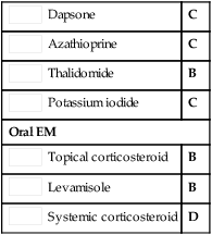
 Antimalarials
 Human immunoglobulin
 Interferon-α
 Tamoxifen
 Zinc sulfate
 Cimetidine
 Cyclosporine
 Pulsed methylprednisolone
