Published on 18/03/2015 by admin
Filed under Dermatology
Last modified 22/04/2025
This article have been viewed 2692 times
Pamela Fiandeiro, Emma Benton and Ian Coulson
Evidence Levels: A Double-blind study B Clinical trial ≥ 20 subjects C Clinical trial < 20 subjects D Series ≥ 5 subjects E Anecdotal case reports
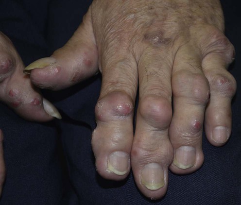
Erythema elevatum diutinum (EED) is a rare neutrophilic dermatosis consisting of violaceous, brown or red papules, plaques, nodules, and occasionally vesicobullous lesions over the extensor surfaces of the joints and buttocks. EED is thought to be a form of immune complex-mediated vasculitis, although its etiology remains unclear. Infections (including HIV and streptococcal), hematologic abnormalities, autoimmune diseases, and other conditions have been associated.
EED is a chronic disease; there are only a few well-documented instances of spontaneous long-term resolution.
Dapsone 100 mg daily remains the initial treatment of choice. The response may be partial and dose dependent. Corticosteroids have also been effective in patients with EED. Topical betamethasone and topical fluocinolone acetonide have been used under occlusion with good effect. In other patients both intralesional and systemic corticosteroids (prednisolone 30–40 mg daily) have produced favorable responses.
Sulfonamides (sulfamethoxypyridazine 500 mg once daily and sulfapyridine 0.5–1 g three times daily), nicotinamide 100 mg three times daily, colchicine 0.5 mg twice daily with 0.5 mg three times daily for 3 to 4 days to abate minor disease flares, and chloroquine 300 mg daily have produced resolution of lesions.
EED has been reported in association with diseases such as HIV, hematological disorders, inflammatory bowel disease, celiac disease, verrucous carcinoma, systemic lupus erythematosus, primary Sjögren syndrome, ophthalmic disorders (peripheral keratitis), pulmonary lymphoepithelioma-like carcinoma (possibly as part of a paraneoplastic syndrome) and evidence of these should be sought. Drug induced EED may result from interferon-β, erythropoietin, antituberculosis chemotherapy, and cisplatin exposure.
Full blood count
Immunoglobulins and serum electrophoresis/ immunofixation electrophoresis
Antineutrophil cytoplasmic antibodies
Antireticulin and antigliadin antibodies
HIV risk factors enquiry
Histology
Lee AY, Nakagawa H, Nogita T, Ishibashi Y. J Cutan Pathol 1989; 16: 211–17.
An electron microscopic study of a patient with EED demonstrating that the characteristic histopathologic changes in early lesions include leukocytoclastic vasculitis and massive dermal infiltrate of neutrophils, histiocytes/macrophages, and Langerhans cells. In late lesions the inflammatory infiltrate is replaced by fibrosis with a dermal infiltrate of lymphocytes, histiocytes/macrophages, and Langerhans cells.
Wahl CE, Bouldin MB, Gibson LE. Am J Dermatopathol 2005; 27: 397–400.
The vascular endothelium of EED stains positive for CD31, CD34, VEGF, and factor VIII, and negative for factor XIIIa, TGFβ, and LANA. This pattern does not distinguish it from similar-appearing lesions. Therefore, the chronic and recurrent nature of EED is the primary means of distinguishing it from entities that are clinically and histologically similar.
Yiannias JA, El-Azhary RA, Gibson LE. J Am Acad Dermatol 1992; 26: 38–44.
Of 13 patients with EED, six had associated hematologic abnormalities, with IgA monoclonal gammopathy occurring most frequently.
Chowdhury MMU, Inaloz HS, Motley RJ, Knight AG. Int J Dermatol 2002; 41: 368–70.
The technique of immunofixation electrophoresis is more sensitive than immunoelectrophoresis and uses a combination of zone electrophoresis and immunoprecipitation with specific antisera to detect monoclonal immunoglobulins or light chains at very low concentrations in serum and urine. This is useful in EED because patients may have associated paraproteinemias, which in some cases may undergo malignant transformation.
The authors note that in monoclonal disorders there is extensive asymptomatic tumor proliferation and possible malignant transformation in 20% of patients during long-term follow-up. It was recommended that there should be lengthy follow-up and monitoring for patients with both EED and IgA paraproteinemia because of the risk of progression to IgA myeloma.
Crichlow SM, Alexandroff AB, Simpson RC, Saldanha G, Walker S, Harman KE. Br J Dermatol 2011; 164: 675–7.
Two studies evaluating the prevalence of antineutrophil cytoplasmic antibodies (ANCAs) in EED. IgA ANCAs were present in all patients with EED.
Muratori S, Carrera C, Gorani A, Alessi E. Br J Dermatol 1999; 141: 335–8.
The largest case series of patients with EED and HIV infection. Streptococcal infection seemed to trigger exacerbations in four of five patients. EED can stimulate Kaposi’s sarcoma and bacillary angiomatosis, which may be particularly confusing in the context of an HIV seropositive patient. Histopathological confirmation of the diagnosis is therefore advocated.
Vaiyavatjamai P, Wattanakrai P. J Eur Acad Dermatol Venereol 2011; 25: 741–2.
Lekhanont K, Patarakittam T, Mantachote K, Waiyawatjamai P, Vongthongsri A. Ophthalmology 2011; 118: 927–33.
Nair SR, Viswanath V, Sonavane AD, Doshi AC, Parab MG, Torsekar RG. Indian J Dermatol Venereol Leprol 2010; 76: 420–2.
Maksimovic L, Duriez P, Lascaux-Cametz A-S, Andre C, Bagot M, Revuz J, Ortonne N. Ann Dermatol Venereol 2010; 137: 386–90.
Chan Y, Mok CC, Tang WYM. Rheumatol Int 2011; 31: 259–62.
The above citations indicate the range of pathologies associated with EED.
Wilkinson SM, English JS, Smith NP, Wilson-Jones E, Winkelmann RK. Clin Exp Dermatol 1992; 17: 87–93.
Dapsone 100 mg daily was the most effective therapy in this series of 13 patients with EED. Responses were often partial and dose dependent. Other effective treatments included sulfa drugs, corticosteroids, and chloroquine.
Dapsone may be ineffective once nodules appear; treatment of associated concomitant conditions is often beneficial to EED outcome.
Maruthappu T, Tharakaram S, Calonje E, Shirlaw PJ, Setterfield J. Br J Dermatol 2012; 167: 222–4.
The authors report a case of EED with classical cutaneous lesions and recurrent oral ulceration. The cutaneous lesions responded rapidly to dapsone 50 mg daily but the oral ulcers took up to 6 weeks before marked improvement was seen.
Seneschal J, Guillet S, Ezzedine K, Taïeb A, Milpied B. Dermatology (Basel) 2012; 224: 115–19.
This patient responded well to dapsone but developed hypersensitivity reaction with DRESS, confirmed in vitro by the presence of circulating dapsone-specific T cells. EED relapsed after discontinuing dapsone. A tolerance induction protocol to dapsone was successfully used.
Smitha P, Sathish P, Mohan K, Sripathi H, Sachi G. Int J Dermatol 2011; 50: 989–91.
This case report of EED in an HIV positive patient was treated with dapsone 100 mg daily along with topical clobetasol propionate and fusidic acid combination cream twice daily to be applied to the lesions. The dapsone was stopped due to a significant drop in hemoglobin. Comment on the extent of resolution of the lesions was not made by the authors.
Shimizu S, Nakamura Y, Togawa Y, Kamada N, Kambe N, Matsue H. Br J Dermatol 2008; 159: 733–5.
Report of a 64-year-old woman with EED and Sjögren syndrome. Administration of oral dapsone for 1 month resolved her symptoms.
Aldave AJ, Shih JL, Jovkar S, McLeod SD. Am J Ophthalmol 2003; 135: 389–90.
Report of a 25-year-old with EED who was diagnosed 15 months later with an inflammatory peripheral keratitis of the left eye (thought to represent an ocular extension of the patient’s cutaneous vasculitis). Dapsone therapy resulted in rapid resolution of both the cutaneous and ocular inflammation.
Di Giacomo TB, Marinho RT, Nico MMS. Int J Dermatol 2009; 48: 290–2.
Report of a 45-year-old woman with cutaneous lesions for 3 years was diagnosed with EED and successfully treated with dapsone 100 mg daily. Residual hyperpigmented macules and fibrotic nodules remained at 3 months.
Suárez J, Miguélez M, Villalba R. Br J Dermatol 1998; 138: 717–18.
Report of a 37-year-old woman with HIV-1 infection and EED whose lesions completely resolved over 3 weeks, after treatment with oral antiretroviral therapy and oral dapsone 100 mg once daily.. The authors concluded that the therapeutic approach to patients with EED associated with HIV infection should focus on reducing circulating P24 antigen with adequate antiretroviral therapy in addition to conventional dapsone treatment.
LeBoit PE, Cockerell CJ. J Am Acad Dermatol 1993; 28: 919–22.
A clinicopathologic study of four patients with HIV infection who had unusual nodular lesions of EED. None of the patients responded to treatment with oral dapsone, and the authors commented that this observed lack of response may reflect the preponderance of fibrosis rather than neutrophils in these advanced lesions.
Takahashi H, Fukami Y, Honma M, Ishida-Yamamoto A, Iizuka H. J Dermatol 2012; 39: 486–7.
In this case of EED with an unusual distribution on the soles, fingers, trunk, and feet accompanied with ulcerations, the patient initially failed to respond to oral dapsone 75 mg daily but improved with the addition of cyclosporine 4 mg/kg/day.
Kohler IK, Lorincz AL. Arch Dermatol 1980; 116: 693–5.
A 60-year-old woman with EED which cleared completely following 4 weeks of treatment with oral nicotinamide 100 mg three times daily and oral tetracycline hydrochloride 250 mg four times daily. Following this, oral nicotinamide alone was sufficient for disease suppression.
Henriksson R, Hofer PA, Hörnqvist R. Clin Exp Dermatol 1989; 14: 451–3.
A case report of a 68-year-old man with EED refractory to treatment with oral dapsone who responded well to treatment with colchicine 0.5 mg twice daily over 6 weeks. Minor flares were abated temporarily increasing colchicine to 0.5 mg 3 times a day for 3 to 4 days without provoking diarrhea.
Tasanen K, Raudasoja R, Kallioinen M, Ranki A. Br J Dermatol 1997; 136: 624–7.
A 47-year-old woman who presented with typical EED in whom previously undiagnosed celiac disease was found. Treatment with dapsone was partially effective, but complete healing of the EED lesions was achieved only after the introduction of a strict gluten-free diet. Maintenance treatment with a gluten-free diet only was required.
Bernard P, Bedane C, Delrous JL, Catanzano G, Bonnetblanc JM. J Am Acad Dermatol 1992; 26: 312–15.
A 69-year-old man with a history of relaping polychondritis developed EED, which responded to treatment with oral cyclophosphamide 100 mg daily and prednisolone 20 mg daily. Cyclophosphamide was discontinued after 2 months and the prednisolone was subsequently tapered to 15 mg daily. This association suggests that EED may represent a cutaneous manifestation of systemic vasculitis.
Chow RK, Benny WB, Coupe RL, Dodd WA, Ongley RC. Arch Dermatol 1996; 132: 1360–4.
Intermittent plasma exchange has been reported to successfully control EED associated with IgA paraproteinemia.
Hernández-Cano N, De Lucas R, Lázaro TE, Mayor M, Burón I, Casado M. Pediatr Dermatol 1998; 15: 411–12.
Lesions of EED resolved following a reduction in cyclosporine dosage in a 10-year-old patient who had previously received a cadaveric hepatic allograft because of Alagille disease.
Buahene K, Hudson M, Mowat A, Smart L, Ormerod AD. Clin Exp Dermatol 1991; 16: 204–6.
A 58-year-old woman developed EED during severe acute exacerbation of ulcerative colitis, which resolved following a colectomy.
Yoshii N, Kanekura T, Higashi Y, Oyama K, Azagami K, Kanzaki T. Clin Exp Dermatol 2007; 32: 211–13.
A 74-year-old man developed EED. The lesions on his limbs responded well to the nicotine patches that released 6.25 mg nicotine applied every 24 hours. The penile lesions remained recalcitrant to the nicotine patches as well as dapsone, requiring treatment with methylprednisolone (40 mg/day).
Liu T-C, Chen I-S, Lin T-K, Lee JY-Y, Kirn D, Tsao C-J. Lung Cancer 2009; 63: 151–3.
A 45-year-old female was diagnosed with pulmonary lymphoepithelioma-like carcinoma and EED. She was treated with platinum-based chemotherapy and thoracic radiotherapy which resulted in complete remission in both the pulmonary lymphoepithelioma-like carcinoma and EED.
Treatment of Skin Disease Comprehensive Therapeutic Strategies 4e
WhatsApp us


 Dapsone
 Dapsone plus antiretrovirals in HIV-associated disease
 Dapsone with cyclosporine
 Corticosteroids
 Sulfonamides
 Chloroquine
 Nicotinamide and tetracycline
 Colchicine
 Dapsone with cyclosporine
 Gluten-free diet
 Cyclophosphamide
 Plasma exchange
 Reduction in cyclosporine dose
 Colectomy
 Transdermal nicotine patches
 Methylprednisolone