Published on 19/03/2015 by admin
Filed under Dermatology
Last modified 22/04/2025
This article have been viewed 3140 times
70
Christine Soon and John Berth-Jones
Evidence Levels: A Double-blind study B Clinical trial ≥ 20 subjects C Clinical trial < 20 subjects D Series ≥ 5 subjects E Anecdotal case reports
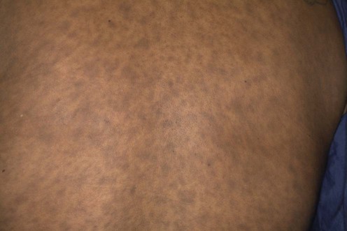
Erythema dyschromicum perstans (EDP) is an acquired, generalized dermal hypermelanosis of unknown etiology. Clinically it presents as asymptomatic, ashen-gray-blue macules of varying sizes, most commonly on the trunk and proximal extremities. Variable components include erythema and papulation. It has been reported most frequently in dark-skinned Latin-American people, although all racial groups can be affected. EDP has similarities to lichen planus pigmentosus and the ‘ashy dermatosis’ of Ramirez, although the precise relationship of these conditions has yet to be established.
No controlled trials have been reported. Although EDP may persist for many years, there have been reports of spontaneous resolution. Camouflage creams can be prescribed for cosmetic purposes. The treatments discussed below have also been tried, with varying success. Treatments that are reportedly ineffective include sun protection, peeling lotions, antibiotics, topical hydroquinone, topical corticosteroid therapy, antimalarials, and griseofulvin.
Biopsy
There is vacuolar degeneration of the basal layer associated with pigmentary incontinence. Dermal vessels are surrounded with an infiltrate of lymphocytes and histiocytes, and there are many melanophages present.
EDP may need to be differentiated from the late stage of pinta. Dark-field examination and serological tests for syphilis should be carried out to exclude this treponematosis in suspected cases.
Idiopathic eruptive macular pigmentation is a similar condition. Histology demonstrates that the pigment is located in the basal layer of the epidermis, and the lichenoid inflammation characteristic of EDP is not present.
Torrelo A, Zaballos P, Colmenero I, Mediero IG, de Prada I, Zambrano A. J Eur Acad Dermatol Venereol 2005; 19: 422–6.
No treatment was used. In six of these cases the eruption cleared or improved during follow-up ranging from one to five years.
Bhutani LK. Br J Dermatol 1979; 100: 473–4.
Lichen planus pigmentosus is considered by some to be the same entity as EDP. Vitamin A was prescribed in pulses of 100 000 units daily for 15 days. Up to 10 such pulses were given. Of the 140 patients, 28 showed a ‘good’ to ‘excellent’ response (50–100% clearance).
Kontochristopoulos G, Stavropoulos P, Panteleos D, Aroni K. Int J Dermatol 1998; 37: 796–8.
Two cases of EDP responded to dapsone 100 mg daily. The duration of therapy ranged from 8 to 12 weeks.
Persechino S, Caperchi C, Cortesi G, Persechino F, Raffa S, Pulcini F, et al. Eur J Dermatol 2011; 21: 261–2.
Three cases demonstrated marked decrease in pigmentation on treatment with dapsone 100 mg daily for 3 months.
Osswald SS, Proffer LH, Sartori CR. Cutis 2001; 68: 25–8.
One case of EDP with active inflammatory areas responded to 3 weeks of oral corticosteroid therapy. The authors do not state the dose.
Baranda L, Torres-Alvarez B, Cortes-Franco R, Moncada B, Potales-Perez DP, Gonzalez-Amaro R. Arch Dermatol 1997; 133: 325–9.
A prospective clinical and immunohistochemical study indicating that clofazimine reduces the inflammatory response in EDP. Four out of six patients treated with clofazimine 100 mg/day showed marked improvement after 3 months of treatment.
Treatment of Skin Disease Comprehensive Therapeutic Strategies 4e
WhatsApp us


 No therapy
 Vitamin A
 Dapsone 100 mg/day for 3 months
 Oral corticosteroid therapy
 Clofazimine 100 mg/day for 3 months