Published on 18/03/2015 by admin
Filed under Dermatology
Last modified 22/04/2025
This article have been viewed 3356 times
Christie G. Regula and Bryan E. Anderson
Evidence Levels: A Double-blind study B Clinical trial ≥ 20 subjects C Clinical trial < 20 subjects D Series ≥ 5 subjects E Anecdotal case reports
Erythema annulare centrifugum
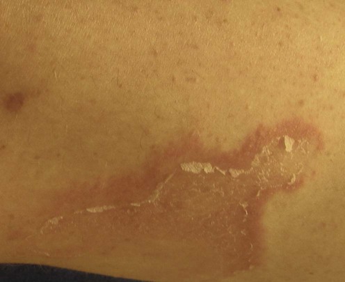
 (EAC) is a gyrate erythema characterized by minimally pruritic, polycyclic, erythematous patches or plaques that may expand up to 2–3 mm/day and clear centrally. There are two forms: the far more common superficial form has trailing scale at the inner borders of the erythema, whereas the deep form has erythematous induration with minimal to no scale. EAC has a mean duration of 2.8 years, and treatment is often difficult. EAC has been seen to persist for decades.
EAC represents a hypersensitivity reaction to a myriad of conditions; therefore, a search for and treatment of an underlying disease is the primary management strategy. Most often an underlying cause is not found.
A concurrent skin infection is the most common underlying association. Fungal, bacterial, viral, mycobacterial, and parasitic pathogens have been reported. Typically, the infection is cutaneous and is at a discrete location from the EAC eruption. Dermatophytosis is implicated in up to 48%. Thus, the skin, especially the feet, groin, and nails, should be carefully examined for tinea. Anecdotal reports of other associated skin infections include molluscum contagiosum, herpes virus infection, and Phthirus pubis infestation. Less commonly the infection is internal and intestinal Giardia or Candida, latent Epstein–Barr virus infection, human immunodeficiency virus (HIV), chronic viral hepatitis, appendicitis, urinary tract Escherichia coli, and nematode infestations have been anecdotally reported. Although EAC is typically associated with an active infection, it has also been reported to occur after reactivation of herpes zoster virus, in corresponding dermatomes.
Rarely, EAC may be associated with either benign or malignant hematologic and solid neoplasms. This paraneoplastic phenomenon is thought to result from hypersensitivity to tumor proteins released by these neoplasms. However, in the absence of strong clinical suspicion, an extensive search for malignancy is not recommended. Should neoplasia be identified, EAC activity correlates with tumor response to treatment.
Medications may be associated with EAC. Anecdotal reports of medications include acetazolamide, amitriptyline, ampicillin, chloroquine, cimetidine, cyclopenthiazide, co-trimoxazole, etizolam, finasteride, gold, hydrochlorothiazide, hydroxychloroquine, ibuprofen, iron, neutradonna (aluminum silicate and belladonna), oxprenolol, piroxicam, salicylates, spironolactone, and thiacetazone. Early reports of antimalarials as a cause of EAC may be debated: what was considered EAC in these reports may actually have been unrecognized forms of subacute cutaneous lupus erythematosus. EAC may also be caused by hypersensitivity to other ingested agents, such as blue cheese Penicillium.
Other conditions associated with EAC include thyroid disease, liver disease, hypereosinophilic syndrome, sarcoidosis, surgical trauma, linear IgA dermatosis, and autoimmune disease such as relapsing polychondritis, rheumatoid arthritis, polyglandular autoimmune disease, and autoimmune hepatitis, and pregnancy. One form of EAC, described as autoimmune progesterone dermatitis, can be reproduced by intradermal and patch testing to progesterone and may involve Th1-type cytokines. Another form of EAC may occur annually and seasonally over 2–40 years, and may be associated with hereditary lactate dehydrogenase deficiency. EAC may even be familial: there has been one report involving identical twins. Rarely, EAC may be a form of contact dermatitis: there is a single report of contact-induced EAC attributed to a hypersensitivity reaction from topical nickel and cobalt exposure.
Once the underlying condition is treated, EAC usually resolves spontaneously. Frequently, however, the cause is elusive, and treatment becomes empiric and temporizing. Spontaneous remission is also possible, making assessments of therapy difficult. Topical steroids may provide symptomatic relief and may improve its appearance. In one report of EAC with unknown etiology, all lesions cleared with topical calcipotriol treatment. Topical tacrolimus can be helpful as well. In another case, EAC remitted after the patient was treated with oral metronidazole given for rosacea. A trial of empiric antimicrobials may be helpful to eradicate an underlying, clinically undetected infection. If these more conservative treatments fail, the patient’s perceived need for treatment should be reassessed. Stronger treatments may be more harmful than the condition itself. Systemic glucocorticoids can usually suppress EAC, but it commonly recurs after the course is completed, and they cannot be routinely recommended. If EAC is very disabling to the patient, other systemic immunomodulators may need to be considered. One patient responded very well to etanercept therapy.
EAC should be distinguished from the following clinical mimickers: tinea corporis, granuloma annulare, sarcoidosis, mycosis fungoides, psoriasis, pityriasis rosea, tinea versicolor, cutaneous lupus, annular erythema of Sjögren’s syndrome, granuloma faciale, necrolytic migratory erythema, bullous pemphigoid, secondary syphilis, Hansen’s disease, annular urticarial and fixed drug reactions, hypereosinophilic dermatitis, annular erythema of infancy, and other reactive erythemas such as erythema multiforme, erythema gyratum repens, erythema migrans, and erythema marginatum. Such clinical possibilities are ruled out by routine histologic examination.
4 mm punch biopsy for histologic examination
– superficial type: focal spongiosis, superficial perivascular lymphocytic infiltrate
– deep type: superficial and deep perivascular lymphohistiocytic infiltrate
Full skin examination for potential skin infections
KOH or culture of suspected EAC lesion, and any sites of potential dermatophyte infection
Wood’s light examination
Consider intradermal trichophyton or candidal skin injection and tuberculin test to test for underlying infection
Review medication list
Systemic work-up: CBC, LFTs, UA, CXR initial screen; if warranted, ANA, TSH, HIV, malignancy work-up including SPEP/UPEP with immunofixation
White Jr JW. Dermatol Clin 1985; 3: 129–39.
Jillson OF. Arch Dermatol Syphilol 1954; 70; 54–8.
Schmid MH, Wollenber A, Sander CA, Beiber T. Acta Derm Venereol 1997; 77: 93–4.
Intradermal trichophyton and candidal skin injection tests may demonstrate a local cutaneous hypersensitivity. These tests may help confirm this reaction pattern and support a trial of empiric antifungals despite an inability to locate the site of a pathogen.
Mahood JM. Clin Exp Dermatol 1983; 8: 383–7.
A basic work-up for internal disease may include a complete blood cell count, liver function tests, urinalysis, and chest radiograph.
Weyers W, Diaz-Cascajo C, Weyers I. Am J Dermatopathol 2003; 25: 451–62.
Kim KJ, Chang SE, Choi JH, Sng KJ, Moon KC, Koh JK. J Dermatol 2002; 29: 61–7.
Gonzalez-Vela MC, Gonzalez-Lopez MA, Val-Bernal JF, Echevarria S, Arce F, Fernandez-Llaca H. Int J Dermatol 2006; 45: 1423–5.
Bessis D, Chraibi H, Guillot B, Guilhou J. Br J Dermatol 2003; 149: 1291.
Burkhart CG. Int J Dermatol 1982; 21: 538–9.
Borbujo J, de Miguel C, Lopez A, de Lucas R, Casado M. Lancet 1996; 347: 897–8.
Lee HW, Lee DK, Rhee DY, Chang SE, Choi JH, Moon KC, et al. Br J Dermatol 2005; 153: 1241–3.
Stokkermans-Dubois J, Beylot-Barry M, Vergier B, Bouabdallah K, Doutre MS. Br J Dermatol 2007; 157: 1045–7.
Panasiti V, Devirgiliis V, Curzio M, Rossi M, Roberti V, Bottoni U, et al. J Eur Acad Derm Venereol 2008; 23: 318–20.
Carlesimo M, Fidanza L, Mari E, Pranteda G, Cacchi C, Veggia B, et al. Acta Derm Venereol 2009; 89: 319–20.
Aygun C, Kocaman O, Gurbuz Y, Celebi A, Senturk O, Hulagu S. Gastroenterol Res 2010; 3: 96–8.
Shelley WB, Shelley ED. Cutis 1985; 35: 53–5.
Ceyhan AM, Akkaya VB, Chen W. Bircan Aust J Dermatol 2010; 52: e11–13.
Thami GP, Sachdeva A, Kaur S, Mohan H, Kanwar AJ. J Dermatol 2002; 29: 347–9.
J Eur Acad Dermatol Venereol 2000; 15: 167–70.
Dippel E, Orfanos CE, Zouboulis C. Ann Dermatol Venereol 2000; 127: 735–9.
Garty B. Cutis 1998; 62: 231–2.
Dogan G. Am J Clin Dermatol 2009; 10(1): 33–5.
Halevy S, Cohen AD, Lunenfeld E, Grossman N. J Am Acad Dermatol 2002; 47: 311–13.
Sambucety PS, Agapito PG, Preto MAR. Contact Dermatitis 2006; 55: 309–10.
Carsuzaa F, Pierre C, Dubegny M. Ann Dermatol Venereol 1987; 114: 375–6 (in French.)
Gupta HL, Sapra SM. J Indian Med Assoc 1975; 65: 307–8.
Al Hammadi A, Asai Y, Patt MI, Sasseville D. J Drugs Dermatol 2007; 6: 460–3.
Hudson LD. Cutis 1985; 36: 129–30.
EAC has been associated with numerous medications. Elimination of the suspected agent may be curative.
Shelley WB. Arch Dermatol 1964; 90: 54–8.
Arch Intern Med 139; 486–7.
Coronel-Perez IM, Morillo-Andújar M. Actas Dermosifiliogr 2010; 101: 177–8.
Two patients, ages 22 and 27 years, each with more than 8 years of EAC, failed treatment with topical corticosteroids. After years of sunlight avoidance, one patient cleared and the other had significant improvement with summer sunlight exposure.
Rao NG, Pariser RJ. J Drugs Dermatol 2003; 2: 421–4.
Two patients with annular erythema of unclear etiology treated selected lesions with topical tacrolimus 0.1% ointment twice daily. Those lesions that were treated resolved within 2 to 6 weeks, whereas other untreated lesions did not respond until they too were treated with tacrolimus.
This suggests that tacrolimus, and not spontaneous remission, was responsible for the improvement.
Seidel DR, Burgdorf WHC. In: Demis, DJ, et al, eds. Clinical Dermatology. Lippincott Williams & Wilkins, Philadelphia, 1999; Ch 7–5, p 1–4.
Gnaiadecki R. Br J Dermatol 2002; 146: 317–9.
A 73-year-old woman had EAC of unknown cause that was resistant to topical and systemic steroids, antifungals, and PUVA treatment. It cleared completely after 3 months of topical, once-daily calcipotriol.
De Aloe G, Rubegni P, Risulo M, Sbano P, Poggiali S, Fimiani M. Clin Exp Dermatol 2005; 30: 583–4.
A 38-year-old man with EAC failed oral antibiotics and antifungals, and topical calcipotriol. Only systemic steroids provided temporary improvement of his skin lesions. Because the patient had rosacea, oral metronidazole 400 mg daily was prescribed for 6 weeks, with resolution of EAC.
Minni J, Sarro R. J Am Acad Dermatol 2006; 54: S134–5.
In a personal communication, one patient with extensive EAC was reported to clear with etanercept 25 mg twice weekly injections. After 4 weeks of treatment the patient was 95% clear, and complete remission was achieved after continued therapy. After 6 months the treatment was discontinued and EAC recurred. The lesions responded again to a repeat treatment regimen.
Treatment of Skin Disease Comprehensive Therapeutic Strategies 4e
WhatsApp us