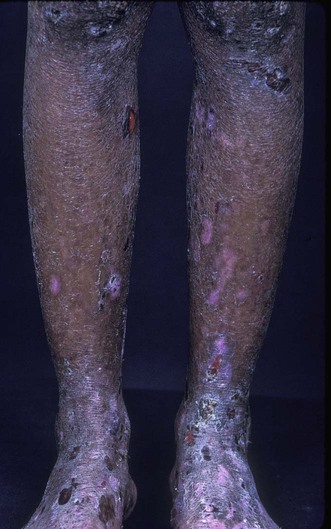
Epidermolysis bullosa acquisita
Specific investigations
 Skin biopsy and serum for direct and indirect immunofluorescence, respectively, to detect IgG or IgA class skin basement membrane-specific autoantibodies
 Serum for ELISA to detect IgG or IgA class type VII collagen-specific autoantibodies
 Gastrointestinal work-up for possible inflammatory bowel disease
First-line therapies
 Systemic corticosteroids
 Mycophenolate mofetil
 Dapsone
Second-line therapies
 Intravenous immunoglobulin
 Colchicine
 Cyclosporine
 Anti-CD20 antibody (rituximab)
