Epidermal nevi

Published on 16/03/2015 by admin

Filed under Dermatology

Last modified 22/04/2025

Print this page

rate 1 star rate 2 star rate 3 star rate 4 star rate 5 star
Your rating: none, Average: 0 (0 votes)

This article have been viewed 1890 times

Epidermal nevi

Jeffrey M. Weinberg

Evidence Levels:  A Double-blind study  B Clinical trial ≥ 20 subjects  C Clinical trial < 20 subjects  D Series ≥ 5 subjects  E Anecdotal case reports

image

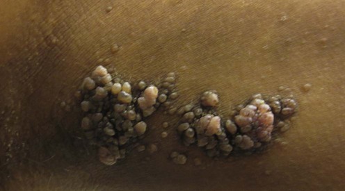
(Courtesy of Neil Fernandes, MD.)

Epidermal nevi are congenital hamartomas of embryonal ectodermal origin classified on the basis of their major component. The components may be sebaceous, apocrine, eccrine, follicular, or keratinocytic. An estimated one-third of individuals with epidermal nevi have involvement of other organ systems. In these cases, the condition is termed epidermal nevus syndrome.

The most common epidermal nevi are verrucous epidermal nevi, which are best treated with an ablative procedure using either surgical or laser technology. Inflammatory epidermal nevi may respond to topical or systemic therapy.

Management strategy

The pluripotential stem cell in the embryonic ectoderm can develop into any of the cell types found within the epidermis and skin adnexae. Therefore, there are many potential nevi that may develop from these cell types. Epidermal nevi may be classified according to the predominant cell type. However, there may be different cell populations, or overlap between different areas within the same nevus.

The focus of this chapter will be on nevi derived from keratinocytes. Of these, the verrucous epidermal nevus is the most common. Other forms include an inflammatory linear verrucous epidermal nevus (ILVEN), an acantholytic or Darier-like nevus, an epidermolytic form, and linear porokeratosis. Very rarely an epidermal nevus may be associated with other birth defects, and a number of epidermal nevus syndromes have been described.

Verrucous epidermal nevi may be localized, segmental, and rarely systematized. The individual lesions are verrucous papules, which may be pink, brown, or gray. These may develop as a result of mosaicism, and, if there is gonadal mosaicism, epidermal nevi may be transmitted to future offspring.

There are very rare case reports of malignant change within epidermal nevi, including squamous cell carcinoma and basal cell carcinoma. The major focus of therapy is improved cosmesis. A possible role for the dermis in the development of epidermal nevi is suggested by the difficulty experienced in ablating such lesions surgically without destroying the underlying dermis. Surgical management of these lesions presents challenges. Superficial treatments, which remove only the epidermis, have a high recurrence rate, whereas excision or more aggressive ablative procedures may produce unacceptable scarring. Laser technology provides the surgeon with more precise tools to maximize efficacy while minimizing scarring. Alternatively, for very widespread lesions, a variety of topical regimens as well as systemic retinoids have been reported to produce some benefit.

ILVEN presents in early childhood as a pruritic, erythematous, linear plaque. It shares many features with psoriasis, and certain cases respond to antipsoriatic therapies such as topical vitamin D analogs, corticosteroids, and dithranol. This has led some authors to suggest that this condition is a nevoid form of psoriasis. Epidermolytic and acantholytic nevi are more likely to respond to treatment with retinoids.

Specific investigations

An epidermal nevus can most often be diagnosed solely on the clinical presentation and distribution of the lesion. A skin biopsy can be used both to confirm the diagnosis if necessary and to determine the predominant cell type and the presence of inflammatory changes, acantholysis, or dysplasia. This can be helpful in determining which therapeutic modality is most likely to succeed. If histopathology demonstrates an epidermolytic nevus, the individual should be counseled that there is a possibility that the mutation could be transmitted to offspring, with the risk that their children may have generalized cutaneous involvement. Biopsy can also indicate the rare occurrence of squamous or basal cell carcinoma, which can develop in epidermal nevi.

Epidermal nevus syndromes refer to the association of epidermal nevi with extracutaneous manifestations involving the central nervous system, eyes, or bones. The evaluation for systemic involvement should be based on the clinical extent of the epidermal nevi, and the presence of any extracutaneous signs and symptoms.

Verrucous epidermal nevi

First-line therapies

image Excision under local anesthetic D
image Shave or curettage under local anesthetic D
image Cryotherapy C

For small verrucous epidermal nevi, excision can be performed with an acceptable cosmetic result. In these cases, this approach is the treatment of choice. However, for larger lesions, or for those in cosmetically sensitive sites, excision may not be appropriate. For larger lesions shave excision can be performed, but recurrence often occurs. Cryotherapy can be used as a destructive method for these lesions, but recurrence is frequent. All these procedures have the benefit of being cost-effective and easily performed.

Second-line therapies

image Laser ablation B
image Dermabrasion E
image Ruby laser E
image Erbium : YAG laser C

A resurfacing procedure, either mechanically by dermabrading, or with laser technology using either the erbium : YAG or the CO2 laser, can produce very acceptable results. However, both these techniques are operator dependent, and in the case of laser therapy access to expensive equipment is necessary. Darkly pigmented nevi may be treated using pigmented lesion lasers such as the ruby laser.

Third-line therapies

image Systemic retinoids D
image Topical retinoids plus 5-fluorouracil E
image Photodynamic therapy E

Systemic retinoids have been shown to reduce hyperkeratosis in very extensive and cosmetically troublesome lesions. However, long-term use is required if the benefit is to be maintained. The topical combination of tretinoin and 5-fluorouracil has also been reported to achieve a significant improvement. Photodynamic therapy has been recently reported in the treatment of verrucous epidermal nevus.

Inflammatory/dysplastic epidermal nevi

Nevi that have inflammatory, epidermolytic, acantholytic, or dysplastic features may respond more effectively to medical therapy than to surgical treatment. Inflammatory linear verrucous epidermal nevus (ILVEN) is a relatively rare entity that presents during childhood and can be difficult to treat.

Second-line therapies

image Topical calcipotriol/tacalcitol D
image Topical retinoids E
image Systemic retinoids E
image Topical dithranol E

ILVEN have been shown to respond to a variety of antipsoriatic therapies, leading some authors to believe that it is a nevoid form of psoriasis.

Third-line therapies

image Pulsed-dye laser E
image CO2 laser E
image Surgical excision D
image Etanercept E
image Photodynamic therapy E

Successful treatment of a widespread inflammatory verrucous epidermal nevus with etanercept.

Bogle MA, Sobell JM, Dover JS. Arch Dermatol 2006; 142: 401–2.

A 55-year-old woman diagnosed with widespread inflammatory epidermal verrucous nevi was presented. She had a history of multiple therapies, including emollients, topical and intramuscular steroids, topical lactic acid, pimecrolimus cream, and isotretinoin. She had minimal improvement with isotretinoin and experienced the largest reduction in pruritus with intramuscular corticosteroid injections.

The authors initiated subcutaneous etanercept therapy at a dose of 25 mg twice weekly. After 1 month, the patient experienced good initial improvement in pruritus and erythema. The etanercept was increased to 50 mg twice weekly, which provided nearly 50% improvement over 3 months. She continued treatment at this dose for a total of 6 months, and achieved almost complete resolution of pruritus and a significant improvement in roughness and erythema. The dose was then reduced to 25 mg twice weekly, and disease activity remained quiescent at follow-up.

Inflammatory linear verrucous epidermal nevus successfully treated with methyl-aminolevulinate photodynamic therapy.

Parera E, Gallardo F, Toll A, Gil I, Sánchez-Schmidt J, Pujol R. Dermatol Surg 2010; 36: 253–6.

The authors report a 67-year-old man with ILVEN, who had failed the following modalities: clobetasol propionate in an occlusive dressing, intralesional triamcinolone acetonide, topical tazarotene 0.1%, calcipotriol 0.005% plus betamethasone propionate, electrodesiccation, and oral antihistamines. Treatment with methyl-aminolevulinate photodynamic therapy (PDT) was performed. Almost complete resolution of the lesions after three PDT sessions (once a week) was observed. Pruritus completely disappeared, although small prurigo-like papules remained at the periphery of the lesion. No recurrence was observed after a follow-up period of 15 months.