Eosinophilic fasciitis

Published on 19/03/2015 by admin

Filed under Dermatology

Last modified 19/03/2015

Print this page

rate 1 star rate 2 star rate 3 star rate 4 star rate 5 star
Your rating: none, Average: 2 (4 votes)

This article have been viewed 3341 times

Eosinophilic fasciitis

Shaheen H. Ensanyat, Amir A. Larian, Brian S. Fuchs and Marsha L. Gordon
Evidence Levels:  A Double-blind study  B Clinical trial ≥ 20 subjects  C Clinical trial < 20 subjects  D Series ≥ 5 subjects  E Anecdotal case reports

image

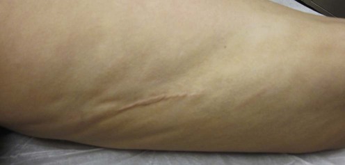
Eosinophilic fasciitis (EF), also known as Shulman syndrome, is a rare fibrosing disorder characterized by the rapid onset of symmetric induration of the extremities. The trunk and neck can be affected, with typical sparing of the hands, feet, and face. Clinically, the progression of EF is marked by edema and erythema followed by dimpling of the skin and a peau d’orange appearance evolving into woody induration and stiffness of the limbs. Laboratory investigations often reveal hypergammaglobulinemia, an elevated erythrocyte sedimentation rate, and peripheral blood eosinophilia, but are not essential in securing a diagnosis, given their transient levels. Histologically, EF presents with severe fascial thickening associated with an inflammatory infiltrate composed of plasma cells, eosinophils, histiocytes, and lymphocytes that can ultimately lead to dermal sclerosis. While the etiology remains unknown, vigorous physical activity has been reported before the onset of EF in many cases, presumably triggering an aberrant immune response.

Management strategy

Whether a distinct entity or variant of scleroderma, EF is differentiated by the relative absence of sclerodactyly, Raynaud phenomenon, and serologic markers. Unlike scleroderma, patients with EF typically demonstrate a more rapid onset with normal nail fold capillaries, infrequent visceral involvement, and response to corticosteroid therapy. EF must also be distinguished from the eosinophilia–myalgia syndrome, which is characterized by diffuse muscle pain and weakness, polyneuropathy, respiratory and pulmonary problems, and most notably a history of contaminated L-tryptophan ingestion.

EF has been reported in association with hematological disorders, including aplastic anemia, hemolytic anemia, thrombocytopenia, leukemias, and lymphomas. In addition, EF has to a lesser extent been linked to administration of simvastatin or exposure to trichloroethylene; these should be checked for in the patient history.

While clinical and laboratory findings help identify EF, diagnosis can only be confirmed with histopathological examination from a full-thickness skin-to-muscle biopsy, which characteristically reveals deep dermal fibrosis and fascial thickening with inflammatory infiltration of lymphocytes, plasma cells, and eosinophils. Magnetic resonance imaging (MRI) has also proven to be a useful non-invasive tool for establishing the diagnosis, choosing the optimal biopsy site, and monitoring the effectiveness of treatment. Findings typically show high signal intensity of the deep and superficial fasciae on T1, T2, and STIR imaging with enhancement after intravenous contrast administration.

Although spontaneous remission of EF has been reported, treatment can help prevent the progression of flexion contractures and limited mobility. Clinical response is usually defined as marked improvement in skin thickening, as fibrosed tissue becomes softer and looser, allowing for increased range of motion. Serum aldolase levels have been proposed as a useful indicator of disease activity.

There is substantial agreement among published reports that moderate- to high-dose corticosteroids serve as the first-line treatment for EF with prednisone as the standard agent, initially at 40–60 mg daily. A clinical response usually is noted within the first few weeks, and the dosage is then tapered slowly over several months to an alternate day regimen. In steroid-refractory patients, the addition of hydroxychloroquine at 200–400 mg daily has also been effective both in combination and as monotherapy.

In patients with partial or no response to corticosteroids, immunosuppressive drugs are frequently introduced as a second-line treatment. Cyclosporine at 3.7 mg/kg daily tapered to 2.5 mg/kg daily has been successful within 1 month at improving clinical symptoms with no relapse after a year. Methotrexate at 15–20 mg/week in conjunction with prednisone has also been commonly used with a favorable response.

Cimetidine at 400 mg every 6 to 12 hours has shown reported success in some cases. Pulsed methylprednisolone 1 g daily for 5 days combined with cyclosporine 150 mg twice daily is another regimen that has led to remission for patients in need of more aggressive therapy. Infliximab, D-penicillamine, ketotifen, chloroquine, psoralen and UVA (PUVA), azathioprine, griseofulvin, dapsone and sulfasalazine have all been reported to have beneficial effects.

Specific investigations