Eosinophilic Cholecystitis

Published on 21/04/2017 by admin

Filed under Pathology

Last modified 22/04/2025

Print this page

rate 1 star rate 2 star rate 3 star rate 4 star rate 5 star
Your rating: none, Average: 2.2 (26 votes)

This article have been viewed 3167 times

 Probably not distinct entity, but rather descriptive designation with associated clinical correlates

Etiology/Pathogenesis

• Majority of cases have no known cause or disease association

• Some associated with hypersensitivity reactions, parasitic infection, other eosinophilic diseases
image Hypersensitivity reaction to bile and bile stones has been hypothesized but never proven

Clinical Issues

• Presenting signs are similar to other forms of cholecystitis
• Peripheral eosinophilia variably present
• Typically acalculous
• Diagnosis virtually always made following resection of gallbladder for symptomatic disease

Macroscopic

• Thickened gallbladder wall, usually without gallstones

Microscopic

• Dense eosinophilic infiltrate of gallbladder ± lymphocytic inflammatory component

image Typically > 50% of inflammatory infiltrate is composed of eosinophils
image So-called lymphoeosinophilic cholecystitis shows significant component of lymphocytes as well
image In “true” or “pure” eosinophilic cholecystitis, close to 100% of inflammatory component is composed of eosinophils
• Specimen should be carefully evaluated for parasites
image
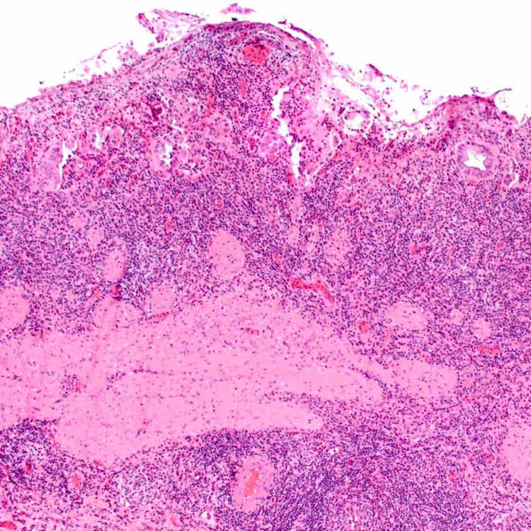
Transmural Eosinophilic Infiltrate
This case of eosinophilic cholecystitis shows a transmural infiltrate consisting of both lymphocytes and a prominent component of eosinophils.

image
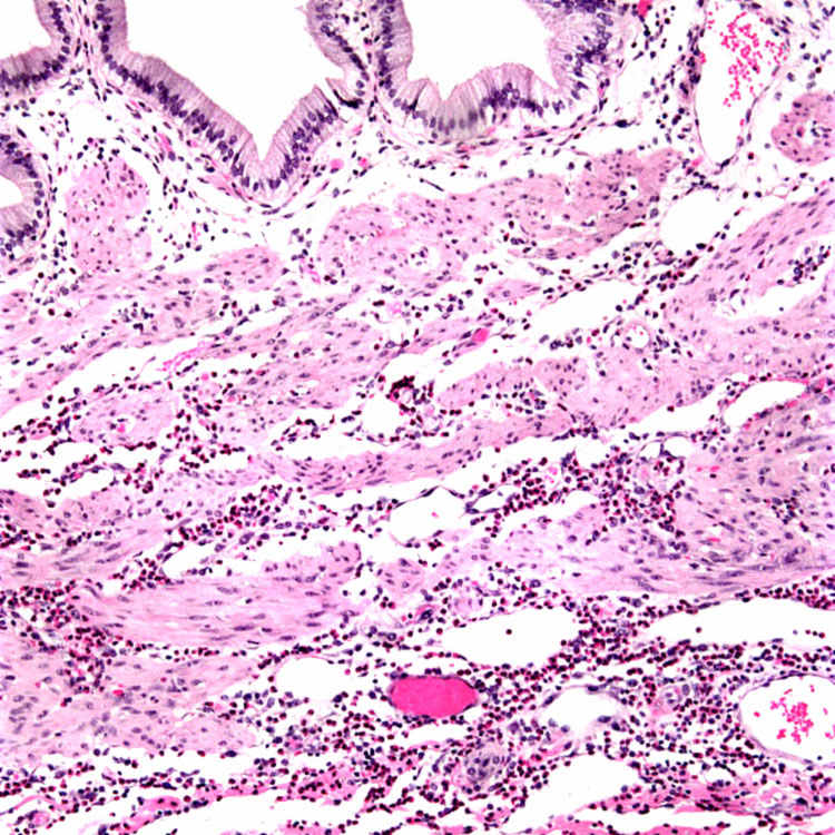
Pure Eosinophilic Infiltrate
This case of “pure” eosinophilic cholecystitis shows an exclusively eosinophilic infiltrate extending into the muscular wall of the gallbladder.
image
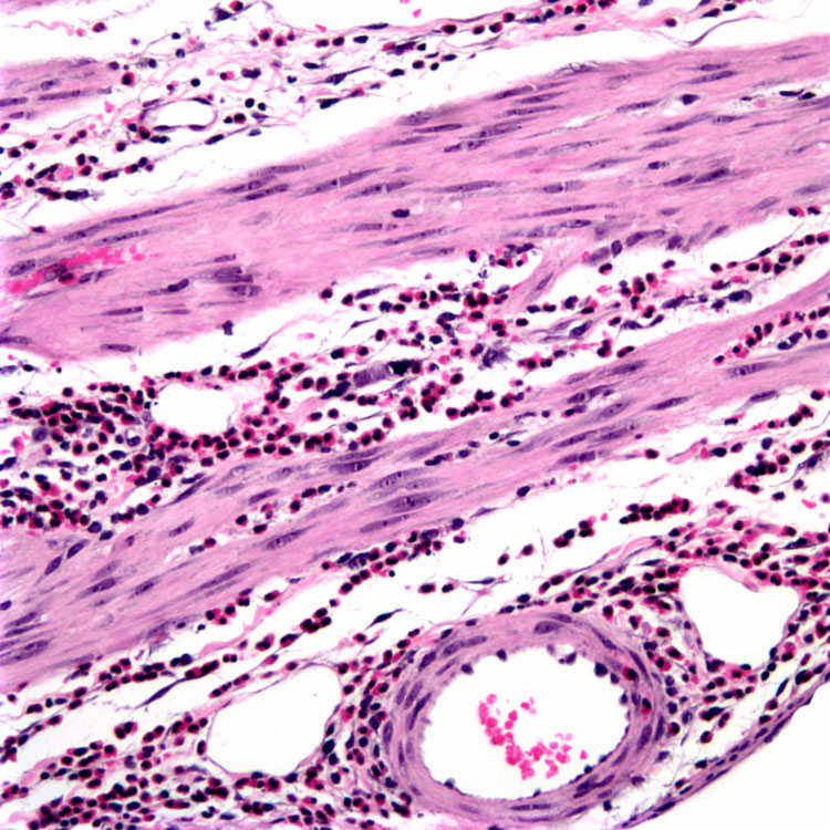
“Pure” Eosinophilic Infiltrate, High Power
This high-power view of “pure” eosinophilic cholecystitis shows the eosinophils within the wall of the gallbladder as well as surrounding vessels. However, unlike eosinophilic vasculitis, the vessels are not significantly infiltrated or damaged.
image
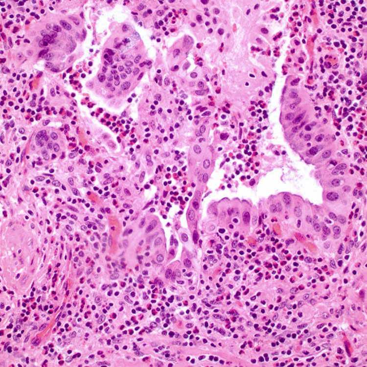
Mixed Lymphocytic/Eosinophilic Inflammation
Some cases of eosinophilic cholecystitis also have admixed lymphocytes in the inflammatory infiltrate, but typically > 50% of the infiltrate is composed of eosinophils.

TERMINOLOGY

Abbreviations

• Eosinophilic cholecystitis (EC)

Definitions

• Inflammatory disease of gallbladder in which inflammatory infiltrate is composed predominantly of eosinophils

image Some advocate for reserving this term for cases in which infiltrate is purely eosinophilic
image Probably not distinct entity, but rather descriptive designation with some associated clinical correlates

ETIOLOGY/PATHOGENESIS

No Specific Cause in Most Cases

• Hypersensitivity reaction to bile and bile stones has been hypothesized but never proven

• Some cases associated with hypersensitivity reaction to drugs, infections ( Echinococcus, Clonorchis sinensis )
• Some cases associated with other eosinophilic diseases
image Eosinophilic gastroenteritis, eosinophilic cholangitis, hypereosinophilic syndrome
• Majority of cases have no specific underlying cause or disease association

CLINICAL ISSUES

Presentation

• Presenting signs are similar to other forms of acute and chronic cholecystitis

image Right upper quadrant pain, biliary colic
• Peripheral eosinophilia

image Frequently but not invariably present
• Typically acalculous cholecystitis

image Although calculi are sometimes seen, stones are less commonly seen in EC than in other forms of acute and chronic cholecystitis

Treatment

• Cholecystectomy

image Diagnosis is invariably made following evaluation of gallbladder specimen
• In rare instances when preoperative diagnosis has been made, steroid therapy is reported to be effective

Prognosis

• Cholecystectomy is curative, and disease does not recur

MACROSCOPIC

General Features

• Thickened gallbladder wall
• Gallstones are usually absent

MICROSCOPIC

Histologic Features

• Thickened and inflamed gallbladder wall

image Predominantly eosinophilic infiltrate
– Typically > 50% of inflammatory infiltrate is composed of eosinophils
– Sheets and clusters of eosinophils infiltrate mucosa, muscularis propria, and subserosa
– Inflammation may preferentially involve one layer of gallbladder wall, or may be transmural
• Variations

image In “true” or “pure” EC, close to 100% of inflammatory component is composed of eosinophils

– More rare than lymphoeosinophilic form
image So-called lymphoeosinophilic cholecystitis shows significant component of lymphocytes as well

DIFFERENTIAL DIAGNOSIS

Chronic Cholecystitis

• Subacute phase of acute cholecystitis may show significant tissue eosinophilia
• Dense sheets and significant clustering of eosinophils are not seen in chronic calculous cholecystitis

Acute and Subacute Cholecystitis With Cholelithiasis

• Frequent admixture of neutrophils and eosinophils, particularly in instances of acute cholecystitis in which cholecystectomy is delayed
• Eosinophils do not typically dominate infiltrate, however

Autoimmune Pancreatitis-Associated Cholecystitis

• Gallbladder inflammation associated with autoimmune pancreatitis is dominated by lymphocytes and plasma cells

image Can show significant numbers of eosinophils
• Elevated numbers of IgG4(+) plasma cells help distinguish this entity from EC

Churg-Strauss Syndrome

• EC lacks significant vascular infiltration by eosinophils, and should not have vascular damage

DIAGNOSTIC CHECKLIST

Pathologic Interpretation Pearls

• Predominance of eosinophils in inflamed gallbladder

• Variably present gallstones
• May be associated with other eosinophilic diseases or peripheral eosinophilia
image Most cases have no specific disease association

SELECTED REFERENCES

1.Lai, CH, et al. Clonorchiasis-associated perforated eosinophilic cholecystitis. Am J Trop Med Hyg. 2007; 76(2):396–398.

2.Shakov, R, et al. Eosinophilic cholecystitis, with a review of the literature. Ann Clin Lab Sci. 2007; 37(2):182–185.

3.Suzuki, M, et al. Churg-Strauss syndrome complicated by colon erosion, acalculous cholecystitis and liver abscesses. World J Gastroenterol. 2005; 11(33):5248–5250.

4.Jimenez-Saenz, M, et al. Biliary tract disease: a rare manifestation of eosinophilic gastroenteritis. Dig Dis Sci. 2003; 48(3):624–627.

5.Dabbs, DJ. Eosinophilic and lymphoeosinophilic cholecystitis. Am J Surg Pathol. 1993; 17(5):497–501.

6.Parry, SW, et al. Acalculous hypersensitivity cholecystitis: hypothesis of a new clinicopathologic entity. Surgery. 1988; 104(5):911–916.

7.Kerstein, MD, et al. Eosinophilic cholecystitis. Am J Gastroenterol. 1976; 66(4):349–352.