Polyps, Endometrial
Synonyms/Description
Etiology
Ultrasound Findings
Differential Diagnosis
Clinical Aspects and Recommendations
Figures
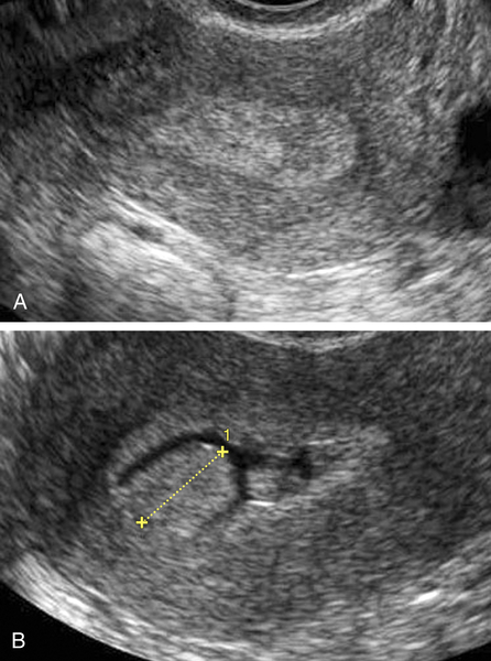
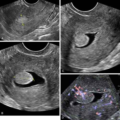
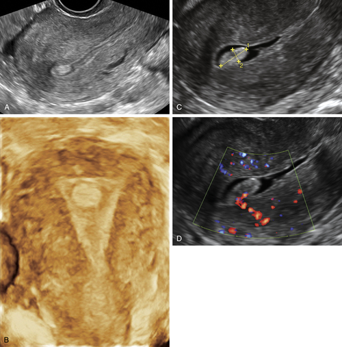
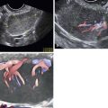
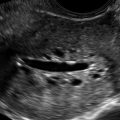
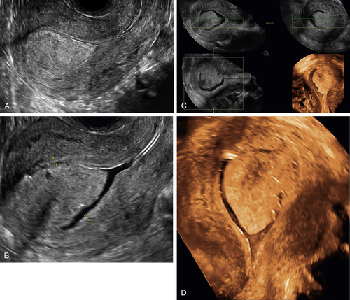
Figure P5-3 Two different patients with small 10-mm polyps seen in 2-D and 3-D at the fundus of the uterus. Note the characteristic smooth, round appearance of the polyps. A and B show the polyps with 2-D and 3-D transvaginal sonography. C and D show the polyp of a different patient using sonohysterography and color Doppler.
Suggested Reading
Fang L., Su Y., Guo Y., Yingpu Sun Y. Value of 3-dimensional and power Doppler sonography for diagnosis of endometrial polyps. Ultrasound Med. 2013;32:247–255.
Fernandez-Parra J., Rodriguez Oliver A., Lopez Criado S., Parrilla Fernandez F., Montoya Ventoso F. Hysteroscopic evaluation of endometrial polyps. Int J Gynaecol Obstet. 2006;95:144–148.
Ferrazzi E., Zupi E., Leone F.P., Savelli L., Omodei U., Moscarini M., Barbieri M., Cammareri G., Capobianco G., Cicinelli E., Coccia M.E., Donarini G., Fiore S., Litta P., Sideri M., Solima E., Spazzini D., Testa A.C., Vignali M. How often are endometrial polyps malignant in asymptomatic postmenopausal women? A multicenter study. Am J Obstet Gynecol. 2009;200:235.
Gerber B., Krause A., Müller H., Reimer T., Külz T., Kundt G., Friese K. Ultrasonographic detection of asymptomatic endometrial cancer in postmenopausal patients offers no prognostic advantage over symptomatic disease discovered by uterine bleeding. Eur J Cancer. 2001;37:64–71.
Goldstein S.R. Sonography in postmenopausal bleeding. J Ultrasound Med. 2012;31:333–336.
Lieng M., Istre O., Qvigstad E. Treatment of endometrial polyps: a systematic review. Acta Obstet Gynecol. 2010;89:992–1002.