Dystonia

Published on 03/03/2015 by admin

Filed under Neurology

Last modified 22/04/2025

Print this page

rate 1 star rate 2 star rate 3 star rate 4 star rate 5 star
Your rating: none, Average: 0 (0 votes)

This article have been viewed 1568 times

40 Dystonia

Dystonia is a hyperkinetic movement disorder characterized by involuntary, sustained muscle contractions that frequently cause twisting and repetitive movements or abnormal postures. Dystonic movements are patterned, meaning they repeatedly involve the same group of muscles. There is simultaneous contraction of agonist and antagonist muscles. In general, the duration of dystonic muscle contractions is longer than other hyperkinesias (i.e., chorea), though sometimes the movements can be rapid enough to resemble repetitive myoclonic jerking. One of the characteristic features of dystonia is that it is often temporarily diminished by tactile sensory tricks (gestes antagonistes). For example, a patient with cervical dystonia may be able to reduce dystonic movements by placing a hand on the chin or the side of the face. Patients with orobuccolingual dystonia often experience improvement by touching the lips or placing an object, such as a toothpick, in the mouth. In some patients, simply thinking about performing the sensory trick diminishes dystonia. The efficacy of sensory tricks can be exploited in the development of therapies. For example, some patients with lower cranial dystonia may benefit from wearing a mouthguard.

The initial presentation of dystonia is usually focal (affecting one body part) and task-specific—the dystonia occurs with a particular action. For example, a subject with foot dystonia may initially note dystonia when walking forward, but not walking backward or running. In the majority of patients, the dystonia remains focal without spreading to other parts of the body. If dystonia spreads, it tends to involve contiguous body parts and becomes a segmental dystonia. In more severe cases, the dystonia can become generalized. As a rule, the younger the age at onset, the more likely the dystonia is to spread. Recent data have also suggested that in the primary dystonias there is a caudal-to-rostral anatomic gradient in the site of onset as a function of age.

As dystonia progresses, it emerges with other actions of the affected body part, therefore becoming less task-specific. For example, the patient with foot dystonia may experience it when walking forwards and backwards, running, or tapping the foot. Further progression can lead to “overflow dystonia,” in which movement of a distant body part elicits the dystonia. As dystonia worsens, it can occur at rest. In the most severe cases of dystonia, contractures may develop.

Dystonia is frequently worsened by fatigue and stress and diminished with relaxation and sleep. Pain is generally uncommon in dystonia except for cervical dystonia, in which ~75% of patients report pain.

Classification of Dystonia

There are several recognized classification schemes for dystonia: (1) age at onset, (2) anatomic distribution, and (3) etiology.

Primary Dystonia

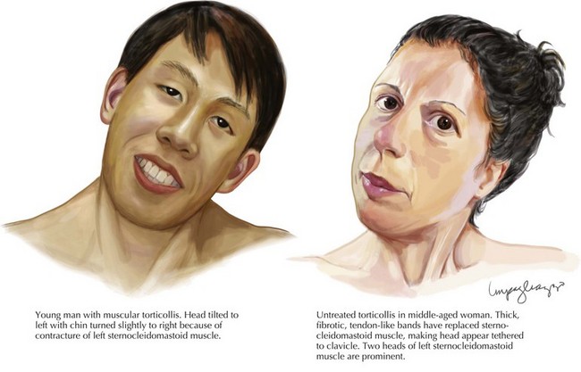
Primary dystonias are characterized by pure dystonia (with the exception that tremor can be present) and they may be sporadic or familial. Most primary dystonias are sporadic with onset in adulthood and a focal or segmental presentation. The most common focal dystonia presenting to movement disorders clinics is cervical dystonia (Fig. 40-1). After cervical dystonia, the most common focal dystonias are blepharospasm, spasmodic dysphonia, oromandibular dystonia, and hand dystonia.

A minority of primary dystonias have an identified genetic etiology (Box 40-1). Perhaps the best studied of the primary dystonias is DYT1 dystonia, or Oppenheim’s dystonia, a generalized torsion dystonia that usually begins in childhood affecting the limbs first. DYT1 dystonia is caused by a deletion in the DYT1 gene, which encodes for the torsin A protein. The disease is inherited in an autosomal dominant fashion and has 30–40% penetrance. Phenotype can vary widely within an affected family. The DYT1 mutation is estimated to account for 90% of limb-onset dystonia cases in the Ashkenazi Jewish population and 50–70% of cases in the non-Jewish population. A number of other primary dystonias (DYT6, DYT7 and DYT13) have had their genetic loci mapped (Table 40-1).

Secondary Dystonia

Secondary dystonia encompasses a broad clinical category that includes inherited degenerative disorders, degenerative disorders of unknown etiology, acquired dystonias (i.e., drug-induced, structural lesions, trauma), and psychogenic dystonia. Patients with secondary dystonia frequently display associated clinical abnormalities including other movement disorders besides tremor (i.e., parkinsonism), dementia, spasticity, ataxia, weakness, reflex changes, eye movement abnormalities, or seizures. Other historical and clinical features that suggest secondary dystonia include history of trauma, perinatal anoxia, toxin or drug exposure, onset of dystonia at rest, and hemidystonia.

A number of heredodegenerative disorders may present with dystonia. Autosomal dominant disorders (Huntington disease, spinocerebellar ataxia type 3, neuroferritonopathy), autosomal recessive disorders (Wilson disease, pantothenate-kinase associated degeneration), X-linked recessive disorders (DYT3 or Lubag), and mitochondrial disorders (Leigh disease) have been described and are listed in Box 40-2.

Dystonia can also be seen in various neurodegenerative parkinsonian syndromes, including Parkinson disease, as well as the atypical parkinsonian syndromes including progressive supranuclear palsy and corticobasal ganglionic degeneration (CBD). Adult-onset focal foot or leg dystonia can be the initial presentation of Parkinson disease (PD). Patients with PD may also develop dystonia as an off-symptom or as part of levodopa-induced dyskinesias. A dystonic, apraxic limb is a hallmark of CBD.

Dystonia-plus syndromes are a rare group of inherited nondegenerative diseases that include dopa-responsive dystonia (DRD), myoclonus–dystonia (MD), and rapid-onset dystonia–parkinsonism (RDP). These diseases are considered neurochemical disorders because they are due to biochemical defects that are not associated with neuronal loss. Dystonia is associated with parkinsonism in DRD and RDP and with myoclonus in MD.

A variety of lesions causing damage to the basal ganglia are associated with dystonia. These include perinatal hypoxia, stroke, head trauma, brain tumor, infection, and autoimmune disorders.

Psychogenic dystonia is perhaps the most diagnostically challenging type of secondary dystonia as illustrated by the record that many now-recognized organic dystonia patients were initially thought to have a primary psychiatric disorder. It is also important to recognize that a minority of patients with organic movement disorder may have a superimposed psychogenic movement disorder including dystonia. Features on history and exam that can suggest a psychogenic dystonia include abrupt onset, spontaneous remission, distractible or entrainable movements, variability and inconsistency of movements (i.e., nonpatterned dystonic movements), false weakness or sensory complaints, multiple somatizations, the presence of secondary gain, and concomitant psychiatric disease.

Treatment

Treatment of dystonia is determined by etiology and anatomic region(s) involved. All children and young adults presenting with dystonia should be given a trial of levodopa to rule out DRD.

The first-line treatment for cervical dystonia, blepharospasm, focal limb dystonia, and spasmodic dysphonia is botulinum toxin. Both serotypes A and B are available in the United States. Botulinum toxin and baclofen can be beneficial for oromandibular dystonia. In addition to medications, physical therapy is an important adjunct in the treatment of dystonia.

For patients with generalized dystonia, trihexyphenidyl at high doses (~90 mg per day) can provide some benefit. Baclofen may also be tried for generalized dystonia and can also be given at high doses. The benzodiazepines may provide adjunctive benefit. Variable results have been found for diphenhydramine, carbamazepine, dopamine agonists and dopamine antagonists. Tetrabenazine can be helpful, especially in tardive dystonia.

For medication-refractory dystonia, surgery can be considered. Peripheral denervation surgeries such as rhizotomy have produced variable results. More recently, deep brain stimulation (DBS) of the globus pallidus has been performed in patients with medication-refractory dystonia. In a recent prospective, double-blind, video-controlled study of patients with primary generalized dystonia (DYT1 and non-DYT1), DBS patients experienced ~50% improvement in motor and disability scores at 1 year. A follow-up study in this group of patients has shown a sustained improvement in both motor and quality of life at 3 years postsurgery. Case series have described benefit of DBS in the treatment of cervical dystonia. DBS also shows promise for treating tardive dystonia.