Diffuse Parenchymal Lung Diseases of Unknown Etiology

Published on 12/06/2015 by admin

Filed under Pulmolory and Respiratory

Last modified 22/04/2025

Print this page

rate 1 star rate 2 star rate 3 star rate 4 star rate 5 star
Your rating: none, Average: 0 (0 votes)

This article have been viewed 2026 times

11

Diffuse Parenchymal Lung Diseases of Unknown Etiology

Approximately 65% of patients with diffuse parenchymal lung disease are victims of a process for which no etiologic agent has been identified, even though a specific name may be attached to the disease entity. Included in this category are idiopathic pulmonary fibrosis, pulmonary fibrosis associated with connective tissue disease, sarcoidosis, pulmonary Langerhans cell histiocytosis, and a variety of other disorders. Many general aspects of these problems were discussed in Chapter 9. This chapter focuses on the specific diseases and their particular characteristics.

Idiopathic Pulmonary Fibrosis

Although the name idiopathic pulmonary fibrosis (IPF) has often been used nonspecifically to describe fibrotic interstitial lung disease without an identifiable diagnosis, most clinicians and investigators believe IPF represents a specific disease entity. This chapter adopts that assumption and considers pulmonary fibrosis associated with an underlying connective tissue disease a separate entity. Other names that have been used interchangeably with IPF are cryptogenic fibrosing alveolitis and usual interstitial pneumonia. The latter term is now generally used as a description of the pathologic pattern associated with IPF, a pattern occasionally seen in clinical settings other than IPF.

As implied by the name, IPF does not yet have a recognizable inciting agent, although most studies demonstrate an association with tobacco smoke exposure. Whether the primary agent, if one exists, reaches the lung via the airways or bloodstream has not been determined. The theory behind the pathogenesis of IPF has changed considerably over the past several years. For many years the prevailing thought was that exposure to an unknown agent (perhaps an antigen leading to formation of antigen-antibody complexes) led to alveolar inflammation, which was perpetuated by release of chemotactic factors from inflammatory cells. The ongoing inflammation was believed to be responsible for subsequent development of fibrosis.

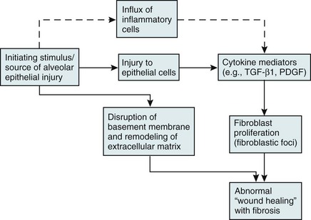
Over the past several years, a newer conceptual framework has emerged. According to the newer theory, alveolar inflammation does not play a critical role in the eventual development of fibrosis. Rather, fibrosis is believed to result directly from alveolar epithelial injury and is thought to be a manifestation of abnormal wound healing within the lung parenchyma. According to the newer paradigm, injury to alveolar epithelial cells (still from an unidentified source or agent) is the primary initiating event. Whereas injury to type I alveolar epithelial cells normally would be followed by a repair process that includes proliferation of type II cells and differentiation into type I cells, this repair process is impaired, at least in part because of disruption of the basement membrane, which normally is important for the reepithelialization process. At the same time, alveolar epithelial cells express a variety of profibrotic cytokines and growth factors, including platelet-derived growth factor (PDGF) and transforming growth factor (TGF)-β1, which enhance fibroblast migration and proliferation. Fibroblastic foci develop at sites of alveolar injury and appear to be responsible for increased extracellular matrix deposition. This process is summarized in Figure 11-1.

Recently, the development of IPF has been associated with gene mutations in two biological pathways known to be related to lung injury and repair. Mutations in the first pathway affect genes encoding surfactant proteins A2 (SFPA2) and C (SFPC), which may act to increase susceptibility to chronic lung injury by causing increased endoplasmic reticulum stress in alveolar type II epithelial cells. Mutations in the second pathway affect genes encoding telomerase (TERT and TERC), the multimeric enzyme system that repairs shortened telomeres. Abnormalities in telomerase function appear to impair wound healing by decreasing replication of progenitor cells. Although these mutations are not present in all patients with IPF, identification of the pathways involved has led to better understanding of the mechanism of disease in IPF, raising hopes of new therapies.

Clinically, the most common age at presentation of patients with IPF is between 50 and 70 years. Disease onset is generally insidious, and symptoms are similar to those of other interstitial lung diseases; dyspnea is the most prominent complaint. In addition to the classic finding of dry crackles or rales on physical examination, patients frequently have evidence of clubbing of the digits.

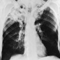
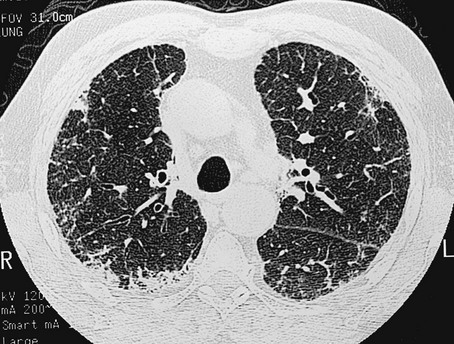
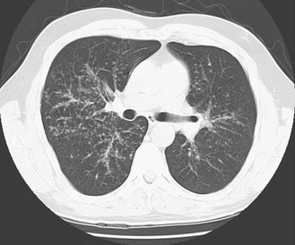
The chest radiograph shows an interstitial (reticular) pattern that is generally bilateral and relatively diffuse but typically is more prominent at the lung bases, particularly in the peripheral subpleural regions (see Fig. 3-6). Neither pleural effusions nor hilar enlargement is common on the radiograph. High-resolution computed tomography (HRCT) scanning often has a characteristic appearance, showing interstitial densities that are patchy, peripheral, subpleural, and associated with small cystic spaces (Fig. 11-2). The pattern of small cystic peripheral abnormalities on HRCT is termed honeycombing and indicates irreversible fibrosis. Many patients have serologic abnormalities, such as a positive test result for antinuclear antibodies, which are generally found in patients with autoimmune or connective tissue disease. However, in the absence of other suggestive clinical features, these abnormalities are thought to be nonspecific and not indicative of an underlying rheumatologic disease.

The diagnosis is definitively made by surgical lung biopsy, but only in the appropriate clinical setting when other etiologic factors for interstitial lung disease cannot be identified. Some patients are too frail for lung biopsy, and if HRCT scan shows the classic pattern of honeycombing, the diagnosis can be made with relative certainty without a lung biopsy. The histologic expression of IPF is in the form of usual interstitial pneumonia (UIP) (see Fig. 9-3), and patients who have a pathologic pattern more compatible with desquamative interstitial pneumonia or nonspecific interstitial pneumonia (see Other Idiopathic Interstitial Pneumonias; also see Chapter 9) should not be considered to have IPF. Granulomas should not be seen on an IPF biopsy specimen. If they are found, granulomas indicate the presence of another disorder.

From the time of clinical presentation, patients have a relatively poor prognosis; mean survival ranges from 2 to 5 years. Although steroids and cytotoxic agents have been used frequently in the past for IPF, newer studies unfortunately demonstrate that these agents are ineffective and appear to be harmful. Older studies suggested that a small group of patients would respond to corticosteroids. However, it is now thought that this subgroup actually had other corticosteroid-responsive diffuse parenchymal lung disease that was misidentified as IPF.

Currently there is no proven effective therapy for IPF. Clinical investigators are now focusing on identifying and testing agents that suppress fibrosis or interfere with mediators involved in the fibrotic process. In some patients with severe IPF, especially those who are younger, lung transplantation is used as the only therapeutic alternative to progressive respiratory failure and death.

Other Idiopathic Interstitial Pneumonias

Several other disorders besides IPF fall under the category of the idiopathic interstitial pneumonias and have often been confused with IPF. Although these disorders are uncommon, some are briefly described here, largely to clarify how their pathologic features differ from UIP and how their clinical features differ from IPF. They are also mentioned in Chapter 9 as part of the discussion on the pathology of the interstitial pneumonias.

Desquamative interstitial pneumonia (DIP) occurs largely in smokers. It generally has a subacute rather than a chronic onset. Imaging studies with chest radiography and HRCT scanning often show a ground-glass (hazy) pattern. Lung biopsy shows a uniform accumulation of intraalveolar macrophages, with little or no fibrosis. The prognosis is better than in IPF, and patients often can improve after cessation of smoking and may respond to corticosteroids.

Nonspecific interstitial pneumonia (NSIP) differs from UIP in its radiographic pattern, histologic appearance, prognosis, and response to treatment. As with DIP, imaging studies often show a ground-glass pattern that usually reflects inflammation rather than fibrosis. Lung biopsy shows a predominantly inflammatory response in the alveolar walls, with relatively little fibrosis. Although NSIP is often idiopathic and not associated with any underlying disease or inciting agent, it can represent the histologic appearance of parenchymal lung disease associated with one of the connective tissue diseases or with drug-induced pulmonary toxicity. The prognosis of NSIP appears to depend on the degree of fibrotic involvement present on both imaging and pathology. If inflammation predominates rather than fibrosis, the prognosis is significantly better than in IPF, and patients often respond to treatment with corticosteroids.

Cryptogenic organizing pneumonia is a disorder characterized by connective tissue plugs in small airways accompanied by mononuclear cell infiltration of the surrounding pulmonary parenchyma. As noted in Chapter 9, the terms cryptogenic organizing pneumonia (COP) and bronchiolitis obliterans with organizing pneumonia (BOOP) have often been used interchangeably, but the term BOOP is best reserved for the pathologic picture rather than the clinical syndrome. Although the histologic picture of BOOP can be associated with connective tissue disease, toxic fume inhalation, or infection, the large majority of cases have no identifiable cause and are considered idiopathic. The term COP is most appropriate for patients who have “idiopathic BOOP”—that is, the histologic pattern of BOOP but no apparent cause for this pattern.

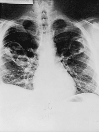
Like chronic eosinophilic pneumonia (see later), COP often has a subacute presentation (over weeks to months) with systemic (constitutional) as well as respiratory symptoms. The chest radiograph shows patchy infiltrates, generally with an alveolar rather than an interstitial pattern, often mimicking a community-acquired pneumonia (Fig. 11-3). Like chronic eosinophilic pneumonia, the response to corticosteroids is often dramatic and occurs over days to weeks. Therapy is usually prolonged for months to prevent relapse.

Acute interstitial pneumonia (AIP) is a more acute or fulminant type of pulmonary parenchymal disease that begins with the clinical picture of acute respiratory distress syndrome (ARDS; see Chapter 28) but without any of the usual inciting events associated with development of ARDS. Imaging studies of AIP typically show features of ARDS, including areas of ground-glass opacification and alveolar filling (as opposed to a purely interstitial pattern). The histologic pattern is that of diffuse alveolar damage, often showing some organization and fibrosis. Although mortality is high overall, a small percentage of patients do well, with clinical resolution of the disease and no long-term sequelae.

One confusing aspect of the nomenclature of the idiopathic interstitial pneumonias is the relationship underlying AIP, UIP (or IPF), and a disorder called Hamman-Rich syndrome. More than 75 years ago, Hamman and Rich described a number of cases of parenchymal lung disease that subsequently were thought to represent the first described cases of IPF, and for many years the term Hamman-Rich syndrome was used synonymously with IPF. However, the cases described by Hamman and Rich now are believed to be cases of AIP rather than IPF, and it is more appropriate that Hamman-Rich syndrome be considered synonymous with AIP rather than either UIP or IPF.

Pulmonary Parenchymal Involvement Complicating Connective Tissue Disease

The connective tissue diseases, also commonly called collagen vascular diseases or systemic rheumatic diseases, include rheumatoid arthritis, systemic lupus erythematosus, progressive systemic sclerosis (scleroderma), polymyositis-dermatomyositis, Sjögren syndrome, and some overlap syndromes that have features of more than one of these disorders. Although they form a diverse group, all are multisystem inflammatory diseases that are mediated immunologically. The organ systems likely involved vary with each disease and are mentioned briefly in the following discussion of each entity.

Each disease is complicated and has been the focus of extensive research into etiology and pathogenesis. However, because none of them primarily affects the lung, they are not considered in detail here. Rather, a brief discussion notes how they affect the respiratory system, particularly with regard to development of parenchymal lung disease. Some clinicians include additional disorders among connective tissue diseases, but this discussion is limited to those in the preceding paragraph, each of which has the potential for pulmonary involvement.

Four assertions are true about each of these disorders. First, although patients generally have evidence of the underlying connective tissue disease before pulmonary manifestations develop, some patients have lung disease as the presenting problem, occasionally predating other manifestations of their illness by several years. Second, detailed histologic, physiologic, or autopsy evaluation of patients with these diseases shows that pulmonary involvement is much more common than clinically suspected. Third, the histopathology of interstitial lung disease associated with connective tissue disorders often is that of UIP and therefore is indistinguishable from the pattern seen in IPF. However, in many cases the histopathology demonstrates NSIP or occasionally COP rather than UIP. Fourth, the interstitial lung disease that may develop with each of these entities preferentially affects the lower rather than the upper lung zones. This fact usually is apparent on examination of the chest radiograph.

Rheumatoid arthritis is a disorder with primary manifestations consisting of inflammatory joint disease. The most common site of involvement within the thorax is the pleura. Involvement takes the form of pleurisy, pleural effusions, or both. The lung parenchyma may become involved, with one or multiple nodules or with development of interstitial lung disease. The latter usually is relatively mild, although severe cases are sometimes seen. Occasionally, patients with rheumatoid arthritis develop airway complications in the form of bronchiolitis (an inflammatory process involving small airways) or bronchiectasis.

Systemic lupus erythematosus is a multisystem disease that primarily affects joints and skin but often has more serious involvement of several organ systems, including kidneys, lungs, nervous system, and heart. Its most frequent presentation within the chest takes the form of pleural disease, specifically pleuritic chest pain, pleural effusion, or both. The lung parenchyma may be involved by an acute pneumonitis in which infiltrates often involve the alveolar spaces as well as the alveolar walls, or less frequently by chronic interstitial lung disease. In the latter, extensive fibrosis usually is not a prominent feature of the histology.

Progressive systemic sclerosis, or scleroderma, is a disease with the most obvious manifestations located in the skin and small blood vessels. Other organ systems, including the gastrointestinal tract, lungs, kidneys, and heart, are involved relatively frequently. Of all the connective tissue diseases, scleroderma is the one in which pulmonary involvement tends to be most severe and most likely associated with significant scarring of the pulmonary parenchyma. Pulmonary fibrosis complicating scleroderma appears to be strongly associated with the presence of a particular serologic marker, an autoantibody to topoisomerase I (antitopoisomerase I, also called Scl70). Another potential pulmonary manifestation of scleroderma is disease of the small pulmonary blood vessels, producing pulmonary arterial hypertension, which is discussed in Chapter 14. This involvement appears to be independent of the fibrotic process affecting the alveolar walls.

In polymyositis-dermatomyositis, muscles and skin are the primary sites of the inflammatory process. The interstitial lung disease of polymyositis-dermatomyositis is relatively infrequent and often has no particular distinguishing features. Patients may have respiratory problems as a result of muscle disease, with weakness of the diaphragm or other inspiratory muscles. Involvement of striated muscle in the proximal esophagus may lead to difficulty in swallowing and recurrent episodes of aspiration pneumonia.

In Sjögren syndrome, a lymphocytic infiltration affects salivary and lacrimal glands and is associated with dry mouth and dry eyes (keratoconjunctivitis sicca). When patients with Sjögren syndrome have pulmonary parenchymal involvement, the histologic appearance is most commonly that of a lymphocytic infiltrate within the alveolar walls (called lymphocytic interstitial pneumonia) rather than UIP or NSIP. Other lymphocytic complications of the lung can develop in patients with Sjögren syndrome, specifically either a localized masslike lesion called a pseudolymphoma or an actual lymphoma.

Finally, a number of overlap syndromes, often called undifferentiated connective tissue disease, have features of several of these disorders, particularly scleroderma, lupus, and polymyositis. Patients may develop any of the complications noted with the more classic individual disorders, including interstitial lung disease, pleural disease, and pulmonary vascular disease.

Sarcoidosis

Sarcoidosis is defined as a systemic disorder in which granulomas, typically described as noncaseating, can be found in affected tissues or organ systems. An important qualification is that these granulomas occur in the absence of any exogenous (infectious or environmental) agents known to be associated with granulomatous inflammation. The lung is the most frequently involved organ, with potential manifestations including parenchymal lung disease, enlargement of hilar and mediastinal lymph nodes, or both.

Sarcoidosis is a relatively common disorder that particularly affects young adults between the ages of 20 and 40 years. It is slightly more common in women than in men. In the United States it is more common in African Americans than in whites, but this racial predilection is not seen throughout the world. The disease is notably prevalent in the white population of Scandinavia. Although a common stereotype in the United States is that the disease is primarily one of young African American women, a substantial number of men and individuals of other ethnic and age groups also are affected. Of all the disorders of unknown cause affecting the alveolar wall, sarcoidosis is the most prevalent.

Despite increasing knowledge about the cells involved in the inflammatory and granulomatous response in sarcoidosis and the identification of multiple cytokines and chemokines that appear to be involved in the pathogenesis of disease, the fundamental etiology of sarcoidosis remains as mysterious as it was when the disease was first described more than a century ago. It has been hypothesized that sarcoidosis represents an immunologic response to an exogenous agent in a genetically susceptible individual. Multiple exogenous antigens and a number of human leukocyte antigens and other candidate genes have been associated with susceptibility to sarcoidosis. However, neither a particular exogenous agent nor a specific genetic susceptibility has been consistently demonstrated. Interest in potential exogenous inciting agents has focused on microorganisms such as viruses, mycobacteria, and other bacteria (e.g., Propionibacterium acnes), as well as inorganic dusts such as silica. However, the identity of a trigger for sarcoidosis remains elusive, and whether such an agent even exists is not known. At present, it seems most likely that sarcoidosis represents a complex interaction among a number of antigens or particles and the effects of multiple genes.

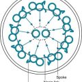
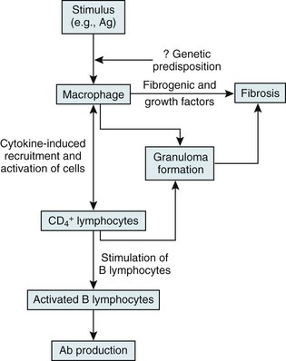
On the other hand, substantial information is available about cells and mediators that appear to be important in the inflammatory and granulomatous tissue reaction in sarcoidosis (Fig. 11-4). The critical cells are macrophages and T lymphocytes. The presumption is that processing of the responsible antigen by alveolar macrophages results in recruitment of helper T lymphocytes (CD4+ cells) with a TH1 profile. A host of proinflammatory cytokines and chemokines, such as interleukin (IL)-2, interferon (IFN)-γ, tumor necrosis factor (TNF)-α, and IL-12, appear to be important in recruiting and activating inflammatory cells, perpetuating the inflammatory response, and inducing the formation of granulomas. Profibrotic cytokines, such as TGF-β, PDGF, and insulinlike growth factor (IGF)-1, subsequently may result in fibrosis as a complication of the initial inflammatory reaction.

Accumulation of CD4+ lymphocytes at sites of active disease appears to result in secondary immunologic phenomena that are well recognized in sarcoidosis. First, presumably because of this concentration of activated lymphocytes in affected tissues, there is a relative depletion of CD4+ cells in peripheral blood. The depletion leads to an apparent depression of cell-mediated immunity, at least as measured by cutaneous delayed hypersensitivity (skin testing). However, patients with sarcoidosis are not unduly susceptible to opportunistic infections that characteristically affect the immunosuppressed host with impaired cellular immunity. Second, T lymphocytes in sarcoidosis nonspecifically activate B lymphocytes and the humoral immune system, leading to production of a variety of immunoglobulins and the common finding of polyclonal hyperglobulinemia.

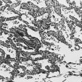
The characteristic histopathologic feature of sarcoidosis is the noncaseating granuloma (see Fig. 9-2). These typically well-formed granulomas show a collection of tissue macrophages (also called epithelioid histiocytes), multinucleated giant cells, and T lymphocytes, particularly toward the periphery or at the rim of the granuloma. The discrete accumulation of tissue macrophages comprising the granuloma does not show evidence of frank necrosis or caseation, as would appear in disorders like tuberculosis and histoplasmosis. In addition to granulomas in the lung parenchyma or intrathoracic lymph nodes, an alveolitis often occurs. The alveolitis is composed of mononuclear cells, including macrophages and lymphocytes, with the latter presumed to be of particular importance in the pathogenesis of disease.

Patients with sarcoidosis most frequently seek consultation either as a result of abnormalities detected on an incidental chest radiograph or because of respiratory symptoms, mainly dyspnea or a nonproductive cough. Because many other organ systems may be involved with noncaseating granulomas, other manifestations occur but are less common. Eye involvement (e.g., anterior uveitis [inflammation in the anterior chamber of the eye]) and skin involvement (e.g., skin papules or plaques) are particularly common extrathoracic manifestations of sarcoidosis, but cardiac, neurologic, hematologic, hepatic, endocrine, and peripheral lymph node findings also may be seen. Although symptoms are often insidious in onset, some patients with sarcoidosis have a more acute presentation called Löfgren syndrome, in which the chest radiographic finding of bilateral hilar lymphadenopathy is accompanied by erythema nodosum (painful red nodules, typically on the anterior surface of the lower legs) and often fever and arthralgias. For unknown reasons, patients who present with Löfgren syndrome typically have an excellent prognosis, with a spontaneous remission rate greater than 80%.

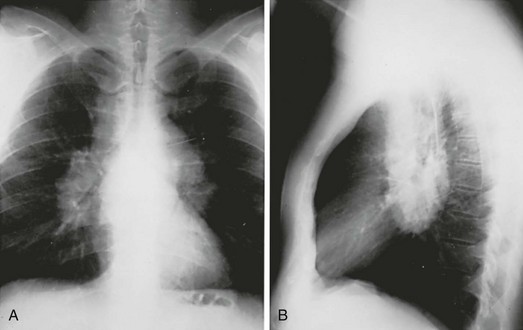
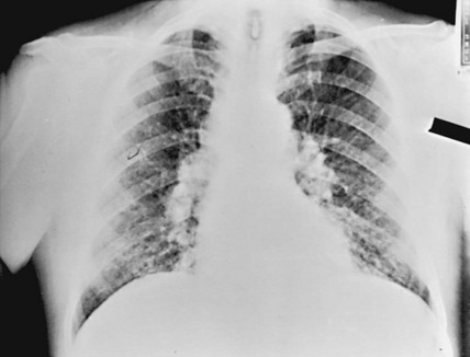
The chest radiograph in sarcoidosis generally shows one of the following patterns: (1) enlargement of lymph nodes, most commonly bilateral hilar lymphadenopathy with or without paratracheal node enlargement (Fig. 11-5); (2) parenchymal lung disease (in the form of interstitial disease, nodules, or alveolar infiltrates); or (3) both adenopathy and parenchymal disease (Fig. 11-6). HRCT scanning, although not generally necessary, is more sensitive than plain chest radiography in detecting parenchymal lung disease. It may show a particularly characteristic pattern of small nodules preferentially distributed along bronchovascular bundles (Fig. 11-7). In addition, HRCT often demonstrates mediastinal lymphadenopathy that cannot be seen on plain chest radiography.

The course of the radiographic findings in sarcoidosis is quite variable. Over time, both the adenopathy and the interstitial lung disease may regress spontaneously. At the other extreme, the interstitial disease may progress to a condition of extensive scarring and end-stage lung disease, at which time the patient has severe respiratory compromise.

Patients often display immune system abnormalities. Clinically, these patients may have anergy, failure to respond to skin tests requiring intact delayed hypersensitivity. They also may have hyperglobulinemia, which is evidence of a hyperactive humoral immune system. Calcium metabolism may be abnormal in sarcoidosis, occurring as a result of increased formation of the active form of vitamin D (1,25-dihydroxy-D3) by activated macrophages in granulomas. Increased amounts of the active form of vitamin D lead to enhanced calcium absorption from the gastrointestinal tract, potentially causing hypercalciuria or, less frequently, hypercalcemia.

The diagnosis of sarcoidosis can be established in several ways. When the clinical diagnosis strongly suggests sarcoidosis, tissue confirmation is sometimes unnecessary. An example of such a presentation is bilateral hilar lymphadenopathy found on an incidental chest radiograph in a symptom-free young African American woman. On the other hand, when the patient has symptoms or when there is a question about the diagnosis, tissue sampling usually is undertaken to look for noncaseating granulomas and rule out other causes. The lung is generally the most appropriate source of tissue. Samples of lung tissue are frequently obtained by transbronchial biopsy through a flexible bronchoscope. Interestingly, even when the chest radiograph shows only hilar adenopathy without obvious parenchymal lung disease, the alveolar walls usually are studded with granulomas that may be seen on transbronchial lung biopsy. Other ways of obtaining tissue include performing a biopsy of a lymph node in the mediastinum (via mediastinoscopy) or a thoracoscopic lung biopsy. In addition, biopsy specimens can be obtained easily from involved areas of skin (when skin involvement is suspected on physical examination). Occasionally they are obtained from a variety of other involved tissues or organs, such as peripheral lymph nodes, conjunctiva, minor salivary glands, or liver.

Elevated serum levels of angiotensin-converting enzyme (ACE) have been found in a large percentage of patients with sarcoidosis. This enzyme, which normally is synthesized by vascular endothelial cells, appears to be produced in the granulomas of sarcoidosis, suggesting it may be useful for diagnosing or following the disease. However, because it is not specific for sarcoidosis and often is normal in the presence of relatively inactive disease, ACE levels are not considered reliable in either diagnosing sarcoidosis or assessing its response to treatment.

The natural history of sarcoidosis is quite variable. In some patients, all clinical and radiographic manifestations resolve within 1 to 2 years. Other patients have persistent radiographic changes, either with or without persisting symptoms. In general, nearly two thirds of patients have spontaneous remissions. A minority of patients (10%-30%) show continued progression of radiographic abnormalities, with or without additional extrathoracic disease, and may have debilitating respiratory symptoms. Clinical factors associated with a worse prognosis include age at onset older than 40 years, African descent, chronic uveitis, chronic hypercalcemia, progressive pulmonary parenchymal fibrosis, and the presence of lupus pernio, a skin lesion affecting the face.

Pulmonary function tests are most useful for quantitating functional impairment. Spirometry, lung volumes, and diffusing capacity all are measured. Techniques for assessing the alveolitis (e.g., gallium scanning, bronchoalveolar lavage) have been investigated but presently are not thought to provide clinically useful information regarding the “activity” of the disease, so are not widely accepted for clinical use in evaluating sarcoidosis.

The initial treatment decision confronting the clinician is whether or not to institute therapy for the patient with sarcoidosis. Many patients do not require treatment, especially when the disease is causing neither significant symptoms nor significant functional organ involvement. The fact that the disease may improve or resolve spontaneously also complicates decisions about instituting therapy. When treatment is indicated because of symptoms and significant tissue involvement affecting organ function, the drug of choice usually is systemic corticosteroids. In patients with refractory disease, a variety of other agents, especially immunosuppressive drugs such as methotrexate and cyclophosphamide, have been used, either with or instead of corticosteroids. More recently, there has been interest in using infliximab, an antagonist of TNF-α, in selected cases, but its ultimate potential role in treating sarcoidosis is uncertain.

Miscellaneous Disorders Involving the Pulmonary Parenchyma

An exhaustive description of all the remaining diseases of unknown etiology affecting the pulmonary parenchyma cannot be presented here. Instead, a brief description of several additional diseases will acquaint the reader with their major features. They include (1) pulmonary Langerhans cell histiocytosis, (2) lymphangioleiomyomatosis, (3) Goodpasture syndrome, (4) Wegener granulomatosis, (5) chronic eosinophilic pneumonia, and (6) pulmonary alveolar proteinosis. For each of these relatively uncommon disorders, certain pathologic, clinical, or radiographic features distinguish them from the diffuse parenchymal lung diseases described earlier in this chapter. However, the defining feature for each of these disorders is a relatively specific pathologic appearance involving various components of the pulmonary parenchyma.

Pulmonary Langerhans Cell Histiocytosis

Pulmonary Langerhans cell histiocytosis, also called eosinophilic granuloma of the lung or pulmonary histiocytosis X, is thought to represent part of a spectrum of disorders involving histiocytic infiltration of one or more organ systems. Whereas multisystem involvement in Langerhans cell histiocytosis or histiocytosis X is typically seen with the childhood disorders called Letterer-Siwe disease or Hand-Schüller-Christian disease (not discussed here), isolated or predominant pulmonary involvement in pulmonary Langerhans cell histiocytosis occurs mainly in young to middle-aged adults.

The responsible histiocytic cell appears to be a type of antigen-presenting dendritic or phagocytic cell called a Langerhans cell. An interesting ultrastructural feature of these cells is the presence of cytoplasmic rodlike structures called X bodies (hence the name histiocytosis X) or Birbeck granules, which can be seen by electron microscopy. Light microscopic examination of the lung, in addition to demonstration of these histiocytes, reveals infiltration by eosinophils, lymphocytes, macrophages, and plasma cells. The process initially involves the lungs in a peribronchiolar distribution and subsequently becomes more diffuse. Although the disease occurs almost exclusively in smokers, the mechanism by which smoking may cause the disease or contribute to its pathogenesis is not well defined.

On chest radiograph, the clinical presentation of pulmonary Langerhans cell histiocytosis typically features a pattern of nodular or reticulonodular disease often accompanied by respiratory symptoms of dyspnea, cough, or both. The radiographic findings tend to be more prominent in the upper lung zones, with HRCT scans showing small cysts in addition to the nodular or reticulonodular changes. The cysts occasionally rupture, leading to a spontaneous pneumothorax, which may be the presenting feature of the disease. In some cases, progression results in a pattern of extensive cystic disease and honeycombing. Unlike the typical restrictive pattern in most of the diffuse parenchymal lung diseases, pulmonary function testing in pulmonary Langerhans cell histiocytosis may show restrictive changes, obstructive changes, or both. The presence of air-filled cysts typically leads to unusually normal or large lung volumes on chest radiography despite the presence of interstitial disease.

The natural history of the disease is variable. In some patients, the disease is self-limited, and the radiographic and functional changes may stabilize over time, especially with cessation of smoking. In other patients, extensive disease and significant functional impairment follow. No clearly effective treatment is available, although corticosteroids are often tried if smoking cessation alone is ineffective.

Lymphangioleiomyomatosis

Lymphangioleiomyomatosis (LAM) is a rare pulmonary disease characterized by proliferation of atypical smooth muscle cells around lymphatics, blood vessels, and airways, accompanied by numerous small cysts throughout the pulmonary parenchyma. LAM occurs almost exclusively in women of childbearing age, suggesting that hormonal influences play a role in the development of disease. In addition to occurring sporadically, LAM also develops in 30% to 40% of female patients with the genetic condition tuberous sclerosis complex (TSC). An interesting aspect is that the pathologic process seen in the lungs in LAM is essentially identical to that seen in multiple organ systems in TSC, suggesting a common pathogenetic mechanism. Germ cell mutations in two genes, TSC1 and TSC2, are associated with TSC, whereas in LAM the abnormal smooth muscle cells have a mutation in the TSC2 gene.

The normal products of TSC1 and TSC2 are proteins that form a complex that acts as a potent suppressor of cell growth and proliferation. Thus, the abnormal proteins lead to loss of this suppressor activity, resulting in uncontrolled growth. Patients with LAM appear to have developed an acquired mutation in smooth muscle cells in the lung, whereas patients with TSC appear to have an inborn genetic error. Furthermore, the gene product of TSC2 also interacts directly with intracellular estrogen receptors to cause inhibition of cell growth. This may account for some of the hormonal influences in LAM.

The clinical manifestations of LAM result from the presence of cysts and involvement of lymphatics, blood vessels, and airways. The overall pathologic process in the pulmonary parenchyma may lead to dyspnea and cough. Vascular involvement may result in hemoptysis, lymphatic obstruction may produce chylous (milky appearing) pleural effusions, and airway involvement may produce airflow obstruction. Rupture of subpleural cysts can lead to development of a spontaneous pneumothorax.

The chest radiograph typically shows a reticular pattern, and cystic changes may be seen. HRCT scanning is much better than plain chest radiography for demonstrating cystic disease throughout the pulmonary parenchyma. The mechanism of cyst formation is thought to be a combination of a ball-valve phenomenon resulting from small airway obstruction by the abnormal smooth muscle proliferation and destruction of tissue attributable to elaboration of metalloproteinases by LAM cells. As is true for pulmonary Langerhans cell histiocytosis, results of pulmonary function testing are not typical of most diffuse parenchymal diseases, because patients may demonstrate obstructive disease, restrictive disease, or both. Similarly, lung volumes on chest radiograph appear normal or increased rather than decreased.

Because of the hormonal dependence of the disease, treatment has focused on hormonal manipulation, most commonly with either oophorectomy or administration of progesterone. However, understanding of the role of the TSC2 gene has led to clinical trials of the drug sirolimus. This drug is an inhibitor of cell growth and proliferation through the same pathway as the TSC2 gene products. Loss of TSC2 gene function in smooth muscle cells leads to unchecked activation of the mammalian target of rapamycin (mTOR) signaling pathway, which regulates multiple aspects of cell growth. Sirolimus (also called rapamycin) blocks mTOR signaling, and thus restores some of the functions of the abnormal TSC2 gene product. Recently, sirolimus has been shown to be effective in stabilizing lung function and improving symptoms and quality of life in patients with LAM.

Goodpasture Syndrome

Goodpasture syndrome is a disease that has become well known not because of its incidence, which is extremely low, but because of its interesting pathogenetic and immunologic features. Two organ systems are involved in this syndrome: the lungs and the kidneys. In the lungs, patients have episodes of pulmonary hemorrhage, and pulmonary fibrosis may develop, presumably as a consequence of the recurrent episodes of bleeding. In the kidneys, patients have a glomerulonephritis characterized by linear deposits of antibody along the glomerular basement membrane. Studies on peripheral blood have demonstrated that patients have circulating antibodies against a component of type IV collagen in their own glomerular basement membrane (GBM), often abbreviated as anti-GBM antibodies. It is believed these antibodies cross-react with the basement membrane of the alveolar wall and that their deposition in the kidney and lung is responsible for the clinical manifestations of the disease.

Why these true autoantibodies develop in patients with Goodpasture syndrome is not clear. In some patients, onset of disease appears to follow influenza infection or exposure to a toxic hydrocarbon. Presumably, injury to basement membranes and release of previously unexposed antigenic determinants are involved, or incidental formation of antibodies (against an unrelated antigen) may cross-react with alveolar and glomerular basement membranes. The disease is associated with certain human leukocyte antigens (HLA-DRw2 and HLA-B7), indicating an underlying genetic susceptibility.

Unlike many diseases associated with autoantibodies, the anti-GBM antibodies are clearly pathogenetic. Therapy for Goodpasture syndrome is based on decreasing the burden of anti-GBM antibodies presented to the lung and kidney. Plasmapheresis is capable of directly removing anti-GBM antibodies from the circulation. Immunosuppressive therapy (e.g., prednisone plus cyclophosphamide), aimed at decreasing the formation of anti-GBM antibodies, usually is given in conjunction with plasmapheresis.

Wegener Granulomatosis

A group of disorders termed the granulomatous vasculitides may affect the alveolar wall as part of a more generalized disease. The most well known of these disorders is Wegener granulomatosis, a disease characterized primarily but not exclusively by involvement of the upper respiratory tract, lungs, and kidneys. The pathologic process in the lungs and upper respiratory tract consists of a necrotizing small-vessel granulomatous vasculitis, whereas a focal glomerulonephritis is present in the kidney. On chest radiograph, patients commonly have one or several nodules (often large) or infiltrates, often with associated cavitation of the lesion(s) (Fig. 11-8). Unlike most of the other disorders of the pulmonary parenchyma discussed in Chapter 10 and this chapter, diffuse interstitial lung disease is not the characteristic radiographic finding in this entity.

Patients with Wegener granulomatosis typically have antibodies in the serum directed against proteinase 3, a serine protease present in the azurophil granules found in the cytoplasm of neutrophils. These antibodies can be detected by immunofluorescent techniques, which demonstrate a coarse, diffuse cytoplasmic pattern of staining when the patient’s serum is incubated with normal neutrophils. The presence of antineutrophil cytoplasmic antibodies (ANCA), specifically with a cytoplasmic staining pattern (c-ANCA), has become an important component of the diagnostic evaluation for Wegener granulomatosis, although the sensitivity of the test has varied considerably in different series. Antibody levels correlate with disease activity, and these antibodies likely play some role in the pathogenesis of disease. However, other factors are probably involved as well.

Although Wegener granulomatosis once was considered an aggressive and fatal disease, its prognosis has improved dramatically since cytotoxic agents, specifically cyclophosphamide, have been used in its treatment. Prednisone is also generally added for the initial period of therapy. Whereas the mean survival time without treatment was 5 months, patients are achieving complete and long-term remissions with institution of appropriate therapy. Some data support use of the combination antibiotic trimethoprim-sulfamethoxazole, either for treatment of selected patients or for prevention of relapse following successful immunosuppressive therapy. The mechanism is uncertain but may involve a reduction in nasal carriage of Staphylococcus, which has been associated with flares. The meaning of this finding in terms of pathogenesis of the disease is unclear, as is the place of this less toxic therapy in the overall strategy for management.

Chronic Eosinophilic Pneumonia

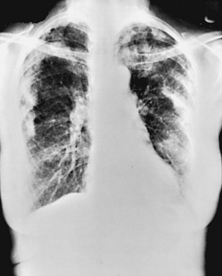
Chronic eosinophilic pneumonia is a disorder in which the pulmonary interstitium and alveolar spaces are infiltrated primarily by eosinophils and, to a lesser extent, by macrophages. The clinical presentation typically occurs over weeks to months, with systemic symptoms such as fever and weight loss accompanying dyspnea and a nonproductive cough. The clues suggesting this diagnosis are often found on the chest radiograph and the routine white blood cell differential count. The radiograph frequently shows pulmonary infiltrates with a peripheral distribution and a pattern more suggestive of alveolar filling than interstitial disease (Fig. 11-9). Because the typical radiographic pattern of pulmonary edema with congestive heart failure has central pulmonary infiltrates with sparing of the lung periphery, the prominent peripheral pattern often seen in chronic eosinophilic pneumonia has been described as the “photographic negative of pulmonary edema.” The majority of patients also have increased numbers of eosinophils in peripheral blood, although this finding is not uniformly present and therefore is not critical for the diagnosis. However, bronchoalveolar lavage typically shows a high percentage of eosinophils, reflecting the pathologic process within the pulmonary parenchyma.

Treatment is gratifying to patient and physician alike because chronic eosinophilic pneumonia characteristically shows a dramatic response to corticosteroid therapy. Clinical improvement and radiographic resolution generally occur within days to weeks, although therapy often must be prolonged for months to prevent recurrence.

Pulmonary Alveolar Proteinosis

Pulmonary alveolar proteinosis (PAP) is a parenchymal lung disease in which the primary pathologic process affects the alveolar spaces, not the alveolar walls. Alveolar spaces are filled with a proteinaceous phospholipid material that represents components of pulmonary surfactant. Accumulation of surfactant components is due to either decreased degradation or surfactant dysfunction. PAP is classified as primary, secondary (usually related to hematologic malignancies), or congenital. Primary PAP is by far the most common of the three types and is discussed here.

Many aspects of the pathogenesis of primary PAP have been elucidated. In most cases, the underlying problem has been identified as production of an autoantibody to granulocyte-macrophage colony-stimulating factor (GM-CSF). The first clues that GM-CSF was involved came from the discovery that GM-CSF knockout mice (in which both alleles for GM-CSF are disabled) consistently develop a pulmonary process with pathology essentially identical to that seen with PAP in humans. GM-CSF, acting through alveolar macrophage-specific transcription factors, affects a number of essential macrophage functions, including regulation of surfactant degradation, intracellular lipid metabolism, and phagocytosis. Thus, inhibiting the activity of GM-CSF via autoantibodies leads to decreased clearance of surfactant from the alveolar spaces.

Patients with alveolar proteinosis present primarily with dyspnea and cough. The chest radiograph is notable for bilateral alveolar infiltrates. HRCT generally shows a distinctive pattern called a crazy paving pattern (produced by thickening of interlobular septa accompanied by ground-glass alveolar filling) that suggests the diagnosis. Patients are susceptible to certain types of superimposed respiratory infections that are uncommon in normal hosts, especially with the organism Nocardia. The susceptibility to unusual pathogens is probably due to abnormal macrophage function as well as to more recently discovered abnormalities in neutrophil function also mediated by GM-CSF.

Treatment of PAP traditionally has been based on whole-lung lavage, which involves washing out the material filling the alveolar spaces while the patient is under general anesthesia. Preliminary studies suggest that administration of exogenous GM-CSF may be an alternative therapy. The prognosis of the disease is generally relatively good, although patients may require additional treatments with whole-lung lavage. The long-term effects of exogenous GM-CSF in this disease are unknown.

References

Idiopathic Pulmonary Fibrosis

Armanios, MY, Chen, JJ, Cogan, JD, et al. Telomerase mutations in families with idiopathic pulmonary fibrosis. N Engl J Med. 2007;356:1317–1326.

ATS/ERS/JRS/ALAT Committee on Idiopathic Pulmonary Fibrosis. An official ATS/ERS/JRS/ALAT statement: idiopathic pulmonary fibrosis: evidence-based guidelines for diagnosis and management. Am J Respir Crit Care Med. 2011;183:788–824.

Fell, CD, Martinez, FJ, Liu, LX, et al. Clinical predictors of a diagnosis of idiopathic pulmonary fibrosis. Am J Respir Crit Care Med. 2010;181:832–837. 15

Gansner, JM, Rosas, IO, Ebert, BL. Pulmonary fibrosis, bone marrow failure, and telomerase mutation. N Engl J Med. 2012;366:1551–1553.

Garcia, CK. Idiopathic pulmonary fibrosis: update on genetic discoveries. Proc Am Thorac Soc. 2011;8:158–162.

Günther, A, Korfei, M, Mahavadi, P. Unravelling the progressive pathophysiology of idiopathic pulmonary fibrosis. Eur Respir Rev. 2012;21:152–160.

Guenther, A, European IPF Network. The European IPF Network: towards better care for a dreadful disease. Eur Respir J. 2011;37:747–748.

Idiopathic Pulmonary Fibrosis Clinical Research Network. Prednisone, azathioprine, and N-acetylcysteine for pulmonary fibrosis. N Engl J Med. 2012;366:1968–1977.

Idiopathic Pulmonary Fibrosis Clinical Research Network Investigators. Acute exacerbations of idiopathic pulmonary fibrosis. Am J Respir Crit Care Med. 2007;176:636–643.

King, TE, Jr., Pardo, A, Selman, M. Idiopathic pulmonary fibrosis. Lancet. 2011;378:1949–1961.

Lawson, WE, Loyd, JE, Degryse, AL. Genetics in pulmonary fibrosis—familial cases provide clues to the pathogenesis of idiopathic pulmonary fibrosis. Am J Med Sci. 2011;341:439–443.

Ley, B, Collard, HR, King, TE, Jr. Clinical course and prediction of survival in idiopathic pulmonary fibrosis. Am J Respir Crit Care Med. 2011;183:431–440.

Maher, TM, Wells, AV, Laurent, GJ. Idiopathic pulmonary fibrosis: multiple causes and multiple mechanisms? Eur Respir J. 2007;30:835–839.

Nathan, SD, Shlobin, OA, Weir, N, et al. Long-term course and prognosis of idiopathic pulmonary fibrosis in the new millennium. Chest. 2011;140:221–229.

Selman, M, Pardo, A. Role of epithelial cells in idiopathic pulmonary fibrosis: from innocent targets to serial killers. Proc Am Thorac Soc. 2006;3:364–372.

Strieter, RM, Mehrad, B. New mechanisms of pulmonary fibrosis. Chest. 2009;136:1364–1370.

Tanjore, H, Blackwell, TS, Lawson, WE. Emerging evidence for endoplasmic reticulum stress in the pathogenesis of idiopathic pulmonary fibrosis. Am J Physiol Lung Cell Mol Physiol. 2012;302:L721–L729.

Vancheri, C, Failla, M, Crimi, N, et al. Idiopathic pulmonary fibrosis: a disease with similarities and links to cancer biology. Eur Respir J. 2010;35:496–504.

Zoz, DF, Lawson, WE, Blackwell, TS. Idiopathic pulmonary fibrosis: a disorder of epithelial cell dysfunction. Am J Med Sci. 2011;341:435–438.

Other Idiopathic Interstitial Pneumonias

American Thoracic Society and European Respiratory Society. American Thoracic Society/European Respiratory Society international multidisciplinary consensus classification of the idiopathic interstitial pneumonias. Am J Respir Crit Care Med. 2002;165:277–304.

Bouros, D, Nicholson, AC, Polychronopoulos, V, et al. Acute interstitial pneumonia. Eur Respir J. 2000;15:412–418.

Caminati, A, Harari, S. Smoking-related interstitial pneumonias and pulmonary Langerhans cell histiocytosis. Proc Am Thorac Soc. 2006;3:299–306.

Collard, HR, King, TE, Jr. Demystifying idiopathic interstitial pneumonia. Arch Intern Med. 2003;163:17–29.

Cordier, JF. Cryptogenic organizing pneumonia. Clin Chest Med. 2004;25:727–738.

Epler, GR. Bronchiolitis obliterans organizing pneumonia. Arch Intern Med. 2001;161:158–164.

Ferguson, EC, Berkowitz, EA. Lung CT: Part 2, The interstitial pneumonias—clinical, histologic, and CT manifestations. AJR Am J Roentgenol. 2012;199:W464–476.

Kinder, BW, Collard, HR, Koth, L, et al. Idiopathic nonspecific interstitial pneumonia: lung manifestation of undifferentiated connective tissue disease? Am J Respir Crit Care Med. 2007;176:691–697.

Visscher, DW, Myers, JL. Histologic spectrum of idiopathic interstitial pneumonias. Proc Am Thorac Soc. 2006;3:322–329.

Young, DA, Cherniack, RM, King, TE, et al. Acute interstitial pneumonitis. Case series and review of the literature. Medicine. 2000;79:369–378.

Pulmonary Parenchymal Involvement Complicating Connective Tissue Disease

Connors, GR, Christopher-Stine, L, Oddis, CV, et al. Interstitial lung disease associated with the idiopathic inflammatory myopathies: what progress has been made in the past 35 years? Chest. 2010;138:1464–1474.

Gochuico, BR. Potential pathogenesis and clinical aspects of pulmonary fibrosis associated with rheumatoid arthritis. Am J Med Sci. 2001;321:83–88.

Grutters, JC, Wells, AU, Wuyts, W, et al. Evaluation and treatment of interstitial lung involvement in connective tissue diseases: a clinical update. Eur Respir Mon. 2006;34:27–49.

Keane, MP, Lynch, JP. Pleuropulmonary manifestations of systemic lupus erythematosus. Thorax. 2000;55:159–166.

Kim, EJ, Collard, HR, King, TE, Jr. Rheumatoid arthritis-associated interstitial lung disease: the relevance of histopathologic and radiographic pattern. Chest. 2009;136:1397–1405.

Strange, C, Highland, KB. Interstitial lung disease in the patient who has connective tissue disease. Clin Chest Med. 2004;25:549–559.

Swigris, JJ, Fischer, A, Gilles, J, et al. Pulmonary and thrombotic manifestations of systemic lupus erythematosus. Chest. 2008;133:271–280.

Westhovens, R, De Keyser, F, van den Hoogen, FHJ, et al. The clinical spectrum and pathogenesis of pulmonary manifestations in connective tissue diseases. Eur Respir Mon. 2006;34:1–26.

Sarcoidosis

Drent, M, Costabel, U. Sarcoidosis. Eur Respir Mon. 2005;10:1–341.

Iannuzzi, MC, Rybicki, BA, Teirstein, AS. Sarcoidosis. N Engl J Med. 2007;357:2153–2165.

Lodha, S, Sanchez, M, Prystowsky, S. Sarcoidosis of the skin: a review for the pulmonologist. Chest. 2009;136:583–596.

Lynch, JP, 3rd., Ma, YL, Koss, MN, et al. Pulmonary sarcoidosis. Semin Respir Crit Care Med. 2007;28:53–74.

Mandel, J, Weinberger, SE. Clinical insights and basic science correlates in sarcoidosis. Am J Med Sci. 2001;321:99–107.

Morgenthau, AS, Iannuzzi, MC. Recent advances in sarcoidosis. Chest. 2011;139:174–182.

O’Regan, A, Berman, JS, Cotton, D, et al. Sarcoidosis. Ann Intern Med.. 156(9), 2012. ITC5-1-ITC5-16

Paramothayan, S, Jones, PW. Corticosteroid therapy in pulmonary sarcoidosis. A systematic review. JAMA. 2002;287:1301–1307.

Weinberger, SE. Clinical crossroads: a 47-year-old woman with sarcoidosis. JAMA. 2006;296:2133–2140.

Zissel, G, Prasse, A, Muller-Quernheim, J. Sarcoidosis—immunopathogenetic concepts. Semin Respir Crit Care Med. 2007;28:3–14.

Miscellaneous Disorders Involving the Pulmonary Parenchyma

Ball, JA, Young, KR. Pulmonary manifestations of Goodpasture’s syndrome. Antiglomerular basement membrane disease and related disorders. Clin Chest Med. 1998;19:777–791.

Carsillo, T, Astrinidis, A, Henske, EP. Mutations in the tuberous sclerosis complex gene TSC2 are a cause of sporadic pulmonary lymphangioleiomyomatosis. Proc Natl Acad Sci U S A. 2000;97:6085–6090.

Collard, HR, Schwarz, MI. Diffuse alveolar hemorrhage. Clin Chest Med. 2004;25:583–592.

Cottin, V, Cordier, JF. Eosinophilic pneumonias. Allergy. 2005;60:841–857.

Doershuk, CM. Pulmonary alveolar proteinosis—is host defense awry? N Engl J Med. 2007;356:547–549.

Frankel, SK, Cosgrove, GP, Fischer, A, et al. Update in the diagnosis and management of pulmonary vasculitis. Chest. 2006;129:452–465.

Frankel, SK, Schwarz, MI. The pulmonary vasculitides. Am J Respir Crit Care Med. 2012;186:216–224.

Greenhill, SR, Kotton, DN. Pulmonary alveolar proteinosis: a bench-to-bedside story of granulocyte-macrophage colony-stimulating factor dysfunction. Chest. 2009;136:571–577.

Henske, EP, McCormack, FX. Lymphangioleiomyomatosis – a wolf in sheep’s clothing. J Clin Invest. 2012;122:3807–3816.

Juvet, SC, McCormack, FX, Kwiatkowski, DJ, et al. Molecular pathogenesis of lymphangioleiomyomatosis: lessons learned from orphans. Am J Respir Cell Mol Biol. 2007;36:398–408.

Lara, AR, Schwarz, MI. Diffuse alveolar hemorrhage. Chest. 2010;137:1164–1171.

Lin, FC, Chang, GD, Chern, MS, et al. Clinical significance of anti-GM-CSF antibodies in idiopathic pulmonary alveolar proteinosis. Thorax. 2006;61:528–534.

Marchand, E, Cordier, JF. Idiopathic chronic eosinophilic pneumonia. Semin Respir Crit Care Med. 2006;27:134–141.

National Institutes of Health Rare Lung Diseases Consortium; MILES Trial Group. Efficacy and safety of sirolimus in lymphangioleiomyomatosis. N Engl J Med. 2011;364:1595–1606.

Review Panel of the ERS LAM Task Force. European Respiratory Society guidelines for the diagnosis and management of lymphangioleiomyomatosis. Eur Respir J. 2010;35:14–26.

Sakagami, T, Uchida, K, Suzuki, T, et al. Human GM-CSF autoantibodies and reproduction of pulmonary alveolar proteinosis. N Engl J Med. 2009;361:2679–2681.

Strizheva, GD, Carsillo, T, Kruger, WD, et al. The spectrum of mutations in TSC1 and TSC2 in women with tuberous sclerosis and lymphangioleiomyomatosis. Am J Respir Crit Care Med. 2001;163:253–258.

Suri, HS, Yi, ES, Nowakowski, GS, et al. Pulmonary Langerhans cell histiocytosis. Orphanet J Rare Dis. 2012;7:16.

Uchida, K, Beck, DC, Yamamoto, T, et al. GM-CSF autoantibodies and neutrophil dysfunction in pulmonary alveolar proteinosis. N Engl J Med. 2007;356:567–579.

Vassallo, R, Schroeder, DR, Decker, PA, et al. Clinical outcomes of pulmonary Langerhans-cell histiocytosis in adults. N Engl J Med. 2002;346:484–490.

Venkateshiah, SB, Yan, TD, Bonfield, TL, et al. An open-label trial of granulocyte macrophage colony stimulating factor therapy for moderate symptomatic pulmonary alveolar proteinosis. Chest. 2006;130:227–237.