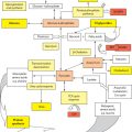
Figure 2.1 Biliary duct development timeline.
Other components of the liver parenchyma have different origins. Thus, Kupffer* cells appear at about 5 weeks’ gestation, apparently from outside of the liver. Mesodermal cells from the early liver anlagen form the mesenchymal framework of the liver, including its perisinusoidal Ito† cell population.
Following these early phases of liver development (induction, migration and formation of hepatocyte cords and hepatic ducts), there is another distinct phase of early postnatal liver development characterised by cell maturation, hepatocyte proliferation and expansion of the liver volume, possibly controlled by activation of the canonical Wnt signalling pathway [13]. During this phase, the intrahepatic bile ducts also elongate from the centre to the periphery.
2.1.1 Haematopoiesis
The liver does not generate haematopoietic cells de novo but is colonised by haematopoietic stem cells, probably from the yolk sac initially, which expand and mature within the developing liver and make anything up to two-thirds of the liver volume within the second trimester. Primitive erythroid cells migrate to the fetal liver and have a dramatic upregulation of adhesion molecules that allows them to bind to fetal liver macrophages. The first haematopoietic stem cells to enter the liver are pluripotent and can form any haematopoietic cell. Their first step in intrahepatic maturation is to commit to a more limited range of lineage options, typically as either an erythromyeloid precursor or a common myelolymphoid progenitor. Nevertheless, erythropoiesis predominates at this stage. This process declines and stops towards the end of gestation with the bone marrow and spleen taking over. At this point, hepatocytes upregulate the expression of metabolic and detoxifying enzymes.
2.1.2 Vascular events
Vascular structures in the developing liver are
1. Vitelline‡ veins (paired): These carry blood from the gut to an evolving sinusoidal plexus and are originally from the yolk sac. These veins initially have a number of extrahepatic interconnections, resembling a stepladder, which then undergoes remodelling to form a single but now S-shaped portal vein, lying behind the distal foregut (Figure 2.2).

Figure 2.2 (a) Development of portal vein. Initial stage of two paired veins (umbilical and vitelline) draining into the sinus venosus. (b) Atrophy of right umbilical vein and vitelline duct. (c) Emergence of sinusoids in liver fed by interconnecting vitelline veins and umbilical vein. (d) Emergence of single portal vein. Umbilical vein connected to suprahepatic veins by ductus venosus.
BOX 2.2: Ductus venosus of Arantius*
This structure is present in many mammalian species, although it has disappeared by term in horses and pigs. In humans, it is a branch of the left portal vein opposite the origin of the umbilical vein and empties into the left hepatic vein (usually). About 20%–30% of umbilical blood flow is shunted through the ductus venosus in the fetus, with progressive diminution as gestational age progresses. The presence of an actual sphincter at its junction with the umbilical vein is controversial [14], although it is capable of dilatation or constriction in response to catecholamines, for instance [15]. The usual reason for this is fetal hypoxia, and diversion of blood away from the liver may be one cause of intrauterine growth retardation. Complete functional closure can be seen in >90% of infants by 2 weeks’ postnatal age as assessed by Doppler scans [20].
Persistence of the ductus venosus has a male predominance, and the resulting portosystemic shunt can be assessed by plasma ammonia and galactose levels and may cause neurocognitive impairment. (See Chapter 22.)
* Giulio Cesare Aranzi (1529–1589), Italian anatomist.
2. Umbilical veins (paired): Carry oxygenated blood from the placenta to the right side of the developing heart at the sinus venosus. The right umbilical vein disappears early in gestation, leaving the other enveloped by the liver. This joins the left portal vein, allowing oxygenated blood into the sinusoids, although the majority of blood flow is directed through a distinct low-pressure venous channel, or ductus venosus, between the left portal vein and the hepatic vein confluence (Box 2.2). This structure is rich in innervated smooth muscle cells and has a condensation of elastin fibres at its origin from the portal sinus, appearing as an ‘hourglass’ in anatomical study and on ultrasound [14]. Although there is no actual sphincter, it does have functionality [15]. There is closure at the time of birth, although it can still be negotiated with catheterisation for a variable time postnatally before subsequent fibrosis and later atrophy. Externally, its course can be appreciated as the site of attachment of the lesser omentum on the undersurface of the anatomical left lobe.

Figure 2.3 Development of pancreas. (a) Initial separation with ventral anlage attached to the developing biliary duct. (b) Rotation of ventral anlage and bile duct behind the duodenum. (c) Fusion of the pancreatic anlagen with crossover of the dorsal duct to now drain through the ventral orifice.
3. Posterior cardinal veins (paired): These carry blood back from the lower half of the body along the posterior body wall. These become the paired azygous* and hemiazygous venous system in the adult.
The larger inferior vena cava is embryologically a much later structure and is formed from many different venous precursors, for example, the subcardinal (to form the prerenal portion) and supracardinal (to form the postrenal portion) veins. The intrahepatic portion itself is an outgrowth from the right subcardinal vein subsumed in the evolving liver tissue (from about the sixth week) to anastomose with the hepatic vein confluence.
The pancreas is derived from ventral and dorsal anlagen,† which arise from the foregut diametrically opposite each other and are recognisable from about 35 days of gestation (Figure 2.3). Some maintain that the ventral bud is divisible into two, with subsequent atrophy of the anterior one. The ventral duct is an offshoot of the bile duct and maintains this connection throughout. The dorsal anlage will give rise to the head, body and tail of the pancreas, while the ventral bud gives rise to the uncinate‡ process. These two buds fuse following a process of rotation of the ventral duct around the foregut at about 50–55 days. From this point, there is an interconnection of the ducts, with the dominant pancreatic drainage from the body and tail of the pancreas being preferentially directed through the ventral duct (of Wirsung§). The entry of the smaller dorsal duct (of Santorini¶) is usually more proximal in the final duodenum and reputedly drains only the uncinate process. Failure of ductal fusion leaves most of the pancreas drained through the entire length of the dorsal duct and is known as the pancreas divisum. This differential heritage can still be evident histologically by staining for pancreatic polypeptide (PP). Thus, PP cells localise to the area derived from the ventral anlagen, while the dorsal pancreas has larger lobules with PP-poor islets.
PDX-1, a member of the ParaHox group of homeodomain transcription factors, and SOX family genes are believed to be the key developmental genes required for normal human pancreas development [16,17]. In contrast to induction of the liver, where BMP signalling is required, development of the pancreas requires inhibition of Bmp signalling. Therefore, differential growth factor signalling within the endodermal cells helps specify the various organ derivatives.
During the last trimester, there is absorption of the junction of bile and pancreatic ducts into the wall of the duodenum so that the final arrangement is a common chamber (the ampulla of Vater) with each duct surrounded by its own sphincter. Failure of this terminal stage leads to an anomalous pancreatobiliary junction or common channel which crucially allows intermixing of pancreatic juice and bile. This defect has been suggested as an aetiological factor for choledochal malformation and also the explanation for the susceptibility to recurrent pancreatitis in this group (see Chapter 7).
The pancreas serves a dual exocrine and endocrine role in the body. Pancreatic acinar tissue, ducts and progenitor endocrine-active cells all appear to arise from the same endodermal rudiment with subsequent differentiation to form two lines of committed endocrine precursor cells. Islets are generated in a multistep process that involves differentiation of progenitor cells from the pancreatic ductal epithelium, migration of these precursors through the basal membrane into the surrounding mesenchyme and subsequent association of hormone-producing cells into mature islets.
Hedgehog, transforming growth factor-β (TGF-β), and Notch signalling are necessary for normal cell differentiation with a complex interplay between them. Notch signalling inhibits endocrine cell differentiation and promotes ductal cell differentiation. All of the endocrine arises from a NeuroD-expressing precursor cell which divides to give rise to α- and γ-cells, which produce glucagon and PPs, respectively, and β- and δ-cells, which secrete insulin and somatostatin, respectively. Each cell type is characterised by a specific combination of transcription factors. Both insulin and glucagon can be detected in the fetal circulation by the fourth or fifth month of fetal development.
2.3 FETAL LIFE AND TRANSITIONAL CIRCULATION
All blood returning passively from the placenta is carried through the umbilical vein, which joins the left portal vein in the liver. Most of the flow then bypasses the sinusoidal network via the ductus venosus to return to the inferior vena cava and right side of the heart. An oxygen saturation of about 80% is observable in the umbilical vein [18].
There is a degree of functional asymmetry during fetal life not seen after birth, with the right side perfused with less well-oxygenated blood derived from portal blood flow, and therefore it has a proportionally greater role in haematopoiesis. The left side, by contrast, receives blood mainly from the umbilical vein and has a higher content of oxygen-dependent enzymes, and is more active in drug binding and metabolism. The blood supply derived from the hepatic arteries is relatively trivial.
At the time of birth, major transitional haemodynamic changes occur, including functional closure of the ductus arteriosus and foramen ovale, reduction in right pulmonary arterial pressures and the cessation of umbilical blood flow. This leads to functional closure of the ductus venosus and separation of the portomesenteric venous systems and a marked increase in well-oxygenated blood flow via the hepatic artery. Not unsurprisingly, there is a measureable increase in portal venous and mesenteric pressure following ductus closure [19], and this has been suggested as one cause of necrotising enterocolitis in susceptible preterm infants. Commencement of enteral nutrition and consequent increases in intestinal perfusion also lead to increased sinusoidal liver perfusion and bile flow.
Crawford LW, Foley JF, Elmore SA. Histology atlas of the developing mouse hepatobiliary system with emphasis on embryonic days 9.5–18.5. Toxicologic Pathology 2010; 38: 872–906.
Larsen’s Human Embryology. 5th ed. Churchill Livingstone, London, 2015.
Strazzabosco M, Fabris L. Development of the bile ducts: essentials for the clinical hepatologist. Journal of Hepatology 2012; 56: 1159–1170.
1. Zhao R, Watt AJ, Li J, et al. GATA6 is essential for embryonic development of the liver but dispensable for early heart formation. Molecular and Cellular Biology 2005; 25: 2622–2631.
2. Fukuda A, Kawaguchi Y, Furuyama K, et al. Loss of the major duodenal papilla results in brown pigment biliary stone formation in pdx1 null mice. Gastroenterology 2006; 130: 855–867.
3. Clotman F, Lannoy VJ, Reber M, et al. The onecut transcription factor HNF6 is required for normal development of the biliary tract. Development 2002; 129: 1819–1828.
4. Coffinier C, Gresh L, Fiette L, et al. Bile system morphogenesis: defects and liver dysfunction upon targeted deletion of HNF1β. Development 2002; 129: 1829–1838.
5. Van Eyken P, Sciot R, Callea F, et al. The development of the intrahepatic bile ducts in man: a keratin immunohistochemical study. Hepatology 1989; 15: 125–135.
6. Kalinichenko VV, Zhou Y, Bhattacharyya D, et al. Haploinsufficiency of the mouse Forkhead Box f1 gene causes defects in gall bladder development. Journal of Biological Chemistry 2002; 277: 12369–12374.
7 Yamashita R, Takegawa Y, Sakumoto M, et al. Defective development of the gall bladder and cystic duct in Lgr4-hypomorphic mice. Developmental Dynamics 2009; 238: 993–1000.
8. Crawford JM. Development of the intrahepatic biliary tree. Seminars in Liver Disease 2002; 22: 213–225.
9. Desmet VJ, Van Eyken P, Sciot R. Cytokeratins for probing cell lineage relationships in developing liver. Hepatology 1989; 15: 125–135.
10. Antoniou A, Raynaud P, Cordi S, et al. Intrahepatic bile ducts develop according to a new mode of tubulogenesis regulated by the transcription factor SOX9. Gastroenterology 2009; 136: 2325–2333.
11. Fischer E, Legue E, Doyen A, et al. Defective planar cell polarity polycystic kidney disease. Nature Genetics 2006; 38: 21–23.
12. Tan CEL, Driver M, Howard ER, Moscoso GJ. Extrahepatic biliary atresia: a first-trimester event? Clues from light microscopy and immunohistochemistry. Journal of Pediatric Surgery 1994; 29: 808–814.
13. Apte U, Zeng G, Thompson MD, et al. beta-Catenin is critical for early postnatal liver growth. American Journal of Physiology and Gastrointestinal Liver Physiology 2007; 292: G1578–G1585.
14. Mavrides E, Moscoso G, Carvalho JS, et al. The human ductus venosus between 13 and 17 weeks of gestation: histological and morphometric studies. Ultrasound in Obstetics & Gynecology 2002; 19: 39–46.
15. Tchirikov M, Schroder HJ, Hecher K. Ductus venosus shunting in the fetal venous circulation: regulatory mechanisms, diagnostic methods and medical importance Ultrasound in Obstetics & Gynecology 2006; 27: 452–461.
16. Habener JF, Kemp DM, Thomas MK. Minireview: transcriptional regulation in pancreatic development. Endocrinology 2005; 146: 1025–1034.
17. Seymour PA. Sox9: a master regulator of the pancreatic program. Reviews in Diabetic Studies 2014; 11: 51–83.
18. Lautt WW. Fetal and neonatal hepatic circulation. In Lautt WW (ed.), Hepatic Circulation: Physiology and Pathophysiology. Morgan & Claypool Life Sciences, San Rafael, CA, 2009. http://www.ncbi.nlm.nih.gov/books/NBK53078/.
19. Sulemanji MN, Azpurua H, Suh M, et al. Ductus venosus closure results in transient portal hypertension – is this the silent trigger for necrotizing enterocolitis? Journal of Pediatric Surgery 2013; 48: 2067–2074.
20. Loberant N, Herskovits M, Barak M, et al. Closure of the ductus venosus in premature infants: findings on real-time gray-scale, color-flow Doppler, and duplex Doppler sonography. American Journal of Roentgenology 1999; 172: 227–229.
* Embryo – from fertilisation to end of week 8 (day 64). This is also the same as the 10th week of gestation, as this is dated from the end of the last period. We then become a fetus until birth.
† CK – cytokeratin, a complex network of proteins forming the intracytoplasmic cytoskeleton. Ranges from CK 1 to 20.
* Joseph Disse (1852–1912), German anatomist.
* Karl Wilhelm von Kupffer (1829–1902), German pathologist; identified cells within liver but never really identified their phagocytic function.
† Toshio Ito (1904–1991), Japanese anatomist who described these fat-storing cells in an abstract published in 1951.
‡ Vitelline – veins of the ‘yolk sac’; derived from Latin vitellus – ‘colour of egg yolk’.
* Azygous – from the Greek azugos, from a, ‘without’, + zugon, ‘yoke’, the vein not being one of a pair.
† Anlage (pl. anlagen) (German) – rudimentary organ or part, especially in an embryo.
‡ Uncinate – from the Latin uncinatus, ‘hook’.
§ Johann Georg Wirsüng (1589–1643), German anatomist who became professor in Padua, Italy. He was reputedly murdered following an argument regarding who the real discoverer of the duct was.
¶ Giovanni Domenico Santorini (1681–1737), Italian anatomist working in Venice.