40
Deposition Disorders
A heterogeneous group of disorders in which there are deposits, usually of endogenous materials, within the skin. Entities belonging to this group that are covered in other chapters include mucinoses (Chapter 38), amyloidosis (Chapter 39), porphyrias (Chapter 41), and calcinosis cutis (Chapter 42).
Gout
• Tophi present as firm skin-colored to white or yellow papules and nodules, usually around joints and on the helices of the ears (Fig. 40.1); in general, they appear ~10 years after the initial episode of acute arthritis and occur in ~10% of patients with gout.
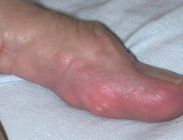
Fig. 40.1 Tophaceous gout of a digit. The deposits create a multilobulated appearance. A small incision over the yellow-white areas followed by microscopic examination of the expressed material would allow a bedside diagnosis.
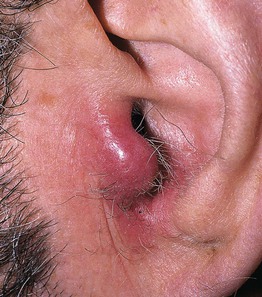
• Tophi can become inflamed (Fig. 40.2), and they may or may not resolve following normalization of serum uric acid levels.
Lipoid Proteinosis
• Skin-colored to yellowish waxy papules and nodules favor the face, including the eyelid margin, and the tongue (Fig. 40.3); atrophic scarring, verrucous changes (especially on the elbows and knees), and a diffuse waxy appearance are additional findings; during infancy, fragility, vesicles and crusting of the face, extremities and mouth can occur.
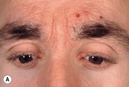
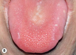
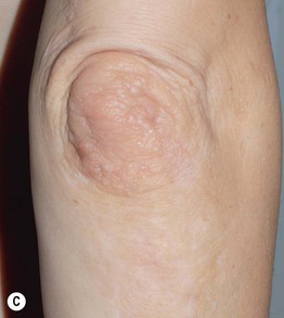
Fig. 40.3 Lipoid proteinosis. A Beaded eyelid papules, hemorrhagic crusts, and confluent waxy papules of the glabella leading to an early leonine facies. B A firm tongue with numerous tiny papules on its dorsal surface. C Skin-colored papulonodules of the elbow as well as irregular hypopigmented scars of the extensor forearm. B, C, Courtesy, Julie V. Schaffer, MD.
Colloid Milium
Mucopolysaccharidoses (MPS)
• The accumulation of GAGs can lead to mental retardation, hepatosplenomegaly, skeletal and joint disease, corneal clouding, and a coarse facies; in addition to thickening of the skin, some patients have hypertrichosis; in Hunter syndrome (MPS II), grouped skin-colored to white papules can develop in the scapular region (‘pebbling’), and in Hurler syndrome (MPS I), patients may have dermal melanocytosis.
For further information see Ch. 48. From Dermatology, Third Edition.


