[level-membership-for-dermatology-category]
Decubitus ulcers
Joseph A. Witkowski, Lawrence Charles Parish, Caren Campbell and Jennifer L. Parish
Management strategy
Prevention


Specific investigations


Staging
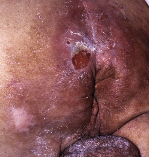
 Stage I: non-blanchable erythema of intact skin
 Stage II: partial-thickness skin loss involving the epidermis and/or dermis


First-line therapies
 Elimination of pressure
 Pressure-reducing and -relieving devices
 Removal of necrotic debris
 Maintenance of a moist wound environment
 Synthetic dressings
 Topical antibacterials
 Nutrition
 Dietary supplements
Eliminating pressure and relieving devices
Debridement
Pressure ulcer treatment guide: quick reference guide for clinicians No. 15.
Bergstrom N, Bennett MA, Carlson CE. Adv Wound Care 1995; 6: 22–44.
Histologic examination of bone biopsy specimens is the gold standard for diagnosing osteomyelitis.
More effective removal of debris can be accomplished by twice-daily wound treatment.
 Hydrocolloid wafer dressing
 Nitroglycerin ointment
 Becaplermin gel
 5-Fluorouracil cream
 Hyperbaric oxygen
[/level-membership-for-dermatology-category][not-level-membership-for-dermatology-category]
Decubitus ulcers
Joseph A. Witkowski, Lawrence Charles Parish, Caren Campbell and Jennifer L. Parish
