Cranial Nerves IX and X

Published on 03/03/2015 by admin

Filed under Neurology

Last modified 22/04/2025

Print this page

rate 1 star rate 2 star rate 3 star rate 4 star rate 5 star
Your rating: none, Average: 3 (1 votes)

This article have been viewed 7774 times

9 Cranial Nerves IX and X

Glossopharyngeal and Vagus

Cranial Nerve IX: Glossopharyngeal Nerve and Swallowing

Clinical Vignette

A 70-year-old man with a history of hypertension and paroxysmal atrial fibrillation presented to the emergency department with acute onset of slurred speech, left-sided paresis, left neglect, and a left inferior quadrantanopsia. MRI revealed a right middle cerebral artery stroke. An initial oral peripheral and cranial nerve examination revealed bilateral depressed gag reflex and diminished soft palate elevation, left central facial weakness, reduced labial retraction, and mild tongue deviation to the right side with protrusion. The patient was to receive nothing per mouth (NPO) and was referred to a speech pathologist.

Clinical swallowing evaluation demonstrated hoarse and moderately dysarthric speech that retained fair intelligibility. Graduated sized boluses of thin liquids, nectar thick liquids, and purees were administered. The patient had significant difficulty with oral containment, bolus formation, and posterior transport through the oral cavity, on the left more than on the right. Posterior placement on the right side facilitated oral swallowing. Reduced orolabial seal benefited from the use of a straw. Attempts to administer a soft solid bolus were unsuccessful because of delayed triggering of oropharyngeal swallow. Mild voice changes after liquids, characterized by a wet vocal quality, indicated laryngeal penetration and possible silent aspiration. The patient was able to clear with cues and to use a throat clear/reswallow strategy. Use of a chin tuck swallowing posture with nectar thick liquids eliminated clinical signs of aspiration.

He was placed on a modified pureed diet with nectar thick liquids, and medications were crushed in applesauce. Aspiration precautions included remaining upright 90° during and 45 minutes after oral intake and single, small boluses. Swallowing strategies included a chin tuck posture and right posterior placement of food in the oral cavity to decrease anterior leakage and to assist in oral transport. A flexible endoscopic evaluation demonstrated bilateral vocal cord movement, but sensory testing revealed severe left laryngopharyngeal sensory deficit and silent aspiration.

Swallowing malfunctioning, or dysphagia, prevents adequate nutrition intake and may predispose to significant aspiration with a risk for potentially fatal pneumonia. Dysphagia occurs in a variety of central and peripheral neurologic disorders and can rarely be the presenting sign of neurologic disease. It is seen with central disorders including acute cerebral infarction, brainstem stroke, Parkinson disease, and demyelinating disease such as multiple sclerosis. Dysphagia may be a prominent and progressive problem in motor neuron disease, syringobulbia, or primary pontomedullary meningeal-based tumors. Peripheral disorders of the nerve (e.g., Guillain-Barré syndrome), neuromuscular junction (e.g., myasthenia gravis), and hereditary or acquired muscle disease (e.g., oculopharyngeal dystrophy, myotonic dystrophy and dermatomyositis) can also compromise swallowing function.

Physiology

Swallowing is a complex process involving motor control with sensory feedback from many anatomic structures within the oral cavity, pharynx, larynx, and esophagus (Fig. 9-1). The trigeminal (CN-V), facial (CN-VII), glossopharyngeal (CN-IX; Fig. 9-2), vagus (CN-X; Fig. 9-3), and hypoglossal (CN-XII) cranial nerves are involved. A “normal swallow” comprises two major components, bolus transport and airway protection. The swallowing process is typically classified into four phases (Fig. 9-4).

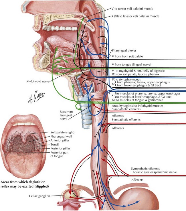
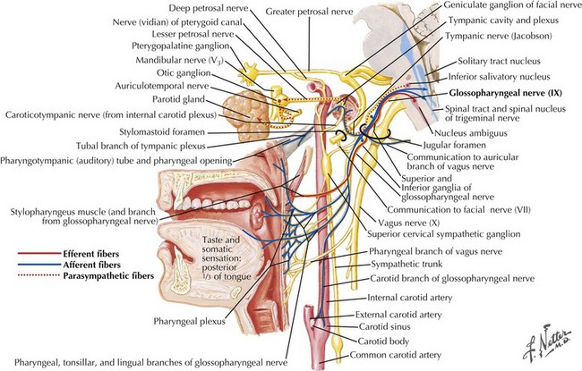
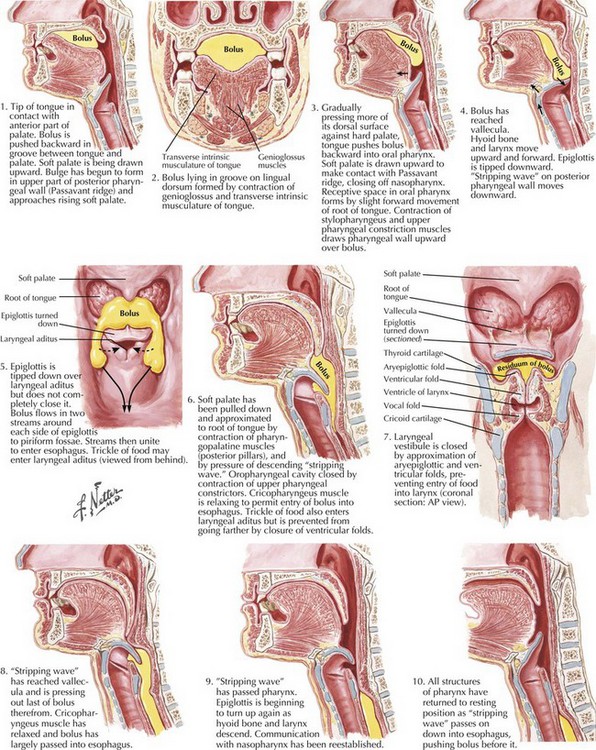
1. The oral preparatory phase involves voluntary motor function during which food or liquid is taken into the mouth, masticated (CN-V), and mixed with saliva to form a cohesive bolus (Fig. 9-4, nos. 1 and 2). This phase requires coordination of several cranial nerves and corresponding structures. Tension in the labial and buccal musculature closes the anterior and lateral sulci (CN-VII) while rotary mandible motion produces chewing (CN-V3). Lateral rolling tongue motion (CN-XII) and bulging of the soft palate forward (widening the nasal airway, and sealing the posterior oral cavity) properly positions the bolus for the swallowing (CN-IX). Tongue mobility is the most important neuromuscular function involved in this first phase. The mid and lower divisions of CN-V provide sensory feedback for positioning the bolus. Saliva derived from the parotid, sublingual, and submandibular glands (innervated by secretomotor fibers of CN-IX and -VII) contain digestive enzymes that act as an emollient to soften and shape the bolus.

2. The oral swallowing phase is initiated when the tongue (CN-XII) sequentially squeezes the bolus posteriorly against the hard palate and initiates propulsion into the oropharynx (Fig. 9-4, nos. 3 and 4). Lips and buccal muscles contract (CN-V and CN-VII) with elevation of the velum (CN-V and CN-X) providing the valving process that generates pressure to seal the nasopharynx, preventing reflux and nasal regurgitation. CN-V is responsible for the afferent (sensory) feedback for the entire oral cavity and tongue. The soft palate (CN-IX), critical to containing the bolus within the oral cavity during the oral preparatory phase, now moves posteriorly to allow the bolus to pass through the faucial arches and simultaneously prevent the bolus from entering the nasopharynx. The swallowing reflex is triggered as the bolus passes the anterior tonsillar pillars, which initiates the pharyngeal phase.

Taste for the anterior two thirds of the tongue is carried by CN-VII, whereas the afferent CN-IX controls taste for the posterior one third of the tongue and the posterior pharyngeal wall. CN-X supplies primary innervation to the palatal muscles, pharyngeal constrictors, laryngeal musculature, and cricopharyngeus. Afferent fibers also provide critical sensory feedback from the larynx and esophageal inlet.

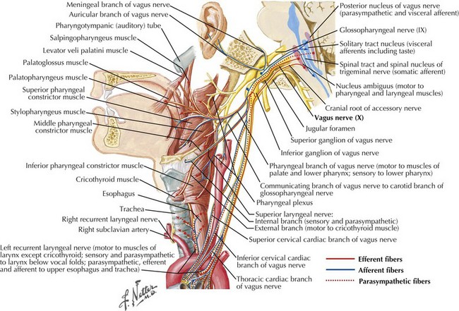
3. The pharyngeal phase begins with the bolus passing into the throat, triggering the swallowing reflex and causing several pharyngeal physiologic actions to occur simultaneously, allowing food to pass into the esophagus (Fig. 9-4, nos. 57). Once pharyngeal swallowing is elicited, essential functions of airway protection occur. Intrinsic laryngeal muscles innervated by CN-X close the larynx at the aryepiglottic, false vocal, and at the true vocal folds, creating a seal that separates the airway from the digestive tract protecting the laryngeal vestibule from foreign material aspiration. The tongue (CN XII) is the major force pushing the bolus through the pharynx. Synergistic actions with CN-X produce pharyngeal peristalsis as it innervates the pharyngeal constrictors and carries afferents from the lower pharynx.

CN-IX mediates the sensory portion of the pharyngeal gag but innervates just 1 muscle, the stylopharyngeus. The absence of the gag reflex is not the sole indicator of a patient’s swallowing abilities. A study of the risk of aspiration in patients with dysphagia and absent gag reflexes demonstrated that the majority could tolerate a modified diet. Additionally, the gag reflex was absent in 10–13% of nondysphagic individuals in the control group.

Poor airway protection and delayed triggering of pharyngeal swallow may cause aspiration. When swallowing is inefficient and aspiration occurs, a reflexive cough needs to occur as a respiratory defense against foreign matter. The cough reflex is induced by irritation of afferent CN-IX and CN-X sensory fibers in the larynx, trachea, and larger bronchi (Figs. 9-2 and 9-3). If a reflexive cough is not elicited in response to foreign material within the airway, silent aspiration results; it is radiographically documented in 50% of aspiration cases.

CN-IX is the primary afferent of the swallowing response, whereas CN-X is the secondary afferent; both nerves terminate in the swallowing center located in the medulla within the nucleus solitarius. Sensory events initiating swallowing occur with stimulation to jaw, posterior tongue, faucial pillars, and upper pharynx and are mediated through CN-V, CN-IX, and CN-X. These afferent fibers converge on the nucleus solitarius in the medulla and communicate with the nucleus ambiguous via interneurons stimulating the motor response.

4. The esophageal phase occurs with the passage of the bolus through the cricopharyngeal sphincter, moving over the closed airway and passing the pharyngoesophageal segment into the esophagus via the cricopharyngeal sphincter at the proximal esophagus (Fig. 9-4, nos. 810). This area contains the cricopharyngeus muscle that normally keeps the esophagus closed. CN-X mediates the action of the cricopharyngeus, which relaxes to allow food to pass from the hypopharynx into the esophagus.

Elevation and anterior movement of the larynx is the significant mechanical force contributing to the opening of the cricopharyngeal sphincter which, in conjunction with the relaxation of the cricopharyngeus muscle, opens the pharyngoesophageal segment, permitting the passage of food into the esophagus. The sphincter must otherwise remain closed to prevent the entrance of air into the stomach and reflux from the esophagus into the hypopharynx. CN-X, specifically the efferent fibers from the dorsal nucleus, innervate the involuntary muscles of the esophagus, stomach, small intestine, and portions of the large intestine.

Diagnostic Approach

Formal assessment of swallowing function through various examinations serves to define the severity of dysphagia and to identify therapeutic strategies to minimize the risk of aspiration. Clinical swallowing evaluation includes the patient history; observations regarding mental awareness and ability to cooperate; oral, peripheral, and cranial nerve examinations; and the overall respiratory status. Various food consistencies are administered with close monitoring of the oral preparatory and pharyngeal phases of swallowing. Based on this initial evaluation, swallowing strategies may be implemented or the need for objective studies may be identified.

Flexible endoscopic evaluation of swallowing with sensory testing (FEES) allows direct evaluation of motor and sensory aspects of the pharyngeal swallow. It requires transnasal passage of a fiberoptic laryngoscope into the hypopharynx to view the larynx and surrounding structures. Laryngeal airway protection and the integrity of the oropharyngeal swallow are assessed by giving various food consistencies tinted with coloring to enhance visualization. Similar to the modified barium swallow (MBS), compensatory strategies and postures are attempted to facilitate improved swallowing function and decrease the risk of aspiration. Velopharyngeal closure, anatomy of the base of the tongue and hypopharynx, abduction and adduction of the vocal folds, pharyngeal musculature, and the patient’s ability to manage secretions are assessed. Laryngopharyngeal reflux can also be visualized.

Modified barium swallow (MBS), also called videofluoroscopy or videopharyngogram, is a functional evaluation requiring active patient participation. Before scheduling MBS, laryngopharyngeal sensation should be evaluated via FEES to assess the risk of barium aspiration. MBS is performed in conjunction with radiologic examination but differs from the standard barium swallow in that patients ingest graduated sized boluses of various consistencies mixed with barium in the upright position. The primary purpose of MBS is to determine appropriate therapeutic intervention strategies to facilitate safe and efficient swallowing function. Aspiration and silent aspiration can also be detected. Images are taken in lateral and anterior–posterior projections to focus on oropharyngeal swallowing anatomy and physiology, and to screen for esophageal motility and pharyngeal reflux. The patient need not be NPO for an MBS.

Clinical Considerations and Outlook

There are three major considerations for resumption of oral intake in dysphagic patients: safety of swallow, ability to maintain oral nutritional support, and quality of life. In patients with central neurologic compromise, safety of swallow is often grossly impaired and the risk of aspiration pneumonia significantly increased. Additionally, many of these patients are bedridden and have cognitive impairment or decreased levels of alertness. In this setting, even small amounts of aspiration cannot be tolerated. Although the majority of stroke patients improve over time and resume oral intake, other neurodegenerative disorders such as ALS have an unrelenting course with progressive dysphagia and increasing risk of aspiration. Others with neuromuscular disorders, such as myasthenia gravis, may present with a good swallow, but fatigue over time with chewing and consecutive swallowing eventually impede proper deglutition and the potential to tolerate a full oral diet. Eating becomes effortful, making consumption of enough calories difficult. Elucidating the exact etiology and pathophysiology for dysphagia in each case helps direct the treatment approach and predict outcome.

Damage to descending corticobulbar fibers can occur from stroke, head injury, or multiple sclerosis. Stroke can result in mild to severe dysphagia depending on the site and size of the lesion and the accompanying deficits. Unilateral hemispheric stroke is a common but usually temporary cause of dysphagia. Delayed oral transit times, delayed or absent triggering of the pharyngeal swallow, and poor pharyngeal bolus propulsion with decreased sensory awareness in the oral and pharyngeal cavities are common sequelae of stroke-induced dysphagia. Apraxia of the swallow mechanism with uncoordinated muscle movements, characterized by reduced bolus formation and inability to manipulate the bolus and trigger timely sequenced swallow, may also occur. If the nucleus ambiguus is damaged in a brainstem stroke, ipsilateral paralysis of the larynx, pharynx, and palate may lead to a severe pharyngeal phase dysphagia. If the nucleus ambiguus is spared but there is unilateral tongue, face, or jaw weakness with concomitant loss of sensation on the affected side of the oral cavity, severe dysphagia could also result.

Optimal management of neurogenic dysphagia requires a multidisciplinary approach and awareness of the natural history of the underlying disorder. Concomitant respiratory disorders and psychosocial aspects of eating should also be considered.

Placement of a percutaneous esophagogastrostomy can be lifesaving in neurologic disorders associated with severe dysphagia that have significant potential for recovery, such as strokes. Even in terminal illnesses such as amyotrophic lateral sclerosis, a percutaneous esophagogastrostomy can provide sustained comfort and maintain nutrition intake while significantly lessening the risk aspiration pneumonia.

Cranial Nerve X, Vagus: Voice Disorders

Clinical Vignette

A 33-year-old female computer programmer with no prior medical problems presented with abrupt onset of a weak, breathy voice. She woke up without a voice 3 days prior to her visit. In addition to the weak voice, she has noted coughing and choking when drinking thin liquids and frequently feels that small particles of food become stuck in the left side of her throat. Physical examination reveals a healthy female with a very breathy, weak voice and asymmetric elevation of the palate. The otolaryngology consultant finds a complete left vocal fold paralysis, with the vocal fold in the paramedian position. Workup, including a chest x-ray and CT of the neck, reveals no lesions or masses along the course of the left vagus nerve. Idiopathic vocal fold paralysis is diagnosed, and the patient defers temporary injection of the vocal fold to improve voice and swallowing. Over the next 12 weeks, the patient notes a slow but steady return of her voice, and reexamination of the larynx 4 months after symptom onset shows near-normal function of the left vocal fold.

Although the larynx is usually considered the source of speech, speech production requires precise coordination of multiple organ systems. Contraction of the abdominal musculature, diaphragm, and chest wall provides a power source for the voice. The larynx acts as a pressure regulator and vibratory source. The pharynx, tongue, nose, and mouth shape these vibrations into recognizable speech and singing. However, the larynx is the most easily injured of these systems, and most vocal problems originate within it.

Anatomy of the Larynx

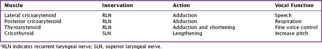
The framework of the larynx consists of thyroid and cricoid cartilages. The arytenoid cartilages articulate with the posterior portion of the cricoid. Vocal ligaments stretch from the arytenoids to the thyroid cartilage. Muscles inserting on the arytenoids move the arytenoids and vocal folds together for speech and swallowing, and apart for respiration. Although the arytenoids’ motion is multidimensional, knowledge of the intrinsic laryngeal muscles and their functions is important for diagnosis (Table 9-1; Fig. 9-3). Note that the cricothyroid muscle is the only intrinsic laryngeal muscle innervated by the superior laryngeal nerve (SLN), and the posterior cricoarytenoid is the only vocal fold abductor.

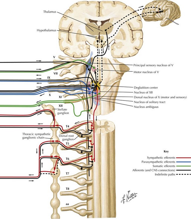
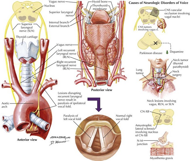
The motor supply of the laryngeal muscles begins in the nucleus ambiguus (see Fig. 9-3). These fibers travel within the vagus nerve (CN-X) as it exits the cranium via the jugular foramen, traveling through the neck within the carotid sheath (Fig. 9-5). High in the neck, the SLN splits from CN-X and travels medially and inferiorly. It splits again into internal and external branches. The internal branch pierces the thyrohyoid membrane and provides sensory innervation to the pharynx and larynx. The external branch travels lower in the neck past the superior pole of the thyroid gland to innervate the cricothyroid muscle.

The recurrent laryngeal nerve (RLN) takes a more tortuous path. It separates from CN-X, loops around the aortic arch on the left and the brachiocephalic artery on the right, and travels back toward the larynx in the tracheoesophageal groove bilaterally. It passes under the thyroid gland and inserts into the larynx under the thyroid cartilage, innervating all other intrinsic laryngeal muscles. Both these nerves are vulnerable to injury and have distinct symptoms when injured.

Disorders of Voice

Recurrent Laryngeal Nerve

Recurrent laryngeal nerve damage usually causes vocal fold immobility on the side of injury. Depending on the position of the vocal fold, symptom severity varies greatly. The most common symptoms are a breathy, hoarse voice and ineffective cough. If the paralyzed vocal fold is in the midline, the only symptom may be vocal fatigue and slight breathiness. Most patients eventually compensate somewhat. The normal vocal fold may cross the midline slightly, or the patient may use muscles around the larynx to squeeze the vocal folds shut. If accessory muscles are used to speak, muscle fatigue and neck pain may develop after prolonged talking.

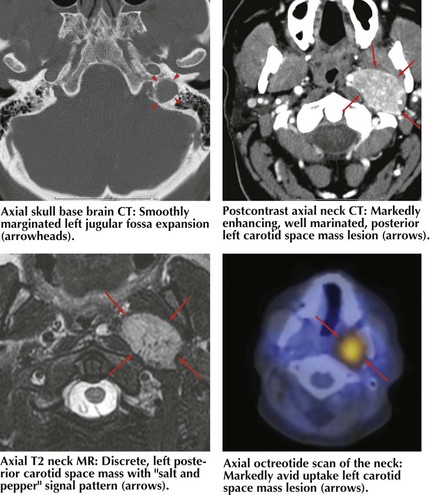
Common causes of vocal fold paralysis include thyroid, lung, or neck tumors; cerebrovascular accidents; CN-X tumors (paragangliomas or glomus vagale [Fig. 9-6]); and surgery near CN-X or the RLN. Less common causes include thyroiditis (causing inflammation of the RLN), infectious diseases, diabetes, or other neuropathies.

Diagnostic evaluation of vocal fold paralysis may include imaging of the brain, neck, and chest, serologic testing, and thyroid function tests; electromyography of the laryngeal muscles may confirm the diagnosis. Even with extensive investigation, the cause of vocal fold paralysis sometimes cannot be determined. Treatment is usually directed at moving the paralyzed vocal fold to the midline. Reinnervation procedures have been described but are not widely used because of inconsistent results. Bilateral vocal fold paralysis is a rare but severe problem and leads to respiratory compromise: the paralyzed vocal folds cannot be abducted and generally move to a medial position. Severe stridor generally results, and tracheostomy is almost always required to allow the patient to breathe.

Additional Resources

Cranial Nerve X

Hill AN, Jankovic J, Vuong KD, et al. Treatment of hypophonia with collagen vocal cord augmentation in patients with parkinsonism. Mov Disord. 2003 Oct;18(10):1190-1192.

Lorenz RR, Esclamado RM, Teker AM, et al. Ansa cervicalis-to-recurrent laryngeal nerve anastomosis for unilateral vocal fold paralysis: experience of a single institution. Ann Otol Rhinol Laryngol. 2008 Jan;117(1):40-45.

Mao VH, Abaza M, Spiegel JR, et al. Laryngeal myasthenia gravis: report of 40 cases. J Voice. 2001;15:122-130. The largest series of patients with laryngeal manifestations of myasthenia gravis

Paniello RC, Barlow J, Serna JS. Longitudinal follow-up of adductor spasmodic dysphonia patients after botulinum toxin injection: quality of life results. Laryngoscope. 2008 Mar;118(3):564-568.

Rosen CA, Gartner-Schmidt J, Casiano R, et al. Vocal fold augmentation with calcium hydroxylapatite: twelve-month report. Laryngoscope. 2009 May;119(5):1033-1041.

Roy N, Smith ME, Dromey C, et al. Exploring the phonatory effects of external superior laryngeal nerve paralysis: an in vivo model. Laryngoscope. 2009 Apr;119(4):816-826.

Rubin AD, Sataloff RT. Vocal fold paresis and paralysis. Otolaryngol Clin North Am. 2007 Oct;40(5):1109-1131. viii-ix

Sulica L. The natural history of idiopathic unilateral vocal fold paralysis: evidence and problems. Laryngoscope. 2008 Jul;118(7):1303-1307. Meta-analysis of 717 cases of idiopathic unilateral vocal fold paralysis regarding duration and outcome of the paralysis