Cranial Nerve V

Published on 03/03/2015 by admin

Filed under Neurology

Last modified 22/04/2025

Print this page

rate 1 star rate 2 star rate 3 star rate 4 star rate 5 star
Your rating: none, Average: 0 (0 votes)

This article have been viewed 3628 times

6 Cranial Nerve V

Trigeminal

Clinical Vignette

A 58-year-old car salesman presented to his physician noting recent onset of a subtle numbness on his left cheek first apparent while shaving or washing his face. He pointed to a “dead area about the size of a dime” over his left zygoma. This patient had a minor pea-sized skin lesion removed from that same area of his face 2 years earlier and the wound healed uneventfully. He otherwise enjoyed perfect health. Twenty months later, he first noted the numbness, describing it as a minor sense of something crawling under his skin. He first mentioned this symptom during a routine checkup with his internist, who could not find any abnormalities such as skin induration, tenderness, or lymphadenopathy. A referral to his previous surgeon also failed to demonstrate any abnormalities; his examination was “totally benign.” For completeness, the surgeon had the tissue pathology reviewed by a pathologist, who reassured that total removal of the tumor was achieved and tumor margins were “clean.”

The area of numbness was described as “dead skin.” These symptoms had an insidious onset. When the patient’s symptoms persisted and became more well defined, he was referred to a neurologist. A very small area of subjective sensory loss, the size of a dime, was noted; the remainder of his neurologic examination was normal. MRI scan of the face and skull base was normal. The patient was advised to return for follow-up examinations every 6 months. Although the small degree of sensory loss persisted, no new clinical or MRI findings were defined during the next 2.5 years. The patient did not return for his 3-year follow-up despite his neurologist advising him to do so. Almost 3.5 years after the onset of his facial numbness, the area of sensory loss enlarged and the patient’s wife felt that his left eye was more prominently seen within the orbit. Repeat neurologic exam confirmed the presence of a larger “quarter-sized” sensory loss. MRI now demonstrated a large infraorbital mass. Biopsy demonstrated metastatic squamous cell skin cancer.

Comment: Invasion of trigeminal nerve perineural spaces is a rare but well appreciated complication of various facial skin malignancies. As this case dramatically illustrates, these can have an insidious onset, with no more than subtle sensory loss at the onset. Excellent but nondiagnostic imaging studies can lead to a false sense of security for the patient and inexperienced clinician alike. Eventually the tumor may metastasize along a peripheral cutaneous nerve per se, in this case the maxillary nerve (second division of the trigeminal), to the base of the brain, where the tumor penetrated the foramen rotundum. This centripetal spread along the trigeminal nerve and its gasserian ganglion is rare. Any epithelial squamous cell carcinoma (SCC) has this discrete potential for a perineural spread with potential central nervous system dissemination. The indolent progression of a cranial nerve palsy in any patient with history of a resected cutaneous SCC of the head and neck must raise clinical suspicion of perineural spread, even in the absence of positive findings on detailed and repeated MRI imaging.

Anatomy

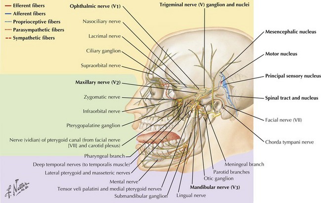
The trigeminal cranial nerve (CN-V) has three major divisions: ophthalmic, maxillary, and mandibular (Figs. 6-1 and 6-2). CN-V is the major sensory nerve of the face, mouth, and nasal cavity. It also supplies motor and proprioceptive fibers to the muscles of mastication. The trigeminal nerve sensory and motor roots are derived from a large sensory and smaller motor nucleus within the pons. These roots converge to emerge at the midlateral pons, then continue toward the trigeminal (Gasserian) ganglion at the base of the middle cranial fossa. General sensory fibers are derived from their cell bodies of origin within this ganglion. Distal to the trigeminal ganglion, three sensory divisions respectively exit the skull through the superior orbital fissure, foramen rotundum, and foramen ovale. The motor component passes through the trigeminal ganglion into the mandibular division.

CN-V Nuclei

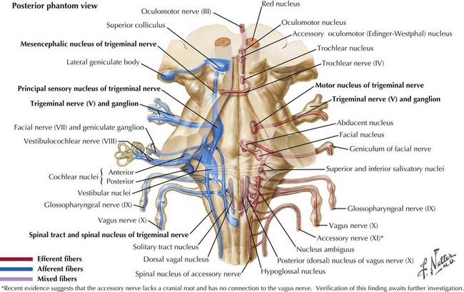
The sensory nucleus is the largest of the trigeminal pontine complex. This begins rostrally within the midbrain, and extends caudally through the pons and medulla into the second segment of the cervical spinal cord (Fig. 6-3). It is subdivided into three portions: (1) the spinal tract nucleus primarily dedicated to pain and temperature fibers; (2) the principal sensory nucleus, the pontine trigeminal portion, which primarily receives tactile stimuli and, therefore, principally subserves light touch; (3) the mesencephalic sensory nucleus, which contains cell bodies of sensory fibers carrying proprioceptive information from the masticatory muscles. Lastly, there is a motor nucleus located within the pons medial to the large sensory nucleus.

Principal Sensory Component

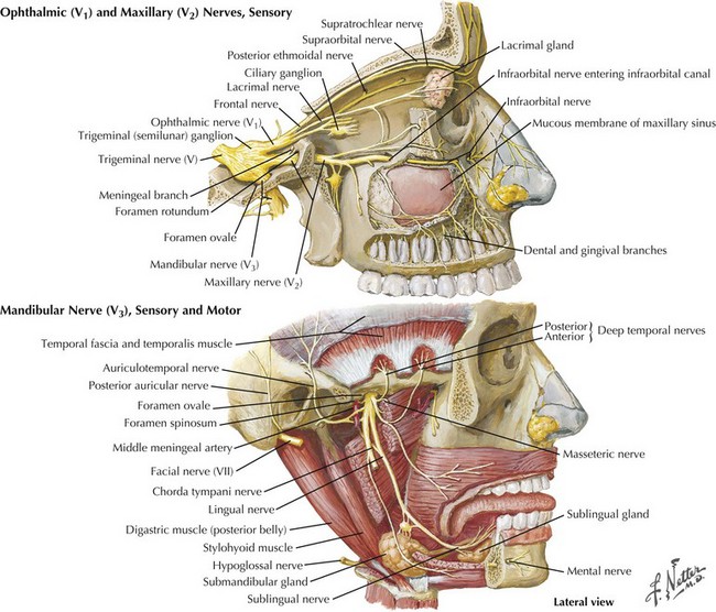
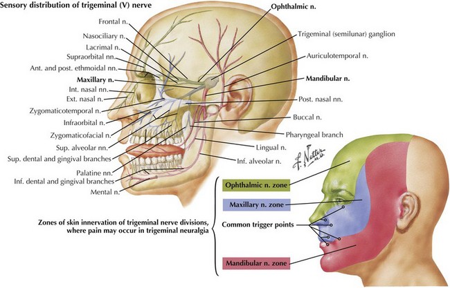
This component of CN-V conveys general sensation from the facial skin and scalp to the top of the head, tragus of the ear, and anterior wall of the external auditory meatus (Fig. 6-4). Also, it provides general sensation from the mouth, including the tongue and teeth, nasal and paranasal sinuses, and meninges lining the anterior and middle cranial fossae.

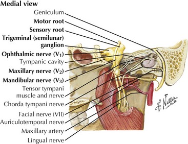
Trigeminal (Gasserian, Semilunar) Ganglion

The cell bodies of almost all sensory CN-V fibers are located within the trigeminal ganglion (Fig. 6-5). This is contained within a skull-based depression, the Meckel cave, that is located in the floor of the middle cranial fossa. Central processes of neuronal cell bodies constitute the large sensory root that enters the pons and projects into the pontine trigeminal main sensory nucleus and spinal tract nucleus. The peripheral processes of this nerve divide into the three sensory divisions that exit the skull through the superior orbital fissure, the foramen rotundum, and the foramen ovale.

CN-V Lesions

Clinical Presentation and Diagnostic Approach

Trigeminal neuralgia is the most common fifth cranial nerve disorder as discussed in Primary and Secondary Headache, Chapter 11. In contrast, a primary trigeminal neuropathy, as illustrated in this chapter’s vignette, is very uncommon.

Facial numbness is the classic symptom of a primary trigeminal (CN-V) neuropathy. Patients with this complaint may have a lesion at any site along the trigeminal nerve sensory pathway, including the nuclei within the brainstem, sensory root, trigeminal ganglion, or primary divisions of CN-V. To determine precisely which portion of the trigeminal nerve complex is affected, the examiner needs to initially test touch, temperature, and pain sensation within the distribution of each of the three major divisions. Examining the corneal reflex provides another useful clinical tool. Application of a wisp of cotton to the cornea normally leads to an eye blink, provided the facial nerve is intact, permitting a blink to occur when the sensory portion of this reflex arc is preserved. When there is a significantly asymmetrical corneal response or actual unilateral loss of this reflex, there is evidence of ophthalmic division CN-V neuropathy.

Both general and special sensation to the tongue and palate are necessary for fully functional taste. Impairment of general sensation from the tongue and palate carried by CN-V may also cause taste disturbances, even though the special sensory fibers providing primary taste sensation are supplied by the facial and glossopharyngeal nerves. Masticatory weakness is often very difficult to test when the change is subtle. Normally both lateral pterygoid muscles pull the jaw anteriorly or forward. When there is a unilateral motor fifth nerve lesion, the healthy unopposed pterygoid pulls the jaw across the midlines, thus leading to a deviation of the jaw to the side of the paretic motor fifth. This occurs when the motor nucleus, root, or mandibular division of the fifth cranial nerve is damaged.

Differential Diagnosis

Facial pain, numbness, or both are the hallmarks of most CN-V lesions. Facial trauma, particularly secondary to vehicular accidents or rarely invasive dental treatments, accounts for the majority of trigeminal nerve injuries. CN-V divisions and branches are exposed to trauma especially from fractures of facial bones within the face and neck and other clinically invasive procedures, particularly from dental work. Typically, these injuries lead to anesthesia within the specific distribution of whichever trigeminal branch is compromised.

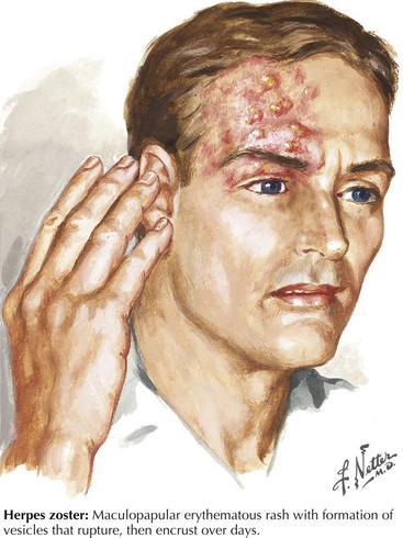
In the Western world, Herpes zoster is the most common infectious cause of a trigeminal neuropathy (Fig. 6-6). Herpes zoster ophthalmicus occurs when latent varicella zoster virus infection within the trigeminal ganglion becomes reactivated, almost always affecting the ophthalmic division. However, very rarely, the maxillary or mandibular divisions may be primarily affected. Most patients present with a characteristic periorbital vesicular rash and severe neuralgic pain within the ophthalmic division (see Fig. 6-6). Similar to other herpes zoster syndromes, the pain may precede the eruption of cutaneous lesions. Permanent visual impairment is the most serious outcome of ophthalmic zoster infection. Antiviral agents such as acyclovir are the main therapy. Corticosteroid eyedrops are shown to decrease pain and hasten corneal healing and are sometimes prescribed by ophthalmologists if there are no other ocular or systemic diseases that might contraindicate their use. Rarely, an ipsilateral middle cerebral artery infarction may subsequently develop in these patients as the virus travels within the trigeminal nucleus and affects the adjacent intracavernous portion of the carotid artery or its branches with a granulomatous angiitis of the brain. CSF varicella zoster virus antibodies are frequently found, and antiviral treatment is felt to be helpful.

Worldwide, leprosy or Hansen disease is a more common cause of CN-V neuropathy. This primarily occurs in economically depressed countries. It generally affects the coolest areas of the skin. Thus, if sensory loss is confined to the tip of the nose or the pinna of the ear, Hansen disease is a primary consideration (Box 6-1).

Trigeminal sensory neuropathy occurs when the trigeminal ganglion cell bodies are the primary pathologic target. Although the pathogenesis of this ganglionopathy is frequently enigmatic, an association with connective tissue disorders, particularly scleroderma or Sjögren syndrome, is recognized. Patients with these disorders typically have numbness that begins around the mouth and spreads slowly over months involving all CN-V divisions, unilaterally or bilaterally, with the ophthalmic division at times spared. In Sjögren syndrome, trigeminal neuropathy is common and is usually representative of a more widespread sensory ganglionopathy. Presumably, circulating autoantibodies attack the ganglion cells, where the blood–brain barrier is more permeable to large molecules than is the blood–brain barrier elsewhere in the peripheral nervous system.

A specific medication reaction is another exceedingly uncommon cause of a trigeminal neuropathy. The two best-recognized offenders are the industrial solvent trichloroethylene and the biological tracer hydroxystilbamidine.

The possibility of a metastatic neoplasm infiltrating a fifth nerve branch must always be included in the differential diagnosis of a persistent, but not always severe, facial numbness or pain. This is very well illustrated in this chapter’s vignette. Tumors involving the face, such as squamous cell carcinoma, microcystic adnexal (sweat gland) carcinoma, and keratoacanthoma, have a proclivity for invading cutaneous nerves because of their innate neurotropism.

Primary trigeminal neurinomas are rare, usually benign, well-demarcated, and slowly growing neoplasms. These comprise 0.2% of intracranial tumors and 2–3% of intracranial neurinomas. Most frequently, these tumors arise near the trigeminal ganglion, usually extending into the middle and posterior cranial fossae. Rarely, they arise exclusively from one of the sensory divisions and spread extracranially. Rare instances of malignant schwannomas originating within the trigeminal ganglion also occur. Most neurinomas have very slow growths, and reach a 2.5 cm diameter before a specific diagnosis is made. Initially, these patients typically report developing numbness and paresthesiae within the CN-V distribution. It is very rare for typical trigeminal neuralgia pain or even intermittent burning pain to be the presenting sign of a primary trigeminal tumor. Rarely, these sensory findings are accompanied by other neurologic symptoms resulting from damage to adjacent structures. For example, tumors growing downward into the posterior fossa may lead to cerebellar ataxia and lesions of CN-VII and CN-VIII, manifesting with facial palsy, tinnitus, or hearing loss. In contrast, neurinomas exert pressure upward on the lateral wall of the cavernous sinus, leading to CN-II, -III, -IV, and -VI lesions.

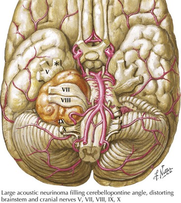
Cerebellopontine angle tumors, typically acoustic neurinomas arising form CN-VIII (Fig. 6-7), may enlarge and compress the trigeminal sensory root and lead to facial numbness or pain with subsequent ipsilateral loss of the corneal reflex. Other neoplasms include meningiomas, epidermoids, lymphomas, hemangioblastomas, gangliocytomas, chondromas, and sarcomas. Similarly, certain skull base tumors, such as nasopharyngeal carcinoma, salivary gland adenocarcinoma, and metastatic disease, may invade various trigeminal divisions. The numb chin syndrome consists of unilateral numbness of the chin and adjacent lower lip. Although seemingly harmless, it is usually an ominous sign of primary or metastatic cancer involving the mandible, skull base, or leptomeninges. Lymphoproliferative malignancies and metastatic breast cancer are the most common etiologies.

Lastly one may find that some primary trigeminal neuropathies defy specific definition and thus are labeled as idiopathic. However, one must emphasize the need for a vigilant approach with frequent follow-up as illustrated in the above vignette, especially with patients with prior known facial malignancies such as SCC or other masses.

Additional Resources

Chan WO, Madge S, Dodd TJ, et al. Multiple cranial nerve palsies as a result of perineural invasion by squamous cell carcinoma. Br J Hosp Med (Lond). 2008 Aug;69(8):476-477.

Elias WJ, Burchiel KJ. Trigeminal neuralgia and other neuropathic pain syndromes of the head and face. Curr Pain Headache Rep. 2002;6:115-124.

Hughes RAC. Diseases of the fifth cranial nerve. In: Dyck PJ, Thomas PK, Griffin JW, et al, editors. Peripheral Neuropathy. 3rd ed. Philadelphia, Pa: WB Saunders Co; 1993:801-817.

Loeser JD. Tic douloureux. Pain Res Manag. 2001;6:156-165.

Maillefert JF, Gazet-Maillefert MP, Tavernier C, et al. Numb chin syndrome. Joint Bone Spine. 2000;67:86-93.

Nager GT. Neurinomas of the trigeminal nerve. Am J Otolaryngol. 1984;5:301-333.

Rosenbaum R. Neuromuscular complications of connective tissue diseases. Muscle Nerve. 2001;24:154-169.

Rowe JG, Radatz MW, Walton L, et al. Gamma knife stereotactic radiosurgery for unilateral acoustic neuromas. J Neurol Neurosurg Psychiatry. 2003;74:1536-1542.

Severson EA, Baratz KH, Hodge DO, et al. Herpes zoster ophthalmicus in Olmsted County, Minnesota: have systemic antivirals made a difference? Arch Ophthalmol. 2003;121:386-390.

Starr CE, Pavan-Langston D. Varicella-zoster virus: mechanisms of pathogenicity and corneal disease. Ophthalmol Clin North Am. 2002;15:7-15.

Sweeney PJ, Hanson MR. The cranial neuropathies. In: Bradley WG, Daroff RB, Fenichel GM, et al, editors. Neurology in Clinical Practice: Principles of Diagnosis and Management. 2nd ed. Boston, Mass: Butterworth-Heinemann; 1996:1721-1732.

Zhu JJ, Padillo O, Duff J, et al. Cavernous sinus and leptomeningeal metastases arising from a squamous cell carcinoma of the face: case report. Neurosurgery. 2004 Feb;54(2):492-498. discussion 498-499