Cranial Nerve I

Published on 03/03/2015 by admin

Filed under Neurology

Last modified 22/04/2025

Print this page

rate 1 star rate 2 star rate 3 star rate 4 star rate 5 star
Your rating: none, Average: 0 (0 votes)

This article have been viewed 3199 times

3 Cranial Nerve I

Olfactory

Clinical Vignette

This 58-year-old lady, a culinary expert, as well as a neurologist, was hiking when she unexpectedly and forcefully struck her forehead on a low-hanging tree limb that she had not seen because of a very low visor on her hat. She immediately fell backwards powerfully striking her occiput, seeing stars but not losing consciousness. She acutely developed terrible vertigo. Profound spontaneous nystagmus was the only abnormality on a neurologic examination shortly after her fall. Within just a few hours, she noted that potato salad had no smell or taste. Her husband, also a neurologic physician, soon tested her olfactory sensation with perfume; he demonstrated that she had total loss of the sense of smell. Brain and skull computed tomographic (CT) scanning failed to demonstrate a skull fracture or hematoma. This lady maintained a total loss of smell for a few years. Gradually, fleeting inappropriate dysosmias occurred, along with some functional return. Eventually, some appropriate olfactory sense returned, particularly for smells of citrus and cucumber. The normally pleasant smell of raspberries is negatively altered. In contrast, car exhaust now has a paradoxically pleasant perfume-like smell. Currently, she notes that the best thing is a return of her smell and taste for garlic. Despite such effects, this epicurean still has significant difficulties cooking. Most interestingly, she perceives “smells” in her dreams and she still misses her spouse’s aroma whenever they are physically close. As a physician, she can no longer appreciate patient odors, such as too much ethanol.

Comment: The olfactory nerves are particularly liable to shearing trauma such as occurs with a closed head injury. Their very thin axons are relatively easily severed. In this instance, the patient experienced two rapidly sequential severe head injuries, first frontal and than occipital. This trauma presumably led to a shearing force totally interrupting the tiny olfactory nerves as they crossed the cribriform plate at the base of the skull prior to entering the olfactory bulb at the base of the frontal lobe. Although there were no overt fractures demonstrated with CT scanning, one can easily suggest that the olfactory nerves were severed at their intracranial point of entry.

The olfactory nerve (CN-I) provides for the sense of smell. This sensory modality provides an important warning system by enabling the identification of spoiled and potentially toxic foods or noxious chemicals. Smell function also contributes to various life qualities as this sensory modality provides awareness of many pleasurable sensations, including appreciation of certain foods and beverages as well as subtle attractions eventually leading to sexual desire and reproduction.

Dysfunction of the olfactory nerve is quite rare, occurring in certain very select circumstances. Examination of olfactory function is not routinely pursued during the average neurologic evaluation (Chapter 1). However, in clinical settings such as in the above vignette, it is essential to routinely evaluate olfactory function by asking the patient to identify a few familiar odors such as coffee, perfumes, tobacco, etc. On occasion, the patient may not be aware of or assign much importance to the loss of his or her smell sensation. This may be particularly true when there is a concomitant neurologic deficit as seen in occasional patients with potentially treatable olfactory groove meningiomas that also compromise frontal lobe function.

Anatomy

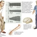
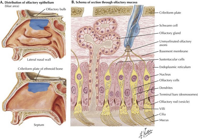
When identifying odors, humans rely on volatile substances entering their nasal cavity to excite receptors. Olfactory receptor cells are bipolar sensory neurons whose dendrites form a delicate sensory carpet on the superior aspect of the nasal cavity (Fig. 3-1). The thin, unmyelinated axons of the bipolar sensory cells collectively form the olfactory nerve. These travel through the cribriform plate into the olfactory bulb at the base of the fronto-orbital lobe. Within the bulb, CN-I fibers synapse with the dendrites of the large mitral cells, whose axons constitute the olfactory tract passing along the base of the frontal lobe and projecting directly into the primary olfactory cortex within the temporal lobe. In contrast to all other sensory modalities, olfactory sensation does not have a central processing site such as within the thalamic nuclei (Fig. 3-2). This direct pathway to the cerebral limbic structures may have had an important evolutionary function in lower animals and, later, primates.

The human primary olfactory cortex includes the uncus, hippocampal gyrus, amygdaloid complex, and entorhinal cortex (Fig. 3-2). Cortical representation of smell is bilateral. Although most of the olfactory tract fibers supply the ipsilateral olfactory cortex, some fibers decussate in the anterior commissure and terminate in the opposite hemisphere. Consequently, a unilateral lesion distal to the decussation rarely produces any olfactory dysfunction.

Differential Diagnosis

Smell dysfunction can be disrupted at any site along the olfactory pathway. Therefore impaired olfaction is not necessarily indicative of first-cranial nerve dysfunction per se. There are some common conditions that interfere with olfactory function without having specific olfactory nerve damage. These particularly include upper respiratory tract infection, especially nasal sinus disease. Primary olfactory bulb, tract, or entorhinal cortex lesions per se are relatively very uncommon. Olfactory impairment is not always apparent to the patient. Instead, he or she may initially complain of a loss of taste because the identification of tasted flavors depends partly on the olfactory system. Disturbances of smell are generally acquired, although there may be a rare patient with a congenital disorder. In general, most patients experiencing olfactory dysfunctions have bilateral loss of function. The rare presence of a unilateral anosmia is an important sign that signals a need for an MRI to exclude an olfactory groove tumor.

Acquired Disorders

Upper respiratory infections are the most frequent causes of olfactory dysfunction. Nasal and paranasal sinus disease account for more than 40% of olfactory disturbances. These intranasal processes mechanically prevent volatile chemical stimuli from reaching the olfactory sensory epithelium and activating the receptors. These are defined as transport or conductive olfactory disorders that are not associated with direct damage to the olfactory nerve pathways. The classic temporal profile, characterized by the intermittent occurrence of conductive olfactory dysfunction, provides the most important clinical clue for the diagnosis of a primary nasal source for these common disorders. A thorough otorhinolaryngologic evaluation is indicated for these patients. Conversely, the presence of persistent smell disturbance is the primary characteristic of direct damage to the olfactory nerve pathways.

Head trauma, as noted in the vignette, is responsible for approximately 20% of all cases of smell dysfunction. This is secondary either to direct damage to primary axons of the first cranial nerve or lesions of the associated frontobasal cerebral cortex responsible for olfactory perception. Depending on the severity of the blunt head injury, the incidence of posttraumatic anosmia varies between 7% and 30%. Direct occipital and side injuries to the head are more dangerous to olfaction than are frontally directed injuries. Posttraumatic olfactory dysfunction typically results from shearing of the olfactory nerve as it passes through the cribriform plate. More substantial damage, such as occurs in severe head trauma with anterior fossa fracture, may lead to a contusion of the olfactory bulb or the cortical–subcortical olfactory brain. Posttraumatic anosmia or hyposmia can be either unilateral or bilateral.

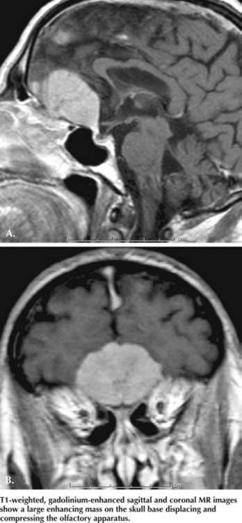
Olfactory groove meningiomas are quite infrequent; however, if these remain undiagnosed, these histologically benign tumors may still lead to significant morbidity unless treated early on. Usually meningiomas are slow growing; olfactory groove lesions comprise 8–18% of all intracranial meningiomas (Fig. 3-3). Although unilateral or bilateral olfactory dysfunction is thought to be their first symptom, very few patients present with just a disturbance in their sense of smell. This is probably because their slow and orderly growth leads to a very gradual decline in olfactory function. Furthermore, as most meningiomas are unilateral, they lead to unilateral anosmia and thus patients still retain olfactory function on the contralateral side. Thus, they are usually unaware of any focal loss. Consequently, most orbital meningiomas are not diagnosed until the tumor is large enough (e.g., >4 cm in diameter) to cause other symptoms resulting from pressure on the frontal lobes and optic tracts. These include headache, visual disturbances, personality changes, and memory impairment. Early diagnosis of olfactory groove meningiomas remains challenging. At times, the behavioral changes can be profound and may create a sense the patient is demented or mentally unbalanced.

Very large olfactory groove tumors, typically meningiomas, rarely lead to the development of Foster–Kennedy syndrome. This is characterized by unilateral optic atrophy and contralateral papilledema. Optic atrophy results from direct pressure of the neoplasm on the optic nerve, whereas increased intracranial pressure produces contralateral papilledema.

Parkinson disease patients often develop olfactory dysfunction as an early clinical feature. In fact, the difficulty with sense of smell may precede the onset of classic striatal motor manifestations by several years. A normal sense of smell in Parkinson disease is such a rare occurrence that its continued normal function should prompt review of this diagnosis. Alzheimer disease and Lewy body disease are other neurodegenerative disorders commonly associated with olfactory dysfunction.

Additional Resources