Congenital pediatric airway problems

Published on 07/02/2015 by admin

Filed under Anesthesiology

Last modified 22/04/2025

Print this page

rate 1 star rate 2 star rate 3 star rate 4 star rate 5 star
Your rating: none, Average: 0 (0 votes)

This article have been viewed 1512 times

Congenital pediatric airway problems

Wayne H. Wallender, DO

The upper airway is more easily compromised in infants and children than in adults. Anatomically, compared with an adult, the tongue of an infant or child is relatively larger within the mouth, the larynx is more cephalad, the glottic opening and airways are narrower, the arytenoid cartilages are more prominent, and the occiput is larger, resulting in greater resistance to airflow. Infants’ and children’s respiratory physiology further complicates the management of the airway. Because infants have higher weight-adjusted basal metabolic rates, O2 consumption and CO2 production rates are higher. In addition, the functional residual reserve of infants, per kilogram, is less than that of adults. Congenital abnormalities associated with airway problems can cause further difficulties with airway management.

Management of the difficult pediatric airway should include spontaneous ventilation with the consideration that direct laryngoscopy has a significant failure rate. As a general approach, these patients can be divided into those who will be difficult to intubate but can be ventilated by mask and those who are difficult or impossible to ventilate by mask. The latter group poses a more difficult anesthetic challenge and may require emergency tracheostomy. If a child can be ventilated by mask, then a number of options can be safely employed until the trachea is successfully intubated. The anesthetic goals are always a safe induction and intubation as well as a safe extubation.

Congenital abnormalities of the airway

Mucopolysaccharidoses

Hurler syndrome is associated with severe mental retardation, gargoyle facies, deafness, stiff joints, dwarfism, pectus excavatum, kyphoscoliosis, abnormal tracheobronchial cartilage, hepatosplenomegaly, severe cardiac valvular disease, and early coronary artery disease. Children with Hunter syndrome often have course facial features, deafness, hypertrichosis, stiff joints, macrocephaly, and carpal tunnel syndrome. Children with Morquio syndrome often appear healthy at birth; however, as the child ages, manifestations may include coarse facial features, prognathism, odontoid hypoplasia, atlantoaxial instability resulting from thoracic or lumbar kyphosis, aortic valve incompetence, hepatomegaly, inguinal hernias, mixed hearing loss, ocular complications, and limb abnormalities.

Upper airway obstruction and difficult intubation are common in children with mucopolysaccharidosis because of infiltration of lymphoid tissue, enlarged tongue, small mouth, and profuse thick secretions. A difficult airway should be expected and, as the infant ages, the airway may become even more difficult to manage.

Cleft lip and palate

Considered together, cleft lip and palate represent some of the most common congenital anomalies and are associated with more than 300 syndromes. The incidence of cleft lip (with or without cleft palate) varies by sex (higher rate in males, with isolated cleft palate higher in females) and ethnicity, with Asian Americans and Native Americans having the highest incidence and African Americans, the lowest. In European Americans, the incidence of cleft lip with or without cleft palate is 1 in 750 births; that of cleft palate alone is approximately 1 in 2500.

These patients may have other associated anomalies. Middle ear infections are quite common in this population. Infants with cleft lip or palate have difficulty swallowing and are at risk for developing pulmonary aspiration.

Anesthetic management depends on the degree of airway abnormality and can be relatively straightforward in uncomplicated cases. Large defects of the palate are usually not associated with airway obstruction, unless the defect is so extensive that the tongue prolapses into the nasopharynx. However, large defects can cause difficulty with intubation if the laryngoscope blade wedges into the cleft, or, if the patient is already intubated, the oral tracheal tube can migrate into the cleft, resulting in extubation. Postoperative airway problems are also common after a palatoplasty. Surgical edema in children with small oral cavities can result in airway obstruction requiring reintubation.

Anesthetic management of the difficult pediatric airway

Regardless of the congenital anomaly that causes an airway that may be difficult to manage, problems should be anticipated and managed expectantly. In most cases, preservation of spontaneous ventilation is strongly recommended, and fiberoptic intubation should be employed only when difficulty is suspected and difficult direct laryngoscopy is anticipated. Suggestions for management include the following:

• The preoperative use of H2-receptor blocking agents may be considered in infants at risk for aspirating.

• Intravenous access should be established either before or as soon as possible after induction.

• The use of intravenously administered sedatives, opioids, or neuromuscular blocking agents should be avoided.

• Atropine should be administered before laryngoscopy is attempted.

• The intravenous administration of lidocaine, 1 mg/kg, before intubation may decrease the risk of laryngeal spasm. Alternatively, topical translaryngeal lidocaine can be given, remembering that transmucosal absorption of lidocaine is close to that of intravenously administered lidocaine.

• Preoxygenation is strongly recommended.

• A variety of laryngoscopy blades, tracheal tubes, and stylets should be readily available.

• Laryngoscopy should be performed under deep anesthesia.

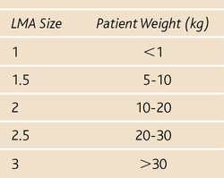
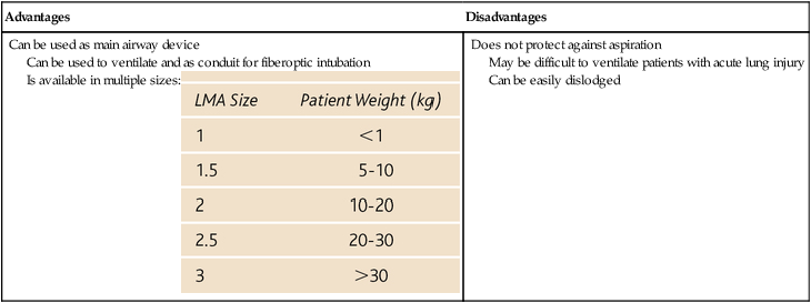
• The use of either mask ventilation or a laryngeal mask airway may be warranted (Box 200-1).

• Several intubation approaches should be considered (e.g., awake, blind nasal, fiberoptic), but alternative methods must be immediately available, including facilities for cricothyrotomy or tracheostomy.

• Induction with sevoflurane via spontaneous ventilation is preferred if awake intubation is not possible. Sevoflurane has a rapid onset of action, provides a smooth induction, and will not sensitize the heart to endogenous catecholamines, such as halothane does (halothane is no longer used in the United States but it may still be used in developing countries by anesthesia providers who provide humanitarian care there to pediatric patients.” Desflurane should not be used for induction because of its pungency and propensity to irritate the airway.

• Spontaneous ventilation should be maintained.

• Fiberoptic intubation is becoming more desirable in pediatric patients as smaller bronchoscopes become available. Currently, pediatric bronchoscopes are available that will fit through a 3.0-mm tracheal tube, although they do not have suction ports.

Because children are very prone to developing laryngospasm at the time of extubation, all equipment for ventilation and reintubation must be available before extubation is attempted. These children should usually not be extubated until awake. Mild laryngospasm can be treated with positive-pressure ventilation with O2 by mask. Severe laryngospasm usually responds to a small dose of succinylcholine (0.3 mg/kg).