95
Common Soft Tissue Tumors/Proliferations
Neural/Neuroendocrine
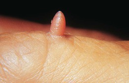
Neurofibroma
• Skin-colored to pink, soft papulonodule, often on the trunk (see Fig. 50.2).
• Usually solitary in most individuals.
• When multiple, need to distinguish linear form (segmental; mosaic) from a generalized distribution pattern (neurofibromatosis type I) (see Chapter 50).
• Histopathology: wavy, delicate spindle cells with tapered nuclei in a pink stroma.
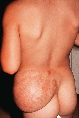
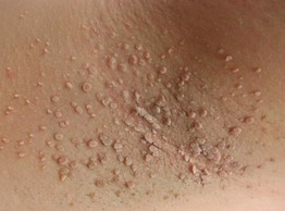
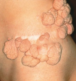
• Plexiform type has been likened to a ‘bag of worms’ (Fig. 95.1); it is generally on the trunk and proximal extremities, highly associated with neurofibromatosis type I, and prone to malignant degeneration (2–13%).
Schwannoma/Neurilemmoma
Granular Cell Tumor
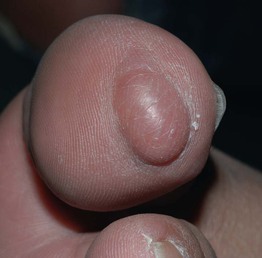
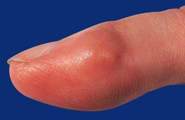
Traumatic Neuroma
• Skin-colored papulonodule(s) at a site of prior trauma.
• Often painful or ‘sensitive’ (Fig. 95.3).
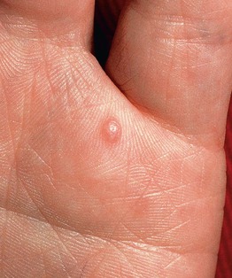
Fig. 95.3 Traumatic neuroma. A painful, firm papule that appeared after a deep puncture injury. Courtesy, Zsolt B. Argenyi, MD.
• Histopathology: haphazardly distributed fascicles of spindle cells with tapered nuclei.
Merkel Cell Carcinoma
• In older adults; solitary, rapidly growing, pink to red to violaceous nodule (Fig. 95.4).
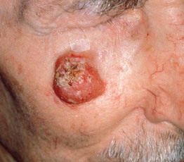
Fig. 95.4 Merkel cell carcinoma (primary cutaneous neuroendocrine carcinoma). Eroded erythematous nodule arising within sun-damaged skin of the cheek. Courtesy, Lorenzo Cerroni, MD.
Fibrous/Fibrohistiocytic
Skin Tag (Acrochordon, Fibroepithelial Polyp, Soft Fibroma)
Angiofibroma (Fibrous Papule)
• Solitary, skin-colored to pink, shiny papule; commonly on the nose (Fig. 95.6).
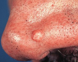
Fig. 95.6 Fibrous papule of the nose. A smooth, dome-shaped, skin-colored papule. Courtesy, Hideko Kamino, MD.
• Histopathology: stellate spindle cells in a hyalinized stroma with dilated vessels.
• DDx: basal cell carcinoma, intradermal melanocytic nevus, adnexal tumors.
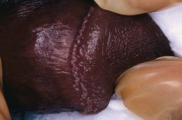
Pearly Penile Papules
Dermatofibroma
• 6- to 10-mm pink (especially in fair-skinned individuals), tan, or brown papule (Fig. 95.8); firm; dimples inward with lateral pressure.
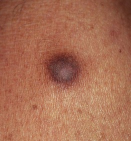
Fig. 95.8 Dermatofibroma. Hyperpigmented firm papule on the lower extremity. Courtesy, Jean L. Bolognia, MD.
Acral Fibrokeratoma
Sclerotic Fibroma
Giant Cell Tumor of Tendon Sheath
Nodular Fasciitis
Connective Tissue Nevus
• Skin-colored to yellow-tan (more yellow when composed predominantly of elastic tissue), firm papulonodules or plaques; solitary or multiple (often grouped) (Fig. 95.11).
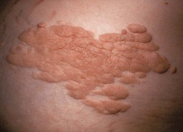
Fig. 95.11 Connective tissue nevus. Coalescence of multiple tan papules and plaques on the lower back. The lesion was firm to palpation and histologically had increased collagen.
Infantile Digital Fibroma
• Firm, skin-colored to pink papulonodule on the fingers or toes (tends to spare the thumb and great toe) (Fig. 95.12).
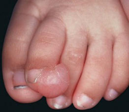
Fig. 95.12 Infantile digital fibroma. Firm skin-colored nodule on the dorsolateral aspect of the second toe in a young child.
• Solitary or multiple; generally present before 1 year of age.
Infantile Myofibromatosis
Fibromatoses
• Five subtypes, four of which are superficial: (1) palmar (Dupuytren’s contracture); (2) plantar (Ledderhose disease); (3) penile (Peyronie’s); and (4) knuckle pads (Fig. 95.13).
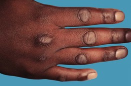
Fig. 95.13 Knuckle pads. Note the localization to the skin overlying the knuckles. Courtesy, Ronald P. Rapini, MD.
• One deep form: extra-abdominal desmoid tumor.
• Slowly growing nodules or plaques or cord-like tumors.
• Palmar fibromatosis can result in flexion contractures, especially of the 4th and/or 5th finger (see Chapter 81).
• Penile fibromatosis can result in pain and erectile dysfunction.
Muscle/Adipose
Leiomyoma
• Solitary or multiple red-brown papules or nodules, often grouped (Fig. 95.14).
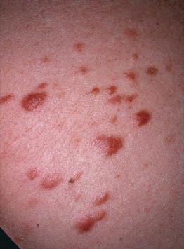
Fig. 95.14 Clustered piloleiomyomas on the back. The trunk is a common location for multiple piloleiomyomas. Patients with this clinical presentation need to be evaluated for the possibility of Reed syndrome.
• Histopathology: fascicles of spindle cells with cigar-shaped nuclei that have perinuclear vacuoles.
Smooth Muscle Hamartoma
• Congenital or acquired, skin-colored to hyperpigmented plaque on the trunk > proximal extremities (Fig. 95.15).
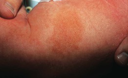
Fig. 95.15 Smooth muscle hamartoma. This infant presented with a firm plaque on the thigh. Courtesy, Ronald P. Rapini, MD.
• Follicular prominence and hypertrichosis may be present.
Lipoma
• Common tumor of mature fat; soft, mobile subcutaneous nodule.
• Generally on the trunk and extremities, but any site possible.
• Multiple lesions may be associated with a lipomatosis (e.g. familial type; Fig. 95.16) or a genodermatosis (e.g. Gardner syndrome, Proteus syndrome).
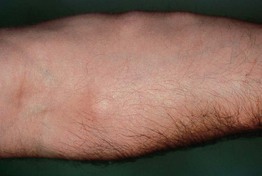
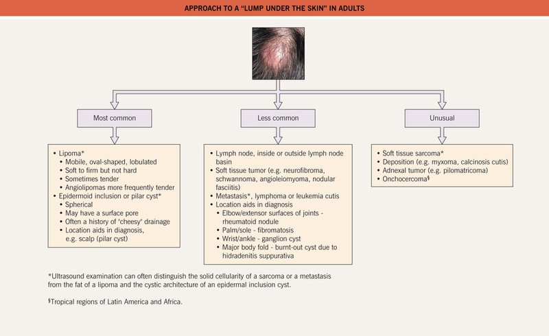
Fig. 95.17 Approach to a “lump under the skin” in adults. Incisional biopsy is preferred for the histopathologic evaluation of subcutaneous nodules while a punch biopsy suffices for dermal lesions. For bedside diagnosis of an epidermal inclusion cyst: after local anesthesia and superficial incision with a #11 blade, keratin is expressed.
Angiolipoma
Nevus Lipomatosus
• Grouped, soft, yellow to skin-colored papulonodules on the hips and/or upper thighs (Fig. 95.18).
Soft Tissue Sarcomas (See Table 95.1)
• Rare in comparison to benign soft tissue tumors.
• Generally presents as a nonspecific, deep-seated nodule.
• Dermatofibrosarcoma protuberans (DFSP) is characteristically multinodular (Fig. 95.19).
Table 95.1
Selected soft tissue sarcomas.
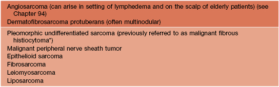
* Some pathologists consider atypical fibroxanthoma (AFX) to be a superficial variant of malignant fibrous histiocytoma (MFH); dermatologists see AFXs more commonly than MFHs.
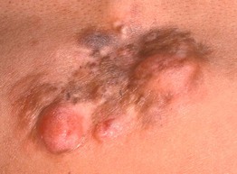
Fig. 95.19 Dermatofibrosarcoma protuberans. A broad, pink-brown, multinodular, firm plaque on the back. Histopathologically, bland spindle cells are arranged in storiform (‘cartwheel’) patterns. Courtesy, Hideko Kamino, MD.
For further information see Chs. 115, 116 and Ch. 117. From Dermatology, Third Edition.


