Chorea

Published on 03/03/2015 by admin

Filed under Neurology

Last modified 22/04/2025

Print this page

rate 1 star rate 2 star rate 3 star rate 4 star rate 5 star
Your rating: none, Average: 0 (0 votes)

This article have been viewed 1421 times

36 Chorea

Clinical Vignette

John is a 56-year-old-right-handed architect who developed tick-like movements in his body within the past 3–4 years and was now “fidgety” all the time. His wife reported intellectual changes in her husband; he seemed withdrawn and forgetful, was occasionally inappropriate in his behavior, had difficulties with his job, and had to retire early due to inability to “concentrate.” He had no other past medical history. He was adopted and there was no health information about his parents. Tourette syndrome was diagnosed by his primary care physician.

When seen in movement disorders clinic, he was unable to sit still. He had constant grimacing in his face, motor impersistence in his tongue, and piano-like playing movements in his hands. Brief, jerky choreiform movements were present in his trunk and extremities. His gait appeared very disorganized and unsteady. He had bradykinesia predominantly with testing of his hands. His demeanor was jovial and he had no signs of depression. The movements were not under any voluntary control and he had no urge to do them.

Because of the combination of cognitive decline and generalized chorea, Huntington disease was suspected. His genetic testing revealed 45 CAG repeats in the HD gene. He was treated with low doses of haloperidol 1 mg daily with substantial improvement in chorea. Recently, an addition of tetrabenazine was initiated, with further improvement of involuntary movements. However, his intellectual and behavioral decline continued.

Chorea (from Latin choreus, dance) is an abnormal involuntary movement usually distal in location, brief, nonrhythmic, abrupt, and irregular, that seems to flow from one body part to another. The movements are random, unpredictable in timing, direction, and distribution. Chorea can be partially suppressed; some patients can incorporate these into semipurposeful movements called parakinesia. Motor impersistence, the inability to maintain a sustained contraction, is a typical feature of chorea.

Athetosis and ballism are sometimes confused with chorea. Athetosis is a slow, writhing, continuous set of involuntary movements, usually affecting limbs distally, but it can involve the axial musculature (neck, face, and tongue). If athetosis becomes faster, it sometimes blends with chorea, that is, choreoathetosis. Ballism is large-amplitude, involuntary movements affecting the proximal limbs, causing flinging and flailing limb movements.

Patients with chorea are often initially unaware of these involuntary movements. The chorea is often first interpreted by observers as fidgetiness. The patients are usually frustrated by their own incoordination or clumsiness.

Etiology

Chorea results from disruption of the basal ganglia’s modulation of thalamocortical motor pathways. Multiple pathophysiologic mechanisms may be implicated. These include neuronal degeneration in selective regions, neurotransmitter receptor blockade, other metabolic factors within the basal ganglia, and exceedingly rarely a structural lesion. Chorea is classified into inherited, primarily Huntington disease (HD), immunologic Sydenham chorea, drug-related, structural, and various miscellaneous etiologies (Table 36-1).

Table 36-1 Causes of Chorea

Type of Chorea
Inherited

Metabolic Immunologic Drug related Structural Miscellaneous

Pathophysiology

The putamen, globus pallidus, and subthalamic nuclei are the key pathologic sites related to the development of chorea. Normal movement patterns depend on the presence of a critical physiologic balance between the direct and indirect motor pathways. Healthy individuals have an excitatory glutamate pathway, arising from within the subthalamic nucleus, that excites the globus pallidus interior and the substantia nigra. Concomitantly, these areas then signal the GABA inhibitory pathway within the thalamus. In a simplified model, the excitatory action of the subthalamic nucleus is reduced or lost, leading to a disinhibition of the pallidothalamic pathway.

The major neurodegenerative pathology in HD occurs within the caudate nuclei and the putamen (striatum). These changes primarily affect medium-sized “spiny” neurons that secrete the inhibitory neurotransmitter GABA. These neurons project from the striatum into the globus pallidus and substantia nigra. Selective loss of these specialized cells theoretically result in decreased thalamic inhibition, that is, leading to an increased activity. Consequently, such an inhibitory cell depletion causes an increased output to the cerebral cortex. The resultant motor activity leads to the disorganized, excessive (hyperkinetic) movement patterns of chorea. Concomitantly, HD patients also have a prominent associated temporal and frontal lobe cerebral cortex neuronal degeneration.

With Sydenham chorea, various streptococcal proteins or antigens (streptococcal M proteins) induce the body’s production of antineuronal IgG antibodies. These antibodies cross-react against the body’s own cells that provide the neuronal antigens within the basal ganglia, such as the caudate nuclei and subthalamic nucleus.

Clinical Presentation
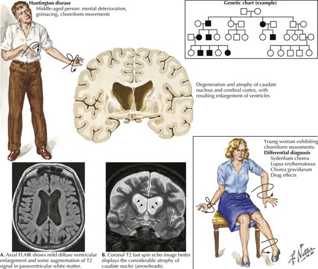
The spectrum of clinical findings in chorea varies, presenting in isolation or with other involuntary movements. At the simplest level, chorea appears as semipurposeful movements resembling fidgetiness. This is exemplified by the flitting movements of the fingers, wrists, toes, and ankles so characteristic of HD. The movements can be focal, as in tardive dyskinesia, where they are more repetitive and stereotypical. They may present as lip pouting or pursing, cheek puffing, lateral or forward jaw movements, or tongue rolling or protruding.

Asymmetric chorea, such as hemichorea, primarily affects the limbs on one side of the body. Sometimes, chorea affects only specific functional muscle groups, such as respiratory chorea. When there is a more diffuse basal ganglia dysfunction, chorea is often accompanied by parkinsonism, tics, and dystonia. Later, chorea can interfere with activities of daily living; for example, limb chorea can cause falls and interfere with dressing and eating. Chorea of the face, jaw, larynx, and respiratory muscles may eventually limit verbal communication.

On neurologic examination, there is altered finger-to-nose testing. Rapid alternating movements are executed with a jerky and interrupted performance. When patients with significant chorea grasp an examiner’s fingers, a squeezing motion called milkmaid’s grip is sometimes noted. This is a sign of motor impersistence. As with other adventitious movements, seen with the various movement disorders, chorea is frequently aggravated while walking. Various oculomotor abnormalities may be observed. These include slow and hypometric saccades and saccadic pursuit, convergence paresis, and gaze impersistence. Parkinsonian features, particularly bradykinesia and dystonia, are sometimes evidenced with more advanced disease.

Huntington Disease

This hereditary, progressive neurodegenerative disorder is the most common cause of chorea. The classic signs of HD include the development of chorea, neurobehavioral changes, and gradual dementia (Fig. 36-1). Symptoms typically become evident during the fourth or fifth decade of life, although onset varies from early childhood to late adulthood. HD symptoms vary among patients in range and severity, as well as by age at onset, and in rate of clinical progression. An early onset is associated with increased severity and more rapid progression. For example, adult-onset HD typically lasts approximately 15–20 years, whereas the course of juvenile HD tends to last approximately 8–10 years.

The initial clinical presentation may be either neurologic or psychiatric. Characteristic early presentations include the gradual onset of subtle personality changes, forgetfulness, clumsiness, and development of choreiform, fidgeting movement of the fingers or toes. Neurobehavioral changes include both emotional and behavioral disturbance. Patients present with increased irritability, suspiciousness, impulsiveness, lack of self-control, and anhedonia. Sometimes anxiety, depression, mania, obsessive–compulsive behaviors, and agitation are seen early in the disease. Later, a severe distortion in thinking and occasionally hallucinations, such as the perception of sounds, sights, or other sensations without external stimuli, may develop. The juvenile form of HD more often presents with dystonia, rigidity, or cerebella ataxia than chorea per se.

Cognitive decline is characterized by progressive dementia or gradual impairment of the mental processes involved in comprehension, reasoning, judgment, and memory. Typical early signs include forgetfulness, inattention, increased difficulty in concentrating, and various forms of disinhibition manifested by emotional outbursts, financial irresponsibility, or sexual promiscuity. Communication difficulties develop, including problems expressing thoughts in words, initiating conversations, or comprehending others’ words and responding appropriately.

Motor disturbances are characterized by the gradual onset of clumsiness, balance difficulties, and fidgeting movements. Early chorea may be limited to the fingers and toes, later extending to the arms, legs, face, and trunk. Eventually, chorea tends to become widespread or generalized. Parkinsonism and dystonia are sometimes seen later in the disease. Many patients with HD develop a distinctive manner of walking that may be unsteady, disjointed, lurching, and dance-like. Eventually, postural instability, dysphagia, and dysarthria appear.

Later disease stages are characterized by severe dementia and progressive motor dysfunction; patients usually become unable to walk, have poor dietary intake, become unable to care for themselves, and eventually cease to talk, leading to a persistent vegetative state. Life-threatening complications may result from serious falls, sometimes even leading to subdural hematomas, poor nutrition, infection, choking, aspiration pneumonia, or heart failure.

Sydenham Chorea

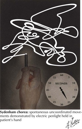
This is the other well-recognized form of chorea. It is related to an autoimmune response to infection with group A beta-hemolytic streptococci leading to acute rheumatic fever (ARF). This is now very uncommon in economically developed countries with the widespread availability of antibiotics for Streptococcus A infection. The initial illness is usually characterized by pharyngitis, followed within approximately 1–5 weeks by the sudden onset of ARF. Chorea primarily occurs in patients between the ages of 5 and 15 years. It usually does not present until 1–6 months after the initial sore throat. Sydenham chorea may occur as an isolated condition or subsequent to other characteristic features of ARF. Initially, these children often are described as unusually restless, aggressive, or “excessively emotional.” The distribution of chorea is usually generalized, and these movements consist of relatively fast or rapid, irregular, uncontrollable, jerky motions that disappear with sleep and may increase with stress, fatigue, and excitement (Fig. 36-2). Occasionally, the choreiform movements are so severe that they have a ballistic character. Some children also evidence emotional and behavioral disturbances.

Typically, in a significant majority of children, Sydenham chorea is a self-limited condition, resolving spontaneously within an average duration of 9 months to 2 years. However, sometimes residual signs of chorea and behavioral abnormalities fluctuate over a year or more. In approximately 20% of patients, Sydenham chorea may recur, usually within approximately 2 years of the initial occurrence. Recurrences are also reported during pregnancy and in association with certain medications in women who had ARF during childhood.

Differential Diagnoses

Diagnostic considerations in a patient presenting with chorea is a broad one (Table 36-1). HD, the most common cause of chorea, is usually easily diagnosed when an adult has the typical triad of chorea, dementia, and family history. Several neurodegenerative disorders, some also having expanded trinucleotide repeats, are phenocopies of HD. These include spinocerebellar atrophy (SCA2, SCA3) and dentatorubral-pallidoluysian atrophy (DRPLA). Additionally, there are some other HD-like diseases (HDL1, HDL2, HDL3) that may present with an HD-like phenotype. Sydenham chorea has an earlier onset, lacks the characteristic mental disturbances, and is usually self-limiting. Chorea with mental dysfunction may also occur as a manifestation of systemic lupus erythematosus (SLE). These patients usually have a more acute onset, with more localized chorea, and the characteristic SLE clinical and serologic abnormalities. There is a prior history of recurrent vascular thromboses or spontaneous abortions and disappearance after therapy with prednisone.

Involuntary movements occurring in psychiatric patients receiving long-term treatment with neuroleptic agents occasionally pose a diagnostic problem when they present with tardive dyskinesia (TD). Usually repetitive, these TD movements contrast with the nonrepetitive and flowing nature of chorea. Patients with TD usually have a predominant oral–lingual–buccal dyskinesia. Unlike in those with HD, these patients’ gait is usually normal. Similar mental dysfunction occurs with some of the dementing disorders, particularly Alzheimer or Pick disease where language is more involved. Myoclonus is more typical than chorea with dementia, especially with spongiform encephalopathies, for example, Creutzfeldt–Jakob disease. Very rarely, a structural basal ganglia lesion, particularly an infarction or hemorrhage or an associated polycythemia rubra vera, leads to an acute focal chorea, or hemiballismus.

If the onset of chorea occurs during childhood, other inheritable disorders including the leukodystrophies and gangliosidosis require differentiation. Neuroacanthocytosis is another hereditary movement disorder also manifested by mild chorea, tics, parkinsonism, and dystonia. Laboratory findings include increased serum creatine phosphokinase and red cell acanthocytes. In all age groups, possible reactions to drugs or toxins must always be investigated.

Diagnostic Evaluation

Treatment

Readily reversible causes of chorea need to be excluded before considering pharmacologic intervention. Therapy depends on the severity of symptoms; mild chorea does not usually require any treatment. Chorea is treated with either dopamine-blocking or dopamine-depleting medications. Dopamine antagonists haloperidol and pimozide are the drugs of choice. Benzodiazepine drugs are another possible therapeutic modality offering a nonspecific means to suppress chorea. With severe chorea, dopamine-depleting agents such as reserpine or tetrabenazine are sometimes considered.

The overall treatment of Huntington disease patients requires an integrated, multidisciplinary approach, including symptomatic and supportive medical management; psychosocial support; physical, occupational, or speech therapy; and genetic counseling. Often more specific additional supportive services are helpful for individual patients and their families. There is no specific treatment available that slows, alters, or reverses the progression of HD. Tetrabenazine is a dopaminergic depleting medication that effectively lessens chorea; it was recently approved for treatment of HD patients.

Sydenham chorea is usually not a disabling disorder; however, the more severely affected patients with more severe chorea, requiring short-time treatment, may respond to dopamine antagonists or valproic acid. Severely affected patients may improve with immunosuppressants, plasmapheresis, or intravenous immunoglobulin. Drug treatment should be withdrawn after a short period because remission invariably occurs. Penicillin prophylaxis for ARF is advisable.

Prognosis depends on the cause of chorea. Drug-induced chorea is usually transient. Patients with a past history of rheumatic chorea are more susceptible to developing chorea during pregnancy or drug-induced chorea, for example, from phenytoin or oral contraceptives.