Chancroid

Published on 19/03/2015 by admin

Filed under Dermatology

Last modified 22/04/2025

Print this page

rate 1 star rate 2 star rate 3 star rate 4 star rate 5 star
Your rating: none, Average: 0 (0 votes)

This article have been viewed 1576 times

Chancroid

Anuradha Lele Mookerjee and Glenn C. Newell

Evidence Levels:  A Double-blind study  B Clinical trial ≥ 20 subjects  C Clinical trial < 20 subjects  D Series ≥ 5 subjects  E Anecdotal case reports

image

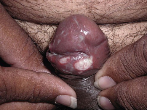
(Courtesy of Dr Shyam Verma, Vadodra, India.)

Chancroid is a genital ulcer disease caused by the Gram-negative facultative anaerobic bacillus Haemophilus ducreyi. It is common in many parts of the world, including Africa, the Caribbean basin, and Southwest Asia. In more developed countries the incidence of chancroid appears to be decreasing. It is often seen in travelers who have had unprotected sex, returning from areas known to have high risk. Chancroid typically is described as a painful, ragged, deep genital ulcer 3–20 mm in diameter. There may be surrounding erythema, and the base is often covered with a yellow-gray exudate. The lesion may be single, but can be multiple as a result of autoinoculation (kissing lesions). Painful lymphadenitis occurs in 30–60% of patients, and approximately one-quarter of patients with lymphadenopathy may develop a suppurating bubo.

Diagnosis and management strategy

Diagnosis on clinical criteria alone is difficult. The painful ulcer of chancroid can easily be confused with genital herpes. Syphilis, especially if secondarily infected, can also mimic chancroid. Co-infection with herpes simplex virus (HSV) or Treponema pallidum occurs in as many as 10% of patients, making the diagnosis more difficult. Extra-anogenital chancroid has been reported and may further represent diagnostic challenge.

The combination of a painful genital ulcer with tender suppurative lymphadenopathy is the only clinical presentation that is nearly pathognomonic. Chancroid has been noted to increase the susceptibility to human immunodeficiency virus (HIV) infection, probably by disrupting mucosal integrity, thereby allowing a portal of entry for HIV.

A definitive diagnosis may be made by culturing the exudates from the ulcer base or by aspiration of a bubo. Gram stain of the ulcer base in chancroid may show Gram-negative coccobacilli in a ‘school of fish’ appearance. Special culture media should be used, and the specimen should be handled by laboratories familiar with H. ducreyi. It should be noted that sensitivity of culture as shown by DNA amplification techniques can be as low as 75%. The newer nucleic acid amplification tests (NAATs) show higher positivity rates than culture and do not depend on live bacteria, making the test especially useful. However, only few laboratories have established NAATs for the diagnosis of chancroid, due to the rare occurrence of H. ducreyi. Polymerase chain reaction testing has the advantage of simultaneous testing of H. ducreyi along with T. pallidum and HSV.

HIV prolongs the incubation period of H. ducreyi and increases the number of genital ulcers. These tend to heal slowly and poorly. Extragenital sites are also frequently found in co-infection with HIV. Notably, there are increased treatment failures with HIV co-infection. Treatment guidelines for patients co-infected with HIV are the same as for those without HIV, but closer follow-up and potentially a longer course of therapy may be recommended.

Chancroid is usually treated on a presumptive basis in endemic areas if clinical features are suggestive. Empiric therapy is also often used if patients fail to respond to treatment of syphilis and/or herpes genitalis.

First-line therapies

image Azithromycin 1 g orally (one dose) A
image Ceftriaxone 250 mg intramuscularly (one dose) A

Second-line therapies

image Ciprofloxacin 500 mg twice a day for 3 days B
image Erythromycin 500 mg orally four times a day for 7 days B

Third-line therapy

image Fleroxacin 400 mg orally (one dose) B
image Spectinomycin 2 g intramuscularly (one dose) B
image Granulated thiamphenicol 5.0 g orally (one dose) B
image Ciprofloxacin 500 mg or 1000 mg orally (one dose) B