Cerebral Venous Thrombosis

Published on 03/03/2015 by admin

Filed under Neurology

Last modified 22/04/2025

Print this page

rate 1 star rate 2 star rate 3 star rate 4 star rate 5 star
Your rating: none, Average: 0 (0 votes)

This article have been viewed 9862 times

56 Cerebral Venous Thrombosis

Clinical Vignette

A 45-year-old man with a history of bipolar disorder and binges of alcohol abuse gradually developed global headaches that suddenly worsened over a 6-day period. He presented to the emergency room reporting excruciating headaches, especially while lying flat or after coughing. He described visual blurring and transient visual dimming while straining or getting up abruptly. He was slightly inattentive but had no focal weakness or numbness on examination. Ophthalmoscopy showed bilateral severe papilledema with peripapillary flame-shaped hemorrhages but no visual field loss. Computed tomographic (CT) scan of the brain showed hyperdensity in the sagittal sinus and the left transverse sinus. Cerebrospinal fluid (CSF) fluid analysis was normal but the opening pressure was elevated. Magnetic resonance imaging (MRI) of the brain showed no acute stroke or hemorrhage, but magnetic resonance venography (MRV) showed partial occlusion of the sagittal sinus, left transverse sinus, and the left jugular vein. He later admitted to drinking heavily and smoking just before his headaches worsened. There was no evidence of malignancy, and initial coagulation studies were normal. He was treated with warfarin and acetazolamide with no evidence of progressive visual loss and ultimately resolution of the headaches. Serial MRVs showed partial recanalization of the occluded cerebral sinuses and he was eventually taken off warfarin. He was admitted about 6 months later with recurrent episodes of shortness of breath and palpitations and was found to have multiple small pulmonary emboli and deep vein thrombosis. A repeat hypercoagulability screen revealed a lupus anticoagulant, and a newly available test showed the presence of a prothrombin gene mutation. He was advised to stay on warfarin life-long.

When venous drainage of the brain is compromised, arterial flow creates back-pressure into tissue capillaries causing capillary congestion, interstitial edema, decreased tissue perfusion, and ultimately ischemia. Eventually capillary rupture causes hematoma formation. This process of cerebral venous congestion followed by infarction (not conforming to strict arterial territories) and hemorrhage is the hallmark of cerebral sinus thrombosis. The causes of cerebral venous thrombosis vary (Box 56-1), but many relate to transient or permanent hypercoagulable states, with dehydration acting as a common precipitating event. A thorough investigation for such etiologies is crucial to directing long-term treatment and anticipating potential comorbidities.

Attention should be given to signs of meningitis, such as fever, stiff neck, and rash. Examining the ears, sinuses, and face for infection or discharge may provide clues to possible septic venous thrombosis. Physical evidence or a history of head or neck trauma is important. Ocular pain, proptosis, and chemosis, often with combinations of cranial neuropathies, are significant signs that may indicate a basal skull or cavernous sinus thrombosis.

Anatomy

Although complex, cerebral venous system anatomy is best considered in three levels: the dural-based posterosuperior group, the dural anteroinferior or basal group, and the deep veins of the brain.

The dura is formed of two layers, one abutting the inner calvarium and the other forming the outer meningeal covering. These layers separate in the midsagittal and transverse planes, forming dural venous sinuses ultimately draining into the jugular veins. A single superior sagittal sinus joins the often asymmetric but paired transverse sinus at the confluence of sinuses or torcular herophili (Fig. 56-1). The transverse sinuses run laterally from the occipital bone to the middle cerebral fossa along the tentorium cerebelli. The right is often larger and is continuous with the superior sagittal sinus whereas the left curves out laterally as an extension of the single midline straight sinus. The straight sinus runs downward from near the splenium of the corpus callosum to the occipital protuberance. The sigmoid sinus curves down toward the skull base from the transverse sinus and joins the inferior petrosal sinus at the jugular foramen to form the jugular vein.

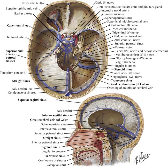
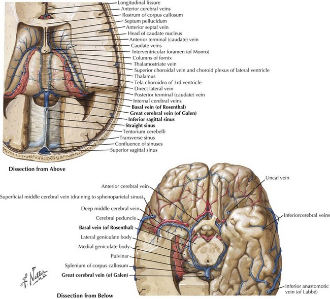
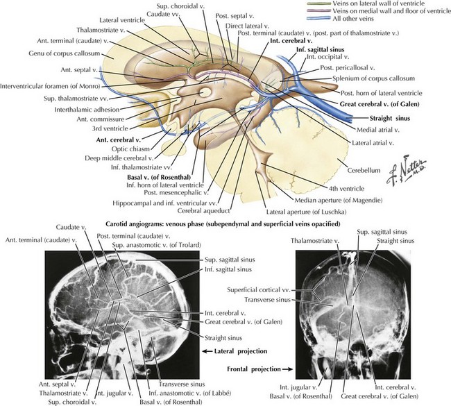
The straight sinus (Figs. 56-1 through 56-4) is formed by the splayed falx layered over the cerebellar tentorium. The inferior sagittal sinus runs in the fold of the lower arch of the falx cerebri and joins the cerebral vein of Galen in the proximity of the posterior horns of the lateral ventricles to form the straight sinus. The superior and inferior sagittal sinuses provide drainage for the cerebral hemispheres.

The great cerebral vein of Galen drains, through paired internal cerebral veins, the brainstem, cerebellum, posterior frontal and anterior parietal lobes, and thalamus; through the paired basal vein of Rosenthal, it drains the limbic system, hippocampus, and mesencephalon.

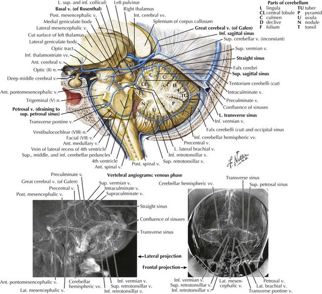
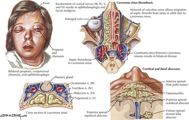
The cavernous sinus runs posteriorly at the brain base from the sphenoid bone in the area of the superior orbital fissure to the petrous temporal bone. Cavernous sinus tributaries include cerebral veins and the ophthalmic vein. The cavernous sinus drains along the medial upper layer of the tentorium and through the superior petrosal sinus, coursing posteriorly to the transverse sinus. The cavernous sinus houses the carotid artery; the oculomotor, trochlear, and abducens nerves; and the ophthalmic division of the trigeminal nerve (Fig. 56-5). A mesh of venous sinuses around the pituitary and the anterior skull base connects the two cavernous sinuses across the midline. The superior petrosal sinus drains the anterior brainstem and the anterior superior and inferior cerebellar hemispheres. Below the tentorium, along the skull base, the inferior petrosal sinus links the cavernous sinus to the sigmoid sinus (Fig. 56-1).

Clinical Presentation

Specific Clinical Presentations

In superior sagittal sinus thrombosis (SSST), increased venous pressure from decreased drainage initially causes generalized headaches with paroxysms of pain occurring with any Valsalva-like maneuver, that is, coughing, sneezing, straining, lifting, or bending. Blurred vision may occur secondary to optic nerve head edema or associated exudates involving the macula. Permanent visual compromise is unusual and only happens when papilledema persists for weeks. Light-headedness, transient blindness, and tinnitus can occur with sudden head elevation from a lying or bending position, similar to pseudotumor cerebri.

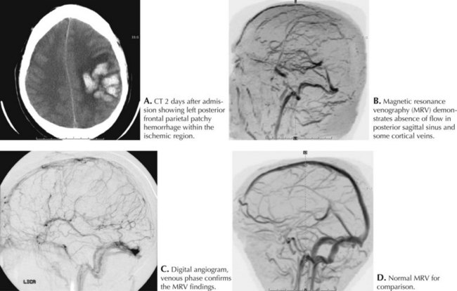
Intracerebral cortically based hemorrhages, common with SSST, are often associated with focal neurologic signs and seizures. Confusion, behavioral changes, somnolence, and coma may occur as thrombosis propagates within the sinus and ICP increases. These signs usually develop after the clot extends into the posterior third of the sinus. In most cases of SSSTs, one of the lateral sinuses is concomitantly involved (Fig. 56-6).

Occasionally, isolated cortical vein thrombosis is seen without sagittal sinus involvement. The clinical picture is again one of headaches, focal neurologic dysfunction, and seizure, however without increased ICP or papilledema. Underlying causes are similar to sagittal sinus thrombosis, and treatment follows the same principles. Neuroimaging shows isolated, often hemorrhagic, ischemic lesions that are not confined to a cerebral artery territory.

Deep cerebral vein thrombosis is present in 40% of superior sagittal sinus cases and is more likely to produce coma, pupillary abnormalities, ophthalmoplegia, and increased ICP than SSST alone. Sole or predominant deep venous system involvement mostly occurs in children but is reported in adults with presentations ranging from isolated drowsiness or obtundation to coma with bilateral posturing and ocular abnormalities. Survivors experience bilateral weakness, rigidity, dystonia or athetosis, memory loss, personality changes, and various neuropsychologic disturbances.

Base of the skull sinus thrombosis has a clinical presentation of painful cranial neuropathies. Cavernous sinus thrombosis is often septic from facial, orbital, or middle ear infections with eye pain, proptosis, and chemosis as frequent features (Fig. 56-7). Varying degrees of ophthalmoplegia are present secondary to involvement of CN-III, -IV, and -VI running through the lateral portion of the cavernous sinus. The ophthalmic division of the trigeminal nerve (V1) also courses through this sinus, and forehead sensory changes are occasionally seen. Inferior petrosal sinus thrombosis, often septic, causes retro-orbital pain, trigeminal V1 sensory changes, and abducens nerve palsy (Gradenigo syndrome; CN-V, -VI). Localized thrombosis involving the internal jugular vein may be an extension of transverse or sigmoid sinus thrombosis or may result from catheterization or trauma. This often presents with CN-IX, -X, and -XI dysfunction (jugular foramen or Vernet syndrome).

Diagnostic Approach

All patients with cerebral venous thrombosis should be examined for a hypercoagulable state. In addition to prothrombin time, partial thromboplastin time and platelet count, blood studies now commonly include protein C and S quantification, lupus anticoagulant, anticardiolipin antibodies, homocysteine levels, and DNA testing for factor V (Leiden factor) and prothrombin gene mutation.

Lumbar puncture often shows an opening pressure greater than 200 mm H2O. The CSF protein is increased but the glucose content, unless there is associated meningitis, is usually normal. CSF RBCs, xanthochromia, and pleocytosis are commonly seen, especially in cases of septic sinus thrombosis and in cases associated with meningitis. Normal CSF analysis, although rare, does not exclude the diagnosis.

Acutely, brain CT without contrast is obtained to assess for intracranial hemorrhage. It may reveal irregularly shaped paramedian cortical venous infarctions that do not conform to defined arterial distributions. The “empty delta sign,” where contrast partially fills the sinus, leaving an unenhanced island of clot within the occipital confluence, occurs in 50% of cases. Over the hemispheric convexities, thrombosed cortical vessels sometimes appear as hyperintense coiled or serpiginous signals. Diffuse edema and narrowed lateral ventricles may be apparent with or without hemorrhagic lesions.

MRI and MRV have largely replaced angiography as standard imaging techniques to confirm cerebral sinus thrombosis (see Fig. 56-6). Cerebral angiography with a prolonged venous phase is now reserved for cases not clearly diagnosed by MRI or CT and for patients requiring intrasinus thrombolysis.

Cerebral sinus thrombosis is often a clinical diagnosis based on a detailed history and corroborating physical findings. Imaging studies, however, have become crucial in the management of these patients from confirming the diagnosis to guiding treatment and to help in predicting the clinical course and outcome.

Treatment

The management of sagittal sinus thrombosis consists of hydration, anticoagulation, and the treatment of any underlying cause. Because dehydration enhances clot propagation, early volume repletion is of utmost importance. Heparin is given to make the partial thromboplastin time double the control value. Anticoagulation is indicated despite hemorrhagic infarctions because the overall outcome is improved and intracranial hemorrhage is rarely worsened. Low-molecular-weight heparin has also been used with safety and efficacy. Close clinical follow-up and repeated brain CT scanning, however, are advised to monitor the size and location of cerebral hemorrhages throughout the course of treatment. Warfarin is given for long-term anticoagulation and is started after 24 hours of intravenous heparin treatment or after the patient is stable. When indefinite anticoagulation is not needed, the duration of warfarin treatment remains unclear; accepted practice is 3–6 months.

After the precipitating cause is resolved, it is best to confirm that headache and papilledema are controlled and that MRV shows, at least, partial recanalization of the sagittal sinus before discontinuing oral anticoagulants. Seizures occur in the acute phase in up to 30% of patients and are usually focal but can be generalized. Recurrent seizures should be treated promptly because they can cause increased intracranial pressure, clinical deterioration, and increased mortality. Up to 10% of patients may experience pulmonary embolism. This is suspected when respiratory deterioration and increased oxygen needs suddenly occur.

If deterioration continues despite IV anticoagulation, many advocate a more invasive approach with in situ clot thrombolysis. A femoral venous catheter thread through the jugular vein to the transverse or sagittal sinus is used. An initial attempt at partial thrombolysis is usually followed by a continuous 12-hour intra-sinus infusion. Numerous case series have shown significant neurologic recovery with only a minor increase in bleeding complications. Monitoring hematomas remains necessary because expanding hemorrhagic infarcts may cause shift and herniation, necessitating acute treatment of increased ICP with osmotic agents or hyperventilation. Surgical evacuation of intracranial hemorrhage is rarely required. To decrease potential bleeding complications, rheolytic mechanical thrombectomy catheters alone or in combination with low doses of thrombolytic agents have been pursued with some success. Numerous cases of thrombolytic treatment have been described where significant clinical improvement occurred even in patients with several hemorrhagic infarcts and days of obtundation or coma.

Prognosis and Long-Term Complications

With anticoagulation, about 80% of patients have good recovery with little or no residual disability. Poor outcomes correlate with rapid deterioration after admission, coma or obtundation on presentation, involvement of the deep venous system, and multiple cerebral hemorrhages, especially if present for days. Before the advent of anticoagulation, the mortality rate was 30–50%. A mortality rate of 6–10% remains in the acute phase. Aggressive treatment with intrasinus thrombolysis, especially in those with evidence of evolving venous infarctions and progressive obtundation, may improve outcomes and decrease the rate of early mortality but, to date, there are no randomized controlled studies to support its routine use.

Long-term complications include focal or generalized seizures, headaches, and papilledema with visual loss, with approximately 10% rate of occurrence for each. Seizures may necessitate continued anticonvulsant therapy despite resolution of all other symptoms and sinus recanalization. Headache usually resolves with increasing recanalization and better venous drainage and often does not necessitate long-term therapy. The recurrence rate of cerebral thrombosis and other thrombotic events, such as deep vein thrombosis or pulmonary embolism, is estimated around 2–5%, with the majority of these patients likely requiring lifelong anticoagulation with warfarin.

Papilledema, when present, should be followed with serial visual fields by an ophthalmologist. If not controlled, progressive visual loss (arcuate mid–peripheral field constriction and central visual loss with widening blind spot) is a danger, secondary to gradual optic nerve atrophy. Treatment of papilledema involves lumboperitoneal shunting, serial spinal taps, carbonic anhydrase inhibitors, or optic nerve fenestration into the orbit to relieve locally increased CSF pressure that otherwise would be transmitted to the optic nerve. Optic nerve fenestration is safe, has few complications, and rarely reoccludes. It is done unilaterally, with positive effects on both eyes. The exact mechanism is unknown but is not thought to relate to general reduction of overall CSF pressure, although many cases still demonstrate high lumbar puncture pressures after fenestration.

Additional Resources

Baker MD, Opatowsky MJ, Wilson JA, et al. Rheolytic catheter and thrombolysis of dural venous sinus thrombosis: a case series. Neurosurgery. 2001;48:487-494.

De Bruijn SF, De Haan RJ, Stam J. Clinical features and prognostic factors of cerebral venous sinus thrombosis in a prospective series of 59 patients. For The Cerebral Venous Sinus Thrombosis Study Group. J Neurol Neurosurg Psychiatry. 2001;70:105-108.

DeBruijn SF, Stam J. Randomized placebo-controlled trial of anticoagulation treatment with low molecular weight heparin for cerebral sinus thrombosis. Stroke. 1999;30:481-482. Of those treated with nadroparin 13% had a bad outcome compared to 21% in the placebo group. There were no new symptomatic intracranial hemorrhagic complications even though many patients in the treatment group initially presented with ICH

Einhaupl KM, Villringer A, Meister W, et al. Heparin treatment in sinus venous thrombosis. Lancet. 1991;338:597-600. Randomized controlled study terminated after 20 patients because of a significant advantage in favor of heparin over placebo in angiographically proven cases of cerebral venous thrombosis

Ferro JM, Canhão P, Stam J, et al. ISCVT Investigators. Prognosis of cerebral vein and dural sinus thrombosis: results of the International Study on Cerebral Vein and Dural Sinus Thrombosis (ISCVT) Stroke. 2004 Mar;35(3):664-670. Large multinational prospective observational study that shows good recovery in 79% of patients. A subgroup of men with venous hematomas, low Glasgow coma scores, deep venous involvement and malignancy tended to do worse

Ferro JM, Lopes MB, Rosas MJ, et al. Long-term prognosis of cerebral vein and dural sinus thrombosis: results of the VENOPORT study. Cerebrovasc Dis. 2002;13:272-278.

Haley EC, Brasmear HR, Barth JT, et al. Deep cerebral venous thrombosis: clinical, neuroradiological and neuropsychological correlates. Arch Neurol. 1989;46:337-340.

Horton JC, Seiff SR, Pitts LH, et al. Decompression of the optic nerve sheath for vision-threatening papilledema caused by dural sinus occlusion. Neurosurgery. 1992;31:203-212.

Mehraein S, Schmidtke K, Villringer A, et al. Heparin treatment in cerebral sinus and venous thrombosis: patients at risk of fatal outcome. Cerebrovasc Dis. 2003;15:17-21.

Preter M, Tzourio C, Ameri A, et al. Long-term prognosis in cerebral venous thrombosis: follow-up of 77 patients. Stroke. 1996;27:243-246.

Wasay M, Bakshi R, Kojan S, et al. Nonrandomized comparison of local urokinase thrombolysis versus systemic heparin anticoagulation for superior sagittal sinus thrombosis. Stroke. 2001;32:2310-2317.