Central nervous system effects of the inhalation agents

Published on 07/02/2015 by admin

Filed under Anesthesiology

Last modified 22/04/2025

Print this page

rate 1 star rate 2 star rate 3 star rate 4 star rate 5 star
Your rating: none, Average: 0 (0 votes)

This article have been viewed 2754 times

Central nervous system effects of the inhalation agents

Michael J. Murray, MD, PhD

Inhalation anesthetic agents induce anesthesia by depressing brain function via a dose-dependent reversible mechanism that is associated with alterations in cerebral metabolic rate (CMR), in cerebral blood flow (CBF), of the electroencephalogram (EEG), and of evoked potentials. The alterations in CMR and CBF can be attenuated to some extent, but not completely, and therefore can adversely affect outcome in patients with neurologic diseases and in patients undergoing neurosurgical procedures.

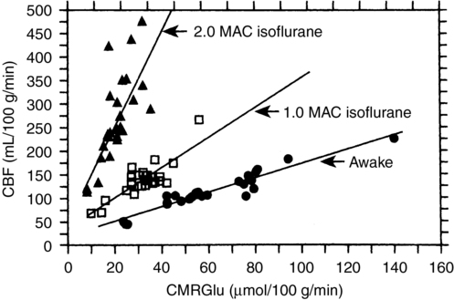
Flow-metabolism coupling is defined as a matching of O2 and glucose delivery to metabolic demand; CBF increases or decreases in concordance with changes in CMR. A misconception about the inhalation anesthetic agents is that, because they increase CBF and decrease CMR, they “uncouple” flow and metabolism. In fact, although increasing concentrations of inhalation anesthetic agents result in a higher CBF for a given CMR, a coupled relationship between these variables persists (Figure 67-1).

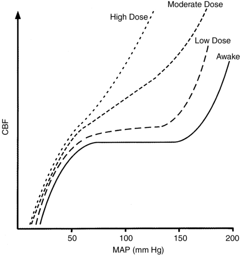
This relationship between CMR and CBF is apparent only if adequate blood pressure is maintained; if blood pressure is allowed to decrease, the increase in CBF will be attenuated or abolished because inhalation anesthesia agents inhibit autoregulation in a dose-dependent fashion (Figure 67-2). However, inhalation anesthetic agents do not inhibit CO2 reactivity and, if anything, may actually exaggerate the response. Thus, in the normal brain, the cerebral vasodilation that occurs in response to an inhalation anesthetic agent can be blunted, abolished, or reversed by decreasing CO2 levels; however, these responses may not apply in the presence of abnormal intracranial anatomy or physiology.

Because the inhalation agents cause an increase in CBF (and in cerebral blood volume [CBV]), the use of these anesthetics in patients at risk for developing or who have increased intracranial pressure (ICP) is a concern. However, numerous studies have confirmed that hypocapnia attenuates or blocks the increase in ICP that otherwise would occur in at-risk patients.

Anesthesia-induced EEG changes follow a common pattern. When anesthesia is induced with an inhalation agent, the frequency and amplitude of the EEG waveforms increase, and the measurements throughout the cortex are more uniform, such that waveforms measured on the EEG appear to synchronize. At about 1 minimum alveolar concentration (MAC), the EEG slows progressively; depending on the anesthetic agent, burst suppression, an isoelectric pattern, or seizures may evolve as the anesthetic concentration increases.

Michenfelder defined critical regional CBF as “that flow below which the majority of subjects develop ipsilateral EEG changes indicative of ischemia within 3 min following carotid occlusion.” Table 67-1 lists the critical CBF rates of various inhalation anesthetic agents.

Table 67-1

Critical Regional Cerebral Blood Flow for Anesthetic Agents in Patients Receiving an Inhalation Anesthetic Agent with N2O

Agent Cerebral Blood Flow Rate
Isoflurane 10 mL • 100 g−1 • min−1
Desflurane ≤10 mL • 100 g−1 • min−1
Sevoflurane 11.5 mL • 100 g−1 • min−1

The inhalation agents also affect evoked potentials, but only minimally so at concentrations below 1 MAC. All anesthetic agents tend to increase latencies and decrease amplitudes of evoked potentials at concentrations greater than 1 MAC. Evoked potentials of cortical origin are particularly sensitive to the effects of inhalation anesthetic agents; brainstem auditory evoked potentials are the most resistant. Although more sensitive to the effects of inhalation anesthetic agents, somatosensory evoked potentials can be adequately monitored at less than 1 MAC of the inhalation anesthetic agent.

Nitrous oxide

Cerebral metabolic rate and cerebral blood flow

Although N2O is perceived to be physiologically and pharmacologically inert, it is a cerebral vasodilator that can significantly increase ICP in patients with increased intracranial elastance. The effect of N2O on ICP is blocked or blunted by opioids, barbiturates, and hypocapnia. Most data suggest that N2O increases CMR.

Pneumocephalus

Pneumocephalus can occur during posterior fossa or cervical spine operations performed with the patient in the sitting position. When the dura is open, gravity can cause the cerebrospinal fluid (CSF) to continuously drain; the CSF is subsequently replaced by air (an effect known as the inverted pop-bottle phenomenon), resulting in a progressive accumulation of air in the ventricles, over the cortical surfaces, or both. If used as part of the anesthetic, N2O will equilibrate with any air-filled space in the body. Because the blood solubility of N2O is 30 times greater than that of nitrogen, a significant, albeit transient, net increase of gas molecules will occur in the air-filled space, and hence, the volume or pressure will increase once the dura is closed. Thus, the use of N2O may cause tension pneumocephalus of sufficient significance to produce major cerebral compromise, manifested by seizures, altered consciousness, or specific neurologic deficits.

If a tension pneumocephalus is suspected, the use of N2O should be discontinued. Patients receiving a second anesthetic within the first 3 weeks after undergoing supratentorial craniotomy are at risk for developing complications if N2O is used because a number of these patients will still have significant intracranial air collection.

Isoflurane

Cerebral metabolic rate and cerebral blood flow

Of the inhalation agents, isoflurane is the least potent cerebral vasodilator. CO2 reactivity and autoregulation are maintained with the use of isoflurane. As do all of the inhalation agents, isoflurane depresses CMR; CMR decreases by 50% at 2.0 MAC of isoflurane, the point at which the EEG becomes isoelectric. Doubling the isoflurane concentration to 4.0 MAC has been shown to cause no further decrease in CMR. There is no evidence of toxicity associated with the use of deep levels of isoflurane anesthesia.

Desflurane

Cerebral metabolic rate and cerebral blood flow

The cerebral metabolic and vascular effects of desflurane are similar to those of isoflurane. Desflurane is a cerebral arteriolar dilator and produces a dose-dependent decrease in cerebrovascular resistance and CMR. Similar to isoflurane, it may be used to induce controlled hypotension, but its use is more often associated with a compensatory tachycardia than is the use of isoflurane.

Sevoflurane

Cerebral metabolic rate and cerebral blood flow

The effects of sevoflurane on CMR and CBF resemble those of isoflurane. In most animal models in which it has been studied, sevoflurane produces little change in global CBF independent of CO2 levels. Cerebral autoregulation and cerebrovascular responsiveness to changes in CO2 are preserved in patients with cerebrovascular disease up to a concentration slightly below 1 MAC.