Heart43
7.2 Arteries43
7.3 Veins43
7.4 Lymph vessels (lymphatics)45
The cardiovascular system consists of a series of closed blood vessels lined by endothelial cells, and a pump: the heart. Arteries may be described as: systemic, taking blood from the left ventricle of the heart to the tissues of the body; and pulmonary, taking blood from the right ventricle to the lungs. Veins may be described as: systemic, conveying blood towards the right atrium of the heart from all tissues of the body except the intestines; pulmonary, conveying blood from the lungs to the left atrium of the heart; and hepatic portal, conveying blood from the intestinal bed to the liver. The lymphatic system conveys tissue fluid back to the venous system: it is important in the spread of disease.
You should:
• describe the basic structure of the arterial system
• describe the basic structure of the venous system.
• list the names of the most commonly palpated arterial pulses
• explain the meaning of: systemic, pulmonary, portal
• describe the clinical importance of the lymphatic system and the positions of the most commonly palpated groups of lymph nodes.
7.1. Heart
The heart is in the thorax and consists of two pumps, right and left, lying side by side in one organ. The left heart (left atrium and left ventricle) receives oxygenated blood from the lungs and pumps it around the body, and the right heart (right atrium and right ventricle) receives deoxygenated blood from the body and pumps it to the lungs for oxygenation. It is described in Chapter 10.
7.2. Arteries (Fig. 7.1)
Arteries convey blood from the heart to the tissues. There are two arterial systems:
• the aorta and its branches – systemic arteries – which convey blood from the left ventricle to the capillary beds; and
• pulmonary arteries, which convey deoxygenated blood from the right ventricle to the lungs.
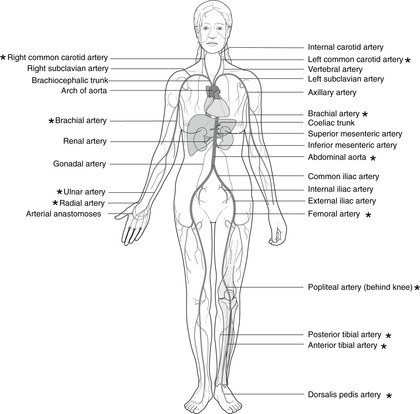
The pulsations of the heartbeat are strongest in large arteries, and palpation of arterial pulses is one of the components of clinical examination of the body. The most commonly palpated pulses are: carotid, radial, aortic, femoral, popliteal, posterior tibial, dorsalis pedis. Other pulses are also used occasionally: brachial, ulnar and anterior tibial. Pulses are marked with an asterisk on Figure 7.1. More precise details of their surface markings are given later in the text.
7.3. Veins (Fig. 7.2)
Veins convey blood from the tissues back to the heart. The arrangement of veins is much more variable than that of arteries and knowledge of only the principal veins is necessary.
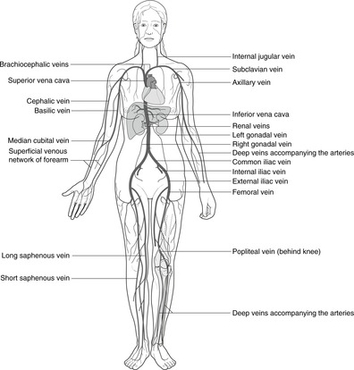
Systemic veins
These convey blood towards the two vena cavas and the heart.
• The superior vena cava receives blood from the upper limbs, head and neck, chest wall and part of the upper abdominal wall.
• The inferior vena cava receives blood from the lower limbs, pelvis, abdominal organs, most of the abdominal wall, and the liver (see below).
Pulmonary veins
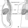
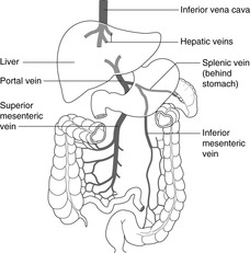
Portal venous system (Fig. 7.3)
Portal veins are veins that connect two capillary beds. All venous blood from the alimentary canal between the lower oesophagus and the anal canal reaches the liver in the hepatic portal vein, usually referred to as simply the portal vein. This with its tributaries (splenic, superior mesenteric, inferior mesenteric veins) constitute the portal system, blockage of which, for example in liver disease, has serious consequences (see later). From the liver, short veins convey blood to the inferior vena cava – the hepatic veins. In certain forms of liver disease in which blood from the intestines is unable to pass through the liver, anastomoses between portal and systemic veins develop or enlarge to allow blood from the intestines to bypass the liver (see portosystemic anastomoses, p. 129).
 |
| Fig. 7.3 |
Superficial and deep veins
Veins that are easily visible under the skin are superficial veins in the superficial tissues of the body: these may be used clinically as access points for injections or for the insertion of catheters etc. Their anatomy is particularly variable. There is also a venous network alongside the arterial tree in the deeper layers: these are the deep veins. Understanding the two groups of veins is particularly important in the lower limb and will be explained in that context. At some point, superficial veins drain into the deep system.
Veins used for clinical procedures
Veins used most frequently for the insertion of catheters and cannulas are: great saphenous vein at the ankle, femoral vein, antecubital veins, cephalic vein, subclavian vein, brachiocephalic vein, scalp veins (particularly in babies).
Valves
Limb veins have valves to ensure blood flows towards the heart; trunk veins do not. Presumably, valves developed in veins of quadruped limbs because of the antigravity blood flow. See Section 13.6.
7.4. Lymph vessels (lymphatics)
These small, thin-walled vessels convey ‘used’ extracellular fluid back to the venous system though various groups of lymph nodes. The clinical importance of lymph nodes and lymphatics is out of all proportion to their insignificant size, since it is by the lymphatics that disease, especially malignant disease, often spreads. The lymphatic drainage of one organ to another may result in disease of the first organ becoming apparent as disease of the second. You therefore need to know the main lymphatic channels and lymph node groups.
From the interstitial spaces of the tissues, lymph passes into small lymphatic channels which join to form progressively larger vessels. They are lined by endothelial cells and, like veins, many lymph vessels contain valves. In general, lymph vessels accompany arteries, and arterial pulsations provide the force, squeezing the lymphatics so that lymph flows through them in the direction permitted by the valves. As with veins, there are superficial and deep lymphatics that eventually unite.
The main lymph channels are shown in Fig. 7.4 and details of the lymph drainage of specific organs are presented in the text as the organs and regions are considered.
• Lymph from the entire body, except the right upper limb and right side of the head, neck and upper thorax, drains to the cisterna chyli in the upper abdomen, then onwards through the thoracic duct to enter the venous system at the junction of the left internal jugular and left subclavian veins as they unite to form the left brachiocephalic vein.
• Lymph from the right upper limb and right side of the head, neck and upper thorax drains to the right lymph duct which enters the venous system at the junction of the right internal jugular and right subclavian veins, which unite to form the right brachiocephalic vein.
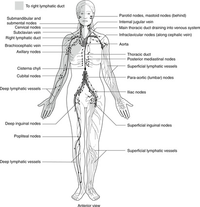
Most commonly palpated lymph node groups
These are: parotid, mastoid, submandibular, deep cervical, supraclavicular, axillary, para-aortic, inguinal.