Cardiac cycle: Control and synchronicity

Published on 07/02/2015 by admin

Filed under Anesthesiology

Last modified 22/04/2025

Print this page

rate 1 star rate 2 star rate 3 star rate 4 star rate 5 star
Your rating: none, Average: 5 (1 votes)

This article have been viewed 6277 times

Cardiac cycle: Control and synchronicity

Brantley D. Gaitan, MD

The cardiac cycle describes the succession of atrial and ventricular events that make up a period of contraction (systole) followed by a period of relaxation (diastole) (i.e., a single heart beat). These periods are further subdivided into phases.

Systole comprises two phases: isovolumic contraction and ejection. On initiation of myocardial contraction, the ventricular pressure rises abruptly, closing the atrioventricular (AV) valves, and continues to increase for approximately 0.03 sec (isovolumic contraction). Once the ventricular pressure sufficiently exceeds the pressure in either the aorta or the pulmonary artery, the semilunar valves open, and ejection occurs.

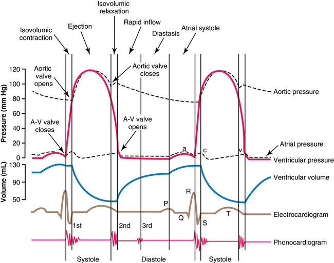
At the end of systole, the second period, diastole, or relaxation of the ventricle—takes place during four phases: isovolumic relaxation, rapid inflow, diastasis, and atrial systole (the final three phases constitute ventricular filling) (Figures 33-1 and 33-2). During isovolumic relaxation, the ventricular pressure rapidly drops below that of the arterial pressure, and the semilunar valves snap shut. For approximately 0.06 sec, the ventricular pressure continues to decrease without any change in ventricular volume. Once the ventricular pressure drops below the atrial pressure, the AV valves open and blood rapidly fills the ventricles. This period of rapid blood inflow is augmented by a negative intracavitary pressure (diastolic suction) created by the rapid myocardial relaxation. Once relaxation is complete and elastic distention of the ventricle begins, the slowing of blood return, termed diastasis, immediately precedes atrial systole. Effective atrial systole contributes up to about 20% of ventricular filling and completes the period of diastole.
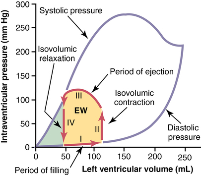
Control

The heart contains a specialized conduction system that comprises foci with automatic rhythmic electrical discharge (action potentials [APs]) that are conducted through both the atria and ventricles, controlling the cardiac cycle from beat to beat. Each normal cardiac cycle is initiated by spontaneous generation of an AP in the sinoatrial (SA) node (located in the posterior wall of the right atrium near the opening of the superior vena) that is conducted first through the atria, then to the ventricular system, resulting in well-coordinated myocardial contraction. Cardiac APs are the voltage changes that result from activation or inactivation of fast sodium channels, slow sodium-potassium channels, and potassium channels at different times that, together, create collective swings in voltage between a hyperpolarized and a depolarized state. APs higher up in the conduction system have a different morphology than do those in the ventricle, which explains why the control of the cardiac cycle resides in the more proximal or cephalad conducting system.

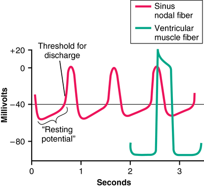
Rhythmicity of the cycle resides within the cells of the SA node because these cell membranes are inherently more “leaky” to Na+ and Ca2+ ions than are cell membranes of the ventricular conduction system. The influx of Na+ and Ca2+ creates a less negative resting membrane potential (−55 mV), a voltage at which many of the fast sodium channels have become inactivated. Therefore, depolarization is a result of the activation of the slow sodium-calcium channels, which results in a slower upslope of depolarization and a slower repolarization period in comparison with APs of ventricular muscle (Figure 33-3). The passive diffusion of Na+ due to its high concentration in the extracellular fluid outside the nodal fibers continues to depolarize the membrane. Once the threshold of −40 mV is reached, the sodium-calcium channels are activated, and an AP is generated. The sodium-calcium channels are quickly inactivated, and potassium channels are opened and allow the positively charged K+ ions to diffuse out of the cell until the resting membrane potential is hyperpolarized again at −55 mV. Finally, potassium channels close, with the inward-leaking Na+ and Ca2+ ions counterbalancing the outward flux of K+ ions. The process repeats itself, eliciting another cycle. The cells in the SA node control the heart rate because they depolarize more rapidly than does the rest of the conducting system (SA node rate, 70 to 80/min; AV node rate, 40 to 60/min; Purkinje fiber rate, 15 to 40/min).

The APs of ventricular myocytes have several key differences when compared with nodal APs. The intracellular potential is significantly more negative at −85 mV; depolarization occurs abruptly as a result of both fast sodium channels and slow sodium-calcium channels opening. Ventricular myocytes also exhibit a plateau phase of about 0.2 sec, which makes the AP last up to 15 times longer than in skeletal muscle. This plateau occurs because of the longer duration of activation of the slow sodium-calcium channels. The membrane of ventricular myocytes also becomes less permeable to K+ ions than that of skeletal muscle cells so that, after activation, the efflux of K+ is slowed and, thus, prevents early return of the AP voltage to its resting level. Once the slow sodium-calcium channels are inactivated at the end of the plateau, the permeability of the membrane to K+ is simultaneously restored, and the AP is abruptly terminated with the return of the membrane potential to its baseline. Thus, the ventricular AP is much more rapid in onset and offset, has a plateau phase, and has a larger variation in membrane potential in comparison with nodal and conducting-system APs.

Physiologic effects on cardiac cycle control

Sympathetic nerves are distributed to all parts of the heart, especially the ventricular myocytes. These nerves (1) increase the rate of SA nodal discharge (chronotropy), (2) increase the rate of conduction plus excitability throughout the heart (dromotropy, synchronicity), and (3) increase the force of contraction of all myocytes (inotropy). (Maximal sympathetic outflow can triple the heart rate and double the strength of contraction.) Norepinephrine released at sympathetic nerve endings is believed to increase membrane permeability to Na+ and Ca2+, increasing the tendency of the membrane potential to drift upward to the threshold for excitation. Increased Ca2+ permeability causes increased inotropic effect.

Parasympathetic preganglionic fibers are distributed mainly to the SA and AV nodes and, to a much lesser extent, the atria and ventricles through the vagus nerve. Vagal stimulation releases acetylcholine from the axonal terminus of the preganglionic fibers, which (1) decreases the rate of SA node discharge and (2) decreases the excitability of AV junctional fibers, thus slowing the impulse transmission into the ventricles. Strong vagal stimulation can completely stop SA node discharge, leading to eventual ventricular escape beats from discharge of Purkinje fibers. Acetylcholine works by increasing the permeability of cells in the SA and AV nodes to K+, thereby producing hyperpolarization (increased negativity of resting membrane potential, −70 to −75 mV); thus, conduction tissue is much less excitable and takes longer to spontaneously reach threshold.

The vasomotor center of the central nervous system (medullary-pontine area) contains neurons that affect chronotropic and inotropic responses from the heart. Vagal activity is reflex in origin and is aroused by impulses from carotid and aortic baroreceptors. The nucleus ambiguus contains vagal motor neurons that travel to the SA and AV nodes. Impulse activity depends mainly on baroreceptor input. Also, phasic input from the inspiratory center causes sinus arrhythmia (increased heart rate with inspiration, decreased heart rate with expiration).

Excess extracellular K+ causes the heart to dilate and become flaccid and slows the heart rate. Larger quantities can cause conduction delays and AV blocks. The mechanism of this effect is that high extracellular K+ concentration will decrease the resting membrane potential in myocytes (the membrane potential becomes less negative), which decreases the intensity of the AP and thereby decreases inotropy. Excess extracellular Ca2+, on the other hand, can cause spastic contraction of the heart through the direct effect of Ca2+ in the contraction process. A deficiency of Ca2+ can cause flaccidity.

Body temperature has an effect on the control of the cardiac cycle as well. Heat increases the permeability of myocyte membranes to ions that control the heart rate; hyperthermia can double the heart rate, whereas hypothermia can slow the heart rate to a few beats per minute. The contractile function of the heart is initially augmented with hyperthermia, but this compensatory mechanism is soon exhausted, and the heart eventually becomes flaccid.

Synchronicity

The AP originating in the SA node spreads through the atrium at 0.3 m/sec; internodal pathways terminate in the AV node (1 m/sec). Delay occurs at the AV node, allowing time for the atria to empty before ventricular contraction begins. The impulse reaches the AV node 0.04 sec after its origin in the SA node. The prolonged refractory period of the AV node helps prevent arrhythmias, which can occur if a second cardiac impulse is transmitted into the ventricle too soon after the first is transmitted. Purkinje fibers lead from the AV node and divide into left and right bundle branches, spreading into the apex of the respective ventricles and then back toward the base of the heart. These large fibers have a conduction velocity of 1.5 to 4 m/sec (6 times that of myocytes and 150 times that of junctional fibers), which allows almost immediate transmission of cardiac impulses through the entire ventricular system. Thus, the cardiac impulse arrives at almost all portions of the ventricle simultaneously, exciting the first ventricular myocytes only 0.06 sec ahead of the last ventricular fibers. Effective pumping by both ventricles requires this synchronization of contraction.

The AP causes myocardial myocytes to contract by a mechanism known as excitation-contraction coupling. The AP passes into the myocytes along the transverse tubules, triggering release of Ca2+ into the cell from the sarcoplasmic reticulum as well as from the transverse tubules themselves. These Ca2+ ions promote the sliding of actin on myosin, which creates myofibril contraction. The transverse tubules can store a tremendous amount of Ca2+; without this store, the sarcoplasmic reticulum of myocytes would not provide an adequate supply of Ca2+ ions for contraction to occur. Availability of extracellular Ca2+ directly impacts the availability of Ca2+ ions for release into the cellular sarcoplasm from the transverse tubules.