Cancer and the Cellular Immune Response

Published on 09/04/2015 by admin

Filed under Hematology, Oncology and Palliative Medicine

Last modified 09/04/2015

Print this page

rate 1 star rate 2 star rate 3 star rate 4 star rate 5 star
Your rating: none, Average: 0 (0 votes)

This article have been viewed 2300 times

Figure 51-1 Toll-like receptor (TLR) family subdivision Phylogenetic analyses of the amino acid sequence and structure of TLRs suggest that they are evolved to recognize a variety of pathogen-derived signals such as microbial nucleic acids, lipids, and proteins.

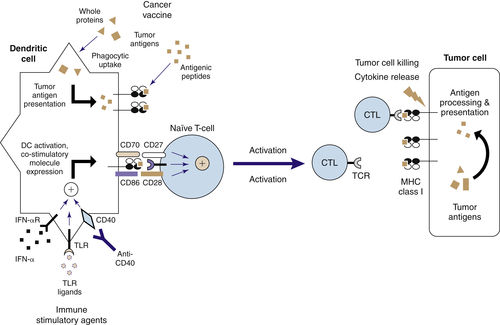
Dendritic Cells Link Innate and Adaptive Immunity

Dendritic cells are specialized antigen-presenting cells, which display an extraordinary capacity to stimulate naïve T cells and initiate primary immune responses. 11,12 This established function of DCs has now offered the hope to apply DC-based immunotherapy for cancers. Recent studies suggest that DCs also play critical roles in the induction of peripheral immunological tolerance, regulate the types of T-cell immune responses, and function as effector cells in innate immunity against microbes. In both humans and mice, DCs can be grouped into two major subsets: the myeloid conventional DCs (mDC) and the plasmacytoid DCs (pDCs). Both display functional plasticity depending on the types of activation signals and also their resident microenvironments. However, mDCs and pDCs display different sets of TLRs and appear to regulate innate and adaptive immunity in different ways (Figure 51-2 ).

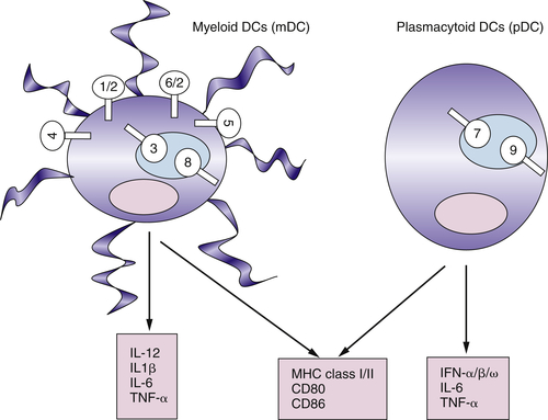
Myeloid DCs and Plasmacytoid DCs Express Different Sets of TLRs

In humans, mDCs can be identified by CD4+CD11c+lineage and pDCs can be identified by CD4+CD11c-lineage-BDCA-2+ILT7+. Strikingly, whereas mDCs express TLR2, 3, 4, 5, 6 and 8, pDCs express only TLR7 and TLR9. In response to the microbial ligands for TLR2, 3, 4, 5, 6 and 8, mDCs produce the TH1 polarizing cytokine IL-12 and cytokines IL-1, IL-6, IL-10, and TNF-α and undergo maturation by upregulation of MHC class I/class II and co-stimulatory cytokines CD80, CD83, and CD86. 1012 In response to microbial ligands for the TLR7 and TLR9 and viral infection, pDCs rapidly produce massive amounts of type 1 IFNs, including IFN-α, IFN-β, and IFN-ω. Therefore, pDCs are also known as specialized type 1 IFN-producing cells (IPC), which represent the key cell type in antiviral innate immunity. pDCs also have the ability to produce IL-6 and TNF-α on activation through TLR7 and TLR9 and undergo differentiation into mature DCs that express high MHC class I/class II and co-stimulatory molecules CD80/CD86 and acquire the ability to prime naïve T-cell activation. 1012

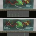
Functional Plasticity of DCs

In humans, DCs were found to display different effector functions in directing T-cell responses that are regulated by the maturation stage of DCs and the maturation signals. 11,12 Whereas mDCs at the mature stage induce TH1 differentiation and strong cytotoxic T lymphocyte (CTL) responses, mDCs at the immature stage induce IL-10–producing CD4+ and CD8+ regulatory T cells. Two groups of signals were shown to stimulate immature mDCs to induce TH1 differentiation: (1) LPS derived from gram-negative bacteria (TLR4-L), gram-positive bacteria Staphylococcus aureus (SAC) (may trigger multiple TLRs), and double-stranded viral RNA (TLR3-L); and (2) T-cell signals such as CD40L and IFN-γ. Several signals were shown to stimulate immature mDC to induce TH2 differentiation, including epithelial cell–derived cytokine TSLP and helminth Schistosoma mansoni egg antigen. 17,18 pDC-derived mature DCs also display different effector functions depending on the types of differentiation factors. Whereas pDCs activated by IL-3 and CD40L preferentially promote TH2 differentiation, pDCs activated through TLR7 or TLR9 prime naïve T cells to produce IFN-γ and IL-10. 12
image
Figure 51-2 Toll-like receptor (TLR) expression in human dendritic cell subsets Myeloid DCs (mDC) and plasmacytoid DCs (pDC) express different sets of TLRs. Whereas mDCs preferentially express TLRs 1, 2, 3, 4, 5, 6, and 8, pDCs preferentially express TLRs 7 and 9. TLR stimulation of both DC subsets leads to the upregulation of MHC class I and class II molecules and T-cell co-stimulatory molecules CD80 and CD86. However, the two DC subsets express distinct cytokine profiles in response to TLR recognition, with mDCs producing IL-12 and pDCs producing primarily type I interferons.

Targeting TLRs on DCs to Induce Effective Antitumor Immunity

A major class of adjuvants for vaccines used in humans or in experimental animal models are killed microbials or microbial-derived products that trigger different TLRs. 19 The current understanding of TLR biology and DC biology reveals that the immune system has been evolved to fight against microbial pathogens, but does not have an optimal system for sensing and effectively responding to cancer. Therefore, a basic principle of developing a cancer vaccine is to instruct DCs to recognize tumor antigens as foreign by introducing microbial-derived adjuvants together with tumor antigens (Figure 51-3 ). In animal models, stimulation of TLR9 mainly expressed on plasmacytoid DCs with CpGs has been shown to increase the immunogenicity of different forms of cancer vaccines, including peptide vaccines, DNA vaccines, tumor cell–based vaccines, and DC-based vaccines. 19 Stimulation of TLR4 (mainly expressed by mDCs) by MPL, BCG, or murine β-defensin can also promote the immunogenicity of cancer vaccines. Several studies suggest that the ability of mDCs to present antigens and activate antigen-specific T cells can be greatly enhanced by activated pDCs through a type-1 IFN-dependent mechanism in both antiviral immune responses and autoimmune responses. 20 We have shown that pDCs activated by CpG promote the ability of mDCs to present melanoma antigens to T cells and induce strong tumor-specific CTL responses in vitro and in vivo. In addition, pDCs activated by CpG also strongly activate NK cells, which kill tumor cells and further enhance the ability of mDCs to take up dead tumor cells and cross-present tumor antigens to CD8+ T cells. 21
image
Figure 51-3 Generation of antitumor immune responses through dendritic cell–mediated T-cell priming Tumor-associated peptide antigens (TAA, red) expressed in the context of surface MHC class I (HLA) molecules on the surface of tumor cells can activate TAA-specific T cells to perform cytolytic functions and/or release inflammatory cytokines to further amplify the adaptive immune response. Generation of such tumor-reactive T cells requires stimulation of naïve T cells by activated dendritic cells (DCs) expressing both the appropriate TAA and co-stimulatory molecules such as CD86 and CD70. These signals together lead to the activation, proliferation, and trafficking of TAA-specific T cells to the tumor site, where they can induce tumor regressions. Cancer vaccines can introduce TAAs into DCs in several ways, including nucleic acids coding for TAAs, tumor cell lysates, whole TAA proteins, or TAA-derived peptides. DCs can then process and present these TAAs to naïve T cells in the context of MHC class I molecules. However, optimal T-cell activation requires the expression by DCs of co-stimulatory ligands for CD27 and CD28, which are known to be upregulated by the combination of TLR stimulation or interferon-α, along with ligation of the CD40 receptor. Thus, the generation of an optimal antitumor T-cell response requires a combination of TAA-specific vaccination with DC activation signals provided by TLR ligands and CD40-specific antibody.

The Nature of Antitumor Immunity

T Cells Can Recognize Self-Antigens Expressed by Tumors

Over the past 10 years, numerous tumor antigens have been described that can be recognized by T cells. 22 These have been identified by two major methods: (1) molecular cloning using tumor antigen–specific T cells derived from cancer patients, and (2) analysis of candidate antigens based on gene expression and molecular profiling of tumors. Many of these antigens are expressed on normal tissues and are therefore considered to be “self” antigens. Examples of this class of antigen include melanocyte differentiation antigens that are expressed on melanoma cells as well as normal melanocytes. These include tyrosinase, MART-1, gp100, and TRP-1. 22 Differentiation antigens, expressed on tumor as well as the normal tissue of origin, can be targeted in immunotherapeutic strategies, as long as the normal tissue is nonessential. Mutated antigens, endogenous retroviral antigens, and antigens expressed in tumor and testis have also been described to be expressed in the context of MHC class I and class II molecules, capable of being recognized by CD8+ and CD4+ T cells, respectively. Although mutated antigens may be more easily recognized than “self” antigens, it is also possible that peripheral tolerance develops against the mutations, because co-stimulation may be absent in tumors, which are often present for many years before clinical diagnosis.

Autoimmune Vitiligo and Response to Immunotherapy

Because many tumor antigens are also expressed by normal host tissues, such as normal melanocytes in the case of melanoma antigens, obtaining an effective antitumor immune response requires overcoming self-tolerance. Indeed, in some settings, effective immunotherapy has been correlated with autoimmune responses against host tissues. IL-2, a cytokine that can stimulate the proliferation of T cells, can result in significant long-lasting regression of disease in some patients with metastatic melanoma and renal cell cancer (see later discussion). Interestingly, Rosenberg found that whereas 0 of 104 renal cancer patients treated with high-dose IL-2 developed vitiligo, the immune destruction of normal melanocytes, 11 of 74 melanoma patients treated with high-dose IL-2 developed vitiligo. 23 Furthermore, vitiligo was seen in 26% of melanoma patients who demonstrated objective tumor response to IL-2, whereas no vitiligo was observed in patients who did not respond to the IL-2. This suggests that tolerance to self-antigens can be overcome in the induction of an effective antitumor immune response.

Cancer Vaccines, Cytokines, and Immunotherapy

Cytokine Therapy of Cancer

Perhaps the strongest evidence that immune responses can result in a significant antitumor effect in patients is the fact that a subset of patients with metastatic melanoma can have complete tumor regressions and long-term survival following the administration of the T-cell growth factor IL-2. Of 270 patients with metastatic melanoma treated with high-dose IL-2, 43 (16%) had an objective response (complete or partial response). 24 More importantly, 60% of those patients who achieved a complete response had prolonged disease-free intervals and long-term survival (more than 10 years). This demonstrates that it is possible, by activation of the immune system, to induce clinically meaningful responses in patients. Future studies are needed to focus on understanding more fully the mechanism of response in patients so that improved strategies can be designed that will result in higher response rates and improved survival.
Another cytokine with significant clinical activity is interferon alpha, a type I interferon that is normally produced by plasmacytoid dendritic cells following viral infections. In randomized trials, high-dose interferon therapy has been shown to decrease tumor recurrence and increase survival in stage III melanoma patients (following surgical resection of tumor-positive lymph nodes). 25 Another study investigated prognostic markers in melanoma patients receiving interferon alpha in the adjuvant setting. Patients who developed autoantibodies during interferon therapy, including antithyroid, antinuclear, or anticardiolipin antibodies, had significantly enhanced survival compared to patients who did not develop signs of autoimmunity. 26 This again highlights the link between immunotherapy and autoimmunity as discussed earlier. In addition, it suggests that although interferon has pleiotropic effects on tumor and host tissues, such as effects on tumor vasculature and direct inhibitory effects on tumor proliferation, the mechanism of action in melanoma patients is by stimulating antitumor immunity by breaking tolerance to self-antigens. This may be due to the effects of type I interferons on antigen-presenting cells or T cells. In addition, interferon alpha can upregulate MHC molecules on tumor cells, thereby rendering them better targets for T cells.

Current Status of Cancer Vaccines

With clear evidence that the immune system can play an important role in mediating clinical responses in cancer patients, current efforts are focused on developing cancer vaccines in order to enhance efficacy as well as specificity, because nonspecific immune stimulation can result in autoimmunity, as discussed. Two major approaches have been pursued in cancer vaccine strategies: the use of whole tumor cells or the use of specific tumor antigens (Table 51-1 ).

Whole-Cell Cancer Vaccine Strategies

Cancer vaccine strategies have been performed using derivatives of both autologous and allogeneic tumor cells. Although the use of autologous tumor cells is more labor intensive, in that vaccines need to be prepared individually for each patient, autologous tumor has the advantage of containing specific mutations for that patient, which may be seen as more foreign compared to shared self antigens. Cell lysates fed to autologous dendritic cells, isolation of heat shock proteins bound to autologous antigens, and gene modification of autologous tumor with immune-enhancing cytokines have been evaluated in clinical trials. In murine models, transduction of tumor cells to express GM-CSF results in enhanced antitumor immune responses against parental non-transduced tumor cells. 27 Antitumor activity of GM-CSF–expressing tumors was found to be dependent on host bone-marrow–derived antigen-presenting cells, CD1d-restricted NK T cells, CD4+ and CD8+ T cells, and antibodies. 28,29

Antigen-Specific Vaccine Approaches
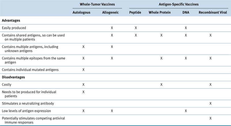
As discussed earlier, numerous tumor antigens have now been identified that can be recognized by T cells in the context of MHC class I and class II molecules. Many of these antigens are shared among specific types of tumors, and represent a feasible target for vaccine development. Antigen-specific vaccine approaches have included the use of specific peptide epitopes, whole proteins, and recombinant DNA and viral vaccines. The advantage of whole protein and recombinant approaches using the entire antigen gene is that multiple class I and class II epitopes may be presented. 30 However, whole proteins have been expensive and challenging to produce clinically, and viral vaccines can induce neutralizing antibodies that prevent the efficacy of serial doses of vaccine. Peptide vaccines, in combination with specific adjuvants, have demonstrated potential clinical efficacy in the case of chronic myelogenous leukemia and have been the most consistent method of inducing high levels of circulating antigen-specific T cells. 31,32 However, in the case of patients with metastatic melanoma, high levels of tumor-specific T cells in the blood do not always result in tumor regression. 33 Therefore, future efforts are focused on stimulating stronger and more effective T-cell priming through the use of specific adjuvants such as TLR agonists, using concepts learned from basic studies of antiviral immunity as described earlier. This may result in T cells with increased affinity and specificity as well as enhanced memory and effector function. One method to more carefully manipulate the specific phenotype of tumor-reactive immune cells is to generate and select specific T cells in the laboratory followed by reinfusion into patients. This is termed adoptive immunotherapy.

Table 51-1

Advantages and Disadvantages of Various Cancer Vaccine Approaches

image

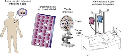
Adoptive Immunotherapy of Cancer

One of the most significant recent advances in clinic immunotherapy has been the adoptive transfer of tumor-reactive lymphocytes. A number of lines of evidence have demonstrated the clinical effectiveness of this approach, including donor-lymphocyte infusion following allogeneic bone marrow transplantation, treatment of metastatic EBV-driven lymphoproliferative tumors, and therapy of metastatic melanoma. 34,35

Tumor-Infiltrating Lymphocytes

In the setting of metastatic melanoma, T cells can be found at the tumor site (tumor-infiltrating lymphocytes, or TILs) that are specific for melanoma antigens, such as MART-1 and gp100 (Figure 51-4 ). Because the tumor is growing, either these T cells are nonfunctional or the tumor is resistant to recognition or lysis. However, when TILs are expanded ex vivo and reinfused, clinical regressions are seen in patients with metastatic disease. A number of reasons may explain the effectiveness of ex vivo expanded lymphocytes compared to endogenous T cells. First, large numbers can be generated in the laboratory that may be difficult to achieve in vivo. Second, expanding the lymphocytes ex vivo takes them out of the suppressive tumor microenvironment. Finally, growth ex vivo may allow reactivation of lymphocytes rendered anergic or nonfunctional by in vivo toleragenic mechanisms. Initially, transfer of tumor-reactive T cells alone resulted in response rates of greater than 20%, but clinical regressions were often transient, and it was clear from gene marking studies that the T cells did not survive long in vivo. 36,37
More recently it was found that transient lymphodepletion using cytoxan and fludarabine before T-cell infusion resulted in improved response rates and T-cell survival. Eighteen of 35 patients (51%) with metastatic melanoma exhibited objective responses following lymphodepletion and adoptive T-cell transfer. 38,39 Substantial numbers of infused lymphocytes were found in the circulation in some patients more than 2 years after infusion. This dramatic improvement following lymphodepletion may be due to a number of potential mechanisms including elimination of regulatory T cells and enhancement of lymphocyte homeostatic proliferation.
image
Figure 51-4 Expansion of tumor-infiltrating lymphocytes (TILs) from melanoma tumors T cells capable of specifically recognizing tumors can be found in some metastatic melanomas. When tumor fragments are cultured in the T-cell growth factor interleukin-2 (IL-2), the T cells expand and destroy the tumor cells in vitro. The expanded TIL can then be reinfused into metastatic melanoma patients in combination with interleukin-2.
Current studies of adoptive immunotherapy are focused on optimizing the generation of T cells ex vivo and their proliferation in vivo. For example, murine models suggest that proliferation of adoptively transferred T cells can be greatly enhanced in vivo by the addition of active immunization, such as dendritic cell vaccines. 40 In addition, significant efforts are focused on the introduction of novel genes into T cells in order to enhance their ability to recognize, migrate to, and eliminate tumor cells. In a recent study, nonspecific peripheral blood T cells were gene modified with a melanoma-specific T-cell receptor and then reinfused into melanoma patients. Two patients (of 17) demonstrated objective clinical responses, demonstrating that gene modification of lymphocytes is a feasible and potentially efficacious maneuver, although future studies are focused on enhancing the overall response rate. 41

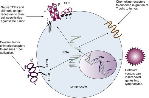
Gene-Modified T Lymphocytes

Because tumor-reactive lymphocytes are naturally found in melanomas, much of the work in adoptive therapy has focused on this disease. Although tumor-reactive lymphocytes are rarely found in other common cancers, antibodies that recognize these tumors in a relatively specific fashion have been described. Therefore, chimeric receptors have been designed using antibody variable regions extracellularly fused to T-cell signaling chains intracellularly (Figure 51-5 ). The initial studies demonstrated the ability to redirect T-cell specificity in vitro against ovarian cancer. 42 Lysis of ovarian cancer cells by human lymphocytes redirected with a chimeric gene composed of an antibody variable region and the Fc receptor gamma chain, but subsequent receptors have been designed that recognize human immunodeficiency virus (HIV) as well as a number of other tumor types. 4345
Besides redirecting T-cell recognition, the introduction of novel genes into effector lymphocytes may be used to enhance other functional properties, such as T-cell migration to the site of tumor and activation status. For example, Kershaw and colleagues introduced the chemokine receptor gene CXCR2 into T cells and demonstrated the ability of these modified cells to migrate toward chemokines produced by tumor cells. 46 In addition, receptor genes have been introduced with signaling chains containing co-stimulatory sequences in order to enhance T-cell activation. 47
In summary, the infusion of tumor-reactive, ex vivo expanded T cells has clearly demonstrated the effectiveness of T-cell–mediated immunity in the treatment of patients with metastatic cancer. Future studies will focus on enhancing response rates through generation of T cells with greater activity and ability to migrate to tumor, durability of response by improved maintenance of T cells in vivo, and the use of T cells in nonmelanoma tumors by redirecting cells with native T-cell receptor or novel chimeric receptor genes.
image
Figure 51-5 Insertion of genes into lymphocytes to enhance antitumor properties Genes can be inserted into T cells using retroviral vectors. These genes can endow the T cells with novel properties. Using genes encoding tumor antigen-specific T-cell receptors (TCRs) or chimeric antibody/T-cell receptor genes, T cells can gain the ability to recognize new targets. Using co-stimulatory receptor genes, T-cell activation can be further enhanced. Finally, introducing chemokine receptor genes into T cells can enhance T-cell migration to tumor sites.

Immune Regulatory Cells and Molecules in Human Cancer

Immunoregulatory cells and molecules are natural mechanisms that the immune system uses to prevent autoimmune destruction. Tumors often exploit these mechanisms to evade antitumor immunity, and several have been reported to be upregulated in human malignancy, including gastric cancer, ovarian cancer, melanoma, Hodgkin’s disease, and kidney cancer (Table 51-2 ).

Regulatory T Lymphocytes in Human Cancer
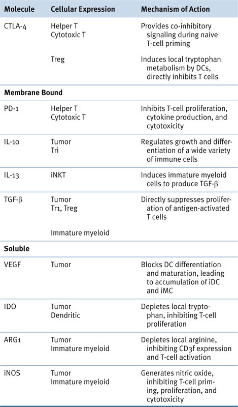
CD4+D25+, naturally occurring regulatory T cells (Treg), constitute 5% to 10% of peripheral CD4+ T cells, which play an essential role in the active suppression of autoimmunity in both humans and rodents. Treg appear to differentiate as a unique T-cell lineage in the thymus from immature T cells expressing T-cell receptor (TCR) with medium to high affinity for self-antigens, which depends on IL-2 and co-stimulatory molecules provided by activated APCs. 4852 Foxp3, a member of the forkhead transcriptional factor family, has been demonstrated to be the master regulator of Treg development in the thymus, as well as Treg suppressive function. Increasing evidence suggests that tumor-specific Treg exist and play an essential role in immune tolerance to tumors and thus represent a major hurdle for antitumor immunotherapy. In addition, the tumor microenvironment appears to be the site where tumor-specific infiltrating T cells are actively converted into tumor-specific Treg. 53

Table 51-2

Potentially Targetable Immunoregulatory Molecules

image

Melanoma is perhaps the most immunogenic of solid tumors, as melanoma-specific CD4+ and CD8+ T cells can be isolated directly from tumor deposits. Yet the tumor metastases grow unabated, thereby implying that immune regulatory mechanisms may be preventing full activation and effector function by tumor-infiltrating T cells. Although it is not clear why melanoma-specific T cells found in tumor deposits are not functioning to eliminate the tumor in vivo, a number of studies have been performed in melanoma patients to help provide some insight into potential regulatory pathways that are active.
Indeed, CD4+CD25+ Treg have been isolated from melanoma deposits, and in fact some Treg lines have been found to be specific for LAGE-1 and ARCT1, expressed by melanoma cells. 54,55 In a DC melanoma vaccine study, Chakraborty and colleagues found that vaccine-induced specific CTL responses declined by day 28, and this was associated with expansion of CD4+CD25+IL-10+ T cells. 56 CD4+CD25+ FoxP3 expressing cells were also found to be overrepresented in melanoma lymph-node metastases. 57 This may represent a mechanism by which tumors escape the immune system by first generating immunosuppression at the local lymph-node site.
Besides CD4+CD25+ Treg, melanoma cells themselves may produce factors or express receptors that can either induce regulatory immune cells or suppress effector T cells directly. For example, melanoma cells have been found to express IL-10, which is capable of inducing Tr-1 cells, another CD4+ regulatory cell type that can induce T-cell anergy and suppression of immune responses, primarily via the production of high levels of IL-10 and TGF-β. 58,59
A significant body of work has been performed in evaluating the cellular infiltrate of ovarian cancer. Zhang and co-workers performed immunohistochemical analysis of 186 advanced-stage ovarian cancer patients and found that the 5-year survival was 38% for patients whose tumors were infiltrated by CD3+ T cells versus 4.5% for patients with an absence of intratumoral T cells. 60 T-cell infiltration was associated with increased interferon-γ, interleukin-2, and specific chemokines within the tumor. However, another group found that not all CD3+ T-cell subpopulations were favorably correlated with outcome in ovarian cancer patients. Accumulation of CD4+CD25+ Treg cells in ovarian cancer was found to predict decreased survival for both stage III and stage IV patients. 61 Wolf and associates confirmed and extended these findings by demonstrating that quantitative FoxP3 levels of ovarian biopsies by real-time PCR could identify a patient subgroup with decreased overall survival. 62 Finally, Sato and colleagues found that ovarian cancer patients with a higher CD8+/Treg ratio in tumor tissue had an increased survival. 63 Together, these studies suggest that effector T cells play an important role in mediating an antitumor immune response in ovarian cancer patients, whereas Treg negatively influences this response. This implies that strategies to enhance tumor-specific CD8+ T cells while decreasing Treg may be effective in improving the outcome for ovarian cancer patients.

Myeloid-Derived Suppressor Cells in Human Cancer

There is accumulating evidence that progressive tumor growth is correlated with an increased frequency of immature myeloid cells (iMC) and immature DCs (iDC) in the tumor microenvironment, which can inhibit the function of tumor-specific T lymphocytes. 6466 These immature myeloid cells can be induced by tumor-derived factors such as vascular endothelial growth factor (VEGF), macrophage colony-stimulating factor, IL-6, granulocyte-macrophage colony-stimulating factor, IL-10, and gangliosides. The proangiogenic cytokine VEGF is a major contributor to immune suppression, and tumors often produce large quantities of VEGF that can be detected in the serum of cancer patients. 67,68 In vitro studies have shown that VEGF can block DC maturation, leading to the production of iMCs; conversely, blocking VEGF promotes normal DC differentiation and enhances antitumor function. 69
Tumors from patients with cancer often contain iDCs with reduced T-cell stimulatory capacity, often expressing low levels of the co-stimulatory molecules CD80 and CD86; further, they fail to upregulate them even in the presence of DC maturation factors. 70,71 Increased levels of circulating immature myeloid cells have also been observed in the peripheral blood of patients with lung, breast, head and neck, and esophageal cancer. 65,72 Importantly, patients with cancer often show a significant reduction in normal circulating levels of mature DCs that can be reversed on surgical removal of tumors. 73,74 Thus, abnormal differentiation and maturation of DCs in vivo, mediated by tumor-derived soluble factors, likely play a substantial role in preventing the effective priming of a productive, T-cell–mediated antitumor immune response. Circulating levels of iMCs have been well correlated with stage of disease and poorer prognosis, and surgical resection of tumors has been shown to decrease the number of peripheral blood iMCs in both human and animal models. 7577
Immature MCs mediate their immunosuppressive activity through the inhibition of IFN-γ production by CD8+ T cells in response to MHC class I–associated peptide epitopes presented on the iMC surface. 78 This effect requires direct cell-to-cell contact and is mediated by reactive oxygen and nitrogen species, such as hydrogen peroxide (H2O2) and nitric oxide (NO), secreted by the iMCs in close proximity to the T cell. 79,80 Although the precise mechanism of action on T cells has yet to be fully elucidated, there is some indication that iMCs act, in part, through downregulation of the CD3ζ chain on responding CD8+ T cells. 81,82 A population of iMCs has been described in the peripheral blood of cancer patients having high arginase-1 activity capable of depleting local arginine levels and down-modulating CD3ζ levels on T cells. Depletion of this iMC subset in vitro restored CD3 expression and normal T-cell responses. 83,84
In summary, there is evidence to suggest that the balance of immature and mature myeloid cells can have a significant effect on both naturally occurring and vaccine-induced antitumor T-cell responses. It is becoming increasingly apparent that effective cancer immunotherapy may require the correction of aberrant myeloid cell differentiation frequently observed in tumor-bearing hosts.

Immune Checkpoint Blockade
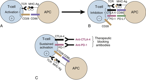
The development of a new class of effective cancer immunotherapy agents has recently become possible because of advances in our understanding of T-cell activation and regulation. T-cell activation is initiated by interaction of the TCR with MHC-bound peptide antigens on APCs (see Figure 51-3). However, effective priming of naïve T cells also requires a second, co-stimulatory signal mediated by the binding of CD28 on the T-cell surface to CD80 or CD86 (Figure 51-6 ). These two signals together allow T cells to proliferate, acquire antitumor effector functions, and eventually migrate to disease sites for tumor-cell killing. 85 T-cell activation is tightly regulated by a number of inhibitory signals in order to avoid prolonged immune responses that can potentially damage normal tissues. Inhibitory signals that are intrinsic to T cells are known as immune checkpoint molecules, with the most well studied being cytotoxic T lymphocyte–associated protein 4 (CTLA-4) or programmed cell death 1 (PD-1). 85,86
image
Figure 51-6 Immune checkpoint blockade therapies (A) T-cell activation is initiated by the interaction of the T-cell receptor (TCR) with major histocompatibility complex (MHC) molecules presenting antigen on an antigen-presenting cell (APC). Optimal activation of the T cell requires additional signals that are provided by the interaction between CD28 and CD86. (B) T-cell activation is naturally attenuated by upregulation of cytotoxic T lymphocyte–associated protein 4 (CTLA-4) on the surface of activated T cells, where it outcompetes CD28 for binding to CD86 on APCs. Additional regulation of T-cell activity is also provided by later inhibitory signals through programmed cell death 1 (PD-1), which binds to PD1 ligand 1 (PD-L1). (C) Strategies to sustain activated tumor-specific T cells include the use of blocking monoclonal antibodies targeting CTLA-4, PD-1, or PD-L1 to neutralize co-inhibitory receptors. Therapeutic antibodies that block intrinsic inhibitory immune checkpoints can allow for sustained T-cell effector responses, including increased production of cytokines and cytotoxic function.

CTLA-4 Blockade

CTLA-4 is expressed by activated CD4 and CD8 T cells. It is a homologue of T-cell co-stimulator CD28 but has a higher binding affinity for its ligands. On T-cell activation, signaling pathways lead to the expression of CTLA-4, which is then mobilized from intracellular vesicles to the cell surface, where it outcompetes co-stimulator CD28 for binding to its ligands. Binding of CTLA-4 to CD86 proteins interrupts CD28 co-stimulatory signals and, as a result, limits T-cell responses (see Figure 51-6). In addition to the intrinsic restriction of effector T-cell responses due to expression of CTLA-4 on effector cells, there can also be extrinsic restriction of effector T-cell responses due to the expression of CTLA-4 on regulatory T cells as well. 8789
Because of the negative regulatory effects of CTLA-4 on T-cell responses, it was hypothesized that blockade of CTLA-4 signaling would potentiate antitumor immune responses. Indeed, anti-CTLA-4 antibodies were able to cause rejection of syngeneic transplanted tumors in mice. 90 These preclinical studies led to the development of an antibody to block human CTLA-4 (ipilimumab), which was shown in Phase III clinical trials to improve overall survival of patients with advanced melanoma. 91 Initial Phase I and Phase II clinical trials in other cancer patients demonstrated that ipilimumab treatment could also induce significant antitumor activity. 92,93 Subsequently, a Phase III randomized trial showed that ipilimumab improved the median overall survival of advanced metastatic melanoma patients by 3.7 months (10.1 vs. 6.4 months; P = .003). 91 However, the most striking feature of this study was the fact that nearly a quarter of the patients survived longer than 4 years. This trial led to the approval of ipilimumab by the U.S. Food and Drug Administration (FDA) in March 2011 for the treatment of patients with metastatic melanoma. Most recently, a second randomized, Phase III clinical trial showed that the addition of ipilimumab to standard dacarbazine chemotherapy significantly improved overall survival by 2.1 months in melanoma patients. 94 More importantly, anti-CTLA-4 therapy has demonstrated the long-awaited promise of immunotherapeutic agents to elicit durable responses in patients with many different types of cancers.

PD-1/PD-L1 Blockade

Another important T-cell checkpoint that can be targeted clinically is the interaction of PD-1 and its ligands (see Figure 51-6). PD-1 is mainly expressed by activated CD4 and CD8 T cells, and it has two ligands, PD-L1 and PD-L2, with distinct expression profiles. 95 PD-L1 is expressed not only on APCs, but also on T cells, B cells, and nonhematopoietic cells, including tumor cells. Expression of PD-L2 is largely restricted to APCs, including macrophages and myeloid DCs. The role of PD-1 as a negative regulator of T and B cells was best demonstrated by the findings that PD-1–deficient mice developed significant autoimmunity. 96,97 Subsequently, blocking antibodies against PD-1 were shown to induce a reduction of tumor growth and metastasis in a number of experimental mouse models. 98,99 Consistent with the immune inhibitory role of PD-1/PD-L1/2 signaling, enforced expression of PD-L1 on tumor cells caused enhanced tumor growth in vivo, which could otherwise be kept in check by T cells. Again, this augmentation of tumor growth could be reversed with the use of blocking antibodies against PD-L1. 100
Consistent with these preclinical studies, a recent Phase I clinical trial using an anti-PD-1 antibody (BMS-936558) showed an 18% to 28% objective response rate in patients with advanced non–small-cell lung cancer (NSCLC), renal call carcinoma (RCC), and melanoma. 101 In addition, another concurrent Phase I trial with anti-PD-L1 antibody demonstrated an objective response rate of 6% to 17% in patients with advanced NSCLC, melanoma, and RCC. 102 These early-phase clinical studies show a very promising potential for employing blocking antibodies against the inhibitory PD-1/PDL-1/2 signaling pathway in anticancer immunotherapy.

Checkpoint Blockade and the Induction of Autoimmunity

As with most experimental cancer therapies, treatment with immune checkpoint blockade agents is sometimes associated with a distinct set of on-target toxicities due to immune-related adverse events. For example, among melanoma patients treated with ipilimumab, up to 60% of patients manifested autoimmune reactions including dermatitis, colitis, hepatitis, and hypopituitarism. 91 Early recognition, diagnosis, and treatment of such drug-induced inflammatory conditions with steroids are critical for minimizing the adverse effects while maximizing the therapeutic benefits of this promising anticancer agent. By contrast, the early Phase I results targeting the PD-1 pathway have shown less overall toxicity in cancer patients, although future studies are required to confirm this. Regardless, investigations into biomarkers/pathways that are associated with or predict clinical benefit or toxicities will be important as the field of cancer immunotherapy moves forward, and recent studies in these areas have been promising. 103,104 Perhaps future strategies combining checkpoint blockade strategies with an effective cancer vaccine may skew the immune response toward tumor-specific antigens and away from normal tissue-associated self-antigens.
In summary, the recent clinical successes with immune checkpoint blockade agents have provided clear data to indicate that the immune system can be harnessed successfully to treat cancer and that the exquisite ability of the immune response to target tumor-specific antigens, as well as generate memory cells to thwart recurrences, can lead to durable clinical benefit in patients. These exciting study results and clinical benefits with anti-CTLA-4 and anti-PD-1 have provided a strong foundation on which to build for even greater success as we determine how to best select patients for treatment and how to combine immunotherapy with other agents to increase the number of patients who benefit.

Future Cancer Immunotherapies Will Use Basic Principles of Cellular Immunity

Although the adoptive transfer of carefully selected T-cell populations or the use of optimized vaccine strategies may allow for the presence of more potent T cells in vivo, this must be coupled with enhanced conditioning of the immunosuppressive tumor microenvironment to allow for both migration and function of specific T cells. In successful immune responses against viruses, a proinflammatory state exists in the infected site by TLR activation through specific viral components, as discussed earlier. This inflammation induces the upregulation of specific adhesion molecules on endothelial cells that will in turn mediate trafficking of primed T cells into the infected site. In addition, the proinflammatory state may enhance effector T-cell function while limiting the effects of immune regulation. Successful immunotherapy will require a better understanding of the interplay between immune-cell subpopulations that results in optimal T-cell induction as well as tumor site conditioning that will allow for enhanced efficacy of the T cells within the tumor microenvironment. Thus, the most effective strategies will likely involve combination approaches that aim to boost antitumor T-cell responses while simultaneously blocking immune inhibitory pathways. A number of immunosuppressive mechanisms are now targetable, including immune checkpoint blockade, depletion of regulatory immune cell subsets, and inhibition of oncogenic signaling. 105 This multidimensional approach will require exploring combinations of new and existing clinical agents to determine which are the most effective.
Finally, although many of the initial principles have been developed using more immunogenic tumors such as melanoma, future goals include the application of these same principles to develop successful immunotherapeutic approaches against other common cancers such as breast, lung, colon, and prostate cancer.
References

1. Pardoll D.M. Cancer vaccines . Immunol Today . 1993 ; 14 : 310 316 .

Buy Membership for Hematology, Oncology and Palliative Medicine Category to continue reading. Learn more here