Published on 19/03/2015 by admin
Filed under Dermatology
Last modified 22/04/2025
This article have been viewed 2639 times
Rajani Nalluri and Ian Coulson
Evidence Levels: A Double-blind study B Clinical trial ≥ 20 subjects C Clinical trial < 20 subjects D Series ≥ 5 subjects E Anecdotal case reports
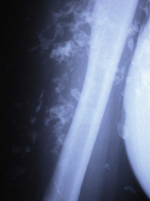
Calcinosis cutis is a rare disease of aberrant calcium deposition in the skin and subcutaneous tissue. There are four major types:
Idiopathic: occurs without tissue injury or metabolic defect (e.g., idiopathic scrotal calcinosis).
Dystrophic: secondary to local tissue damage or alterations in collagen, elastin or subcutaneous fat but normal calcium and phosphate levels (e.g., in connective tissue diseases, post trauma or infection).
Metastatic: abnormal calcium and/or phosphate metabolism leading to precipitation of calcium salts in normal tissue.
Iatrogenic: secondary to a treatment or procedure (such as extravasation of calcium or phosphate infusions).
Other rare variants of calcinosis cutis that have been described include calcinosis cutis circumscripta, calcinosis universalis, tumoral calcinosis, transplant-associated calcinosis cutis, and milia-like idiopathic calcinosis cutis (usually associated with Down syndrome). Firm white or yellow dermal lesions may ulcerate and extrude a gritty material. Stiffening of the skin can limit joint mobility and function, and fingertip lesions may be painful.
The first step in management is to identify any underlying cause. Dystrophic calcification occurs in up to 10% of patients with scleroderma and 10–40% of patients with juvenile dermatomyositis, but is rare in systemic lupus erythematosus. Examination and investigations for connective tissue disease are therefore strongly recommended. Skin biopsy can help to distinguish cutaneous calcification from ossification.
A number of malignancies have been implicated in causing metastatic calcification (e.g., leukemia and multiple myeloma). However, successful treatment of the underlying cause does not always have an impact on calcinosis cutis, which frequently requires other treatment modalities. There are no large studies for the treatment of calcinosis cutis, and most therapies are based on case reports.
Spontaneous extrusion of calcium salts may occur; this may need surgical encouragement. Intralesional corticosteroids, aluminum hydroxide supplements, bisphosphonates, diltiazem, colchicine, and probenecid have shown success, mostly in calcinosis associated with dermatomyositis. Low-dose minocycline has been reported to reduce the frequency of ulceration and inflammation associated with cutaneous calcinosis in patients with limited systemic sclerosis. The mechanism of action may be mainly through inhibition of matrix metalloproteinases and anti-inflammatory effects.
Warfarin has been advocated in both dermatomyositis and systemic sclerosis-associated calcinosis for small calcified deposits. Carbon-dioxide laser vaporization, extracorporeal shock wave lithotripsy and intravenous immunoglobulin are recent approaches that have been tried in cutaneous calcinosis secondary to CREST syndrome.
Full blood count
Urea and creatinine
Serum calcium and phosphate
Parathyroid hormone levels
Vitamin D levels
Serum electrophoresis
Creatine kinase
Autoantibodies
Skin biopsy
Plain radiographs
Bone scintigraphy
Pitt AE, Ethington JE, Troy JL. Cutis 1990; 45: 28–32.
A case of dystrophic calcinosis following trauma that resolved over 8 weeks with spontaneous transepidermal elimination.
Park YM, Lee SJ, Kang H, Cho SH. J Korean Med Sci 1999; 14: 589–92.
A 22-year-old woman with systemic lupus erythematosus developed soft tissue calcification with ulceration, infection, and abscess formation. She was treated with aluminum hydroxide 600 mg three times a day with reduction in size and softening of the deposits after 9 months.
Nakagawa T, Takaiwa T. J Dermatol 1993; 20: 558–60.
A case of calcinosis cutis in juvenile dermatomyositis was successfully treated with oral aluminum hydroxide; near complete clearance was observed after 8 months of therapy.
The use of these agents in patients with renal insufficiency may result in aluminum toxicity.
Hazen PG, Walker AE, Carney JF, Stewart JJ. Arch Dermatol 1982; 118: 366–7.
An 83-year-old woman with scleroderma had areas of calcinosis with ulceration on the face and neck. Four to eight weekly treatments with 1 mL per area of intralesional triamcinolone acetonide (20 mg/mL) resulted in improvement in 3 months and almost complete resolution in 12 months.
Balin SJ, Wetter DA, Andersen LK, Davis MD. Arch Dermatol 2012; 148: 455–62.
Nine of 17 patients with autoimmune connective tissue disorders showed partial response to treatment with diltiazem at the dose of <480mg/day. This is recommended as first line treatment. Eight patients were treated with colchicine (<1.2 g/day) resulting in one patient showing complete response and two patients showing partial response. Six patients received minocycline (200 mg/day). Only one patient showed partial response, two patients did not respond, and the response was unknown in three patients. Four patients were treated with warfarin with only one patient partially responding. Out of the11 patients who received surgical excision alone, all 11 responded with eight having a complete response. Another 17 patients had surgical and medical treatment with complete response in 14 patients, partial response in two patients and no response in one patient.
Reiter N, El-Shabrawi L, Leinweber B, Berghold A, Aberer E. J Am Acad Dermatol 2011; 65: 15–22.
Calcinosis cutis in dermatomyositis has been successfully treated with diltiazem (2–4 mg/kg/day), with lower doses being ineffective. Surgical excision or curettage is the treatment of choice in idiopathic calcinosis cutis, especially scrotal calcinosis. It is also effective for small, digital calcified skin lesions. A 16-year-old boy with morphea profunda developed small calcified deposits on both arms, his chest, and left thigh. He was treated with ceftriaxone 2 g/day intravenously for 20 days and the calcification diminished within weeks.
Jiang X, Yi Q, Liu D, Wang S, Li L. Int J Dermatol 2011; 50: 74–7.
A 12-year-old boy with calcinosis secondary to juvenile dermatomyositis received diltiazem at a dose of 30 mg/day resulting in obvious softening and radiological regression and functional improvement at 4 months.
Marco Puche A, Calvo Penades I, Lopez Montesinos B. Clin Exp Rheumatol 2010; 28: 135–40.
Three children with juvenile dermatomyositis received treatment with intravenous pamidronate at 1 mg/kg/day on 3 consecutive days every 3 months. In all three cases calcinosis significantly decreased and even totally cleared in one of them.
Mukamel M, Horev G, Mimouni M. J Pediatr 2001; 138: 763–6.
A 6-year-old boy with dermatomyositis and ectopic calcification leading to fixed contractures of all joints showed dramatic clinical improvement after treatment with alendronate (10 mg/day). During the 1-year treatment period, the tissue softened and range of motion of the joints improved so that he was able to run and swim again.
Nakamura H, Kawakami A, Ida H, Ejima E, Origuchi T, Eguchi K. J Rheumatol 2006; 33: 1691–3.
An 11-year-old boy with juvenile dermatomyositis developed calcinosis of both legs. Probenecid was used to reduce calcinosis, resulting in remarkable improvement of calcinosis accompanied by normalization of serum phosphorus level.
Eddy MC, Leelawattana R, McAlister WH, Whyte MP. J Clin Endocrinol Metab 1997; 82: 3536–42.
A 19-year-old man with extensive subcutaneous calcification secondary to juvenile dermatomyositis failed treatment with aluminum hydroxide, intravenous immunoglobulin and daily hydroxychloroquine. Treatment with probenecid was started increasing the dose from 250 mg/day to 500 mg three times a day with remarkable physical and radiographic improvement.
Robertson LP, Marshall RW, Hickling P. Ann Rheum Dis 2003; 62: 267–9.
In an open-label study, eight out of nine patients with limited cutaneous systemic sclerosis prescribed minocycline 50 or 100 mg daily showed definite improvement. The frequency of ulceration and inflammation associated with the calcinosis deposits decreased with treatment. A reduction in calcinosis size was evident but less dramatic with improvement occurring by 5 months. The mean duration of treatment was 3.5 years.
See also Balin et al. above re colchicine and minocycline.
Cukierman T, Elinav E, Korem M, Chajek-Shaul T. Ann Rheum Dis 2004; 63: 1341–3.
Three patients with disseminated subcutaneous calcinosis were treated with low doses of warfarin (1 mg/day) for 1 year. Two patients (relatively small lesions up to 2 cm in diameter) had complete resolution within 2 months. The other patient (larger and longer-standing lesions reaching up to 5 cm) did not respond to treatment. None of the patients showed a prolongation of prothrombin time, partial thromboplastin time, or an increased tendency for bleeding.
See also Reiter et al. above re ceftriaxone.
Daoussis D, Antonopoulos I, Liossis SN, Yiannopoulos G, Andonopoulos AP. Semin Arthritis Rheum 2012; 41: 822–9.
This patient with frequently ulcerating and painful extensive CREST-related calcinosis received two rituximab courses (consisting of four weekly infusions, 375 mg/m2 each); calcinosis significantly improved and pain disappeared.
Peñate Y, Guillermo N, Melwani P, Martel R, Hernández-Machín B, Borrego L. J Am Acad Dermatol 2009; 60: 1076–7.
A 55-year-old female with amyopathic dermatomyositis and progressive dystrophic calcinosis on the limbs with ulceration and pain failed to respond to various immunosuppressants and diltiazem. She had treatment with intravenous immunoglobulin (IVIG) (2 g/kg/month) at a dose of 0.4 g/day for 5 consecutive days, in combination with a reduced dose of prednisolone. After five courses of IVIG the dermal calcifications reduced both clinically and radiologically and became asymptomatic.
Schanz S, Ulmer A, Fierlbeck G. Arch Dermatol 2008; 144: 585–7.
A 56-year-old woman with CREST syndrome presented with a large calcified deposit on a finger. Treatment with IVIG at a dose of 2 g/day on a 4-day protocol once a month resulted in reduction in pain and inflammation after two courses and complete resolution of symptoms after three courses.
Raffaella C, Annapaola C, Tullio I, Angelo R, Giuseppe L, Simone C. Pediatr Dermatol 2009; 26: 311–15.
A 5-year-old boy with T-cell acute lymphoblastic leukemia developed soft tissue calcification with motility impairment at sites of intravenous 10% calcium gluconate infusion. Treatment with intravenous sodium thiosulphate 435 mg/kg three times a week for 3 months resulted in massive reduction of soft-tissue calcification and functional recovery of affected limbs.
Bair B, Fivenson D. J Drugs Dermatol 2011; 10: 1042–4.
A 74-year-old lady with pseudohypoparathyroidism developed ulcerative calcinosis cutis on her right lower leg. Treatment with topical sodium thiosulphate 100% powder mixed 1 : 4 (25%) in zinc oxide applied twice daily to the wound base and peri-wound skin with elastic wraps for compression and leg elevation resulted in marked improvement after 5 weeks with good re-epithelialization and complete healing after 15 weeks of continued treatment.
See also Reiter et al. above re surgical excision.
Saddic N, Miller JJ, Miller OF 3rd, Clarke JT. Arch Dermatol 2009; 145: 212–13.
A 58-year-old woman with CREST syndrome underwent 2 mm curettage. There was immediate pain relief and 7 months after the procedure, calcinosis had not recurred.
See also Balin et al. above. Surgical excision of large, discrete and symptomatic lesions can be beneficial to patients.
Chamberlain AJ, Walker NP. Dermatol Surg 2003; 29: 968–70.
Six affected digits in a patient with CREST syndrome, having failed various medical therapies, received a single treatment with carbon dioxide laser vaporization using the Sharplan 1040 SilkTouch system. Treated digits healed over a 6-week period and led to a significant remission in symptoms, with an average remission time of at least 3 years, allowing the patient to remain in full-time employment.
Meissner M, Ochsendorf F, Kaufmann R. Dermatol Surg 2010; 36: 727–8.
Patients with subcutaneous nodules smaller than 2 cm in diameter were given local anaesthesia, then the skin over the mass was opened using a focused erbium-doped yttrium aluminum garnet (Er : YAG) beam (5 J/cm2, 5 mm diameter, 5 Hz). The chalky material was then removed with a swab or curette. Re-epithelialization and cosmetic recovery were seen at 2 to 3 weeks and 14 weeks, respectively.
Sultan-Bichat N, Menard J, Perceau G, Staerman F, Bernard P, Reguiaï Z. J Am Acad Dermatol 2012; 66: 424–9.
A single center study including eight consecutive patients (with ten calcinosis cutis (CC) lesions) who underwent three extracorporeal shock-wave lithotripsy sessions at 3-week intervals for 6 months. At the end of this treatment period the median area had decreased by more than 50% in three CC lesions, pain scores decreasing significantly in five patients and analgesia consumption decreasing in three patients with no difference in results according to the underlying causal disease.
Treatment of Skin Disease Comprehensive Therapeutic Strategies 4e
WhatsApp us


 No treatment/self-healing
 Aluminum hydroxide
 Intralesional corticosteroid
 Diltiazem
 Bisphosphonates
 Probenecid
 Colchicine
 Minocycline
 Warfarin
 Ceftriaxone
 Rituximab
 Intravenous immunoglobulin
 Intravenous Sodium thiosulphate
 Topical sodium thiosulphate
 Surgery
 Carbon dioxide laser
 Extracorporeal shock wave lithotripsy