Brucella

Published on 08/02/2015 by admin

Filed under Basic Science

Last modified 22/04/2025

Print this page

rate 1 star rate 2 star rate 3 star rate 4 star rate 5 star
Your rating: none, Average: 0 (0 votes)

This article have been viewed 1623 times

Brucella

The family Brucellaceae comprises three genera: Ochrobactrum, Mycoplana, and Brucella. Brucella spp. are discussed in this chapter. Brucellae are free-living organisms that are subcategorized into nine recognized species. Six of the species are terrestrial, and four of those have been associated with human disease: B. abortus (seven biovars), B. melitensis (three biovars), B. suis (five biovars), and B. canis. Of the four species capable of causing human infection, all but B. canis are considered potential agents of bioterrorism.

Epidemiology and Pathogenesis

The disease brucellosis occurs worldwide, especially in Mediterranean and Persian Gulf countries, India, and parts of Mexico and Central and South America. The organisms are capable of survival for extended periods (e.g., soil, 10 weeks; aborted fetuses, 11 weeks; bovine stool, 17 weeks, milk and ice cream, 3 weeks); they can survive in fresh cheese for several months. Brucellosis is a zoonosis and is recognized as a cause of devastating economic loss among domestic livestock.

Each of the four Brucella spp. that are pathogenic for humans has a limited number of preferred animal hosts (Table 36-1). In the host, Brucella tend to localize in tissues rich in erythritol (e.g., placental tissue), a four-carbon alcohol that enhances their growth. Humans become infected by four primary routes:

TABLE 36-1

Brucella spp. and Their Respective Natural Animal Hosts

Organism Preferred Animal Host
B. abortus Cattle
B. melitensis Sheep or goats
B. suis Swine
B. canis Dogs
B. ovis Rams (not associated with human infection)
B. neotomae Desert and wood rats (not associated with human infection)

Rare cases of transmission by blood and bone marrow transplantation and by sexual intercourse, in addition to neonatal brucellosis, have been reported. Individuals considered at risk for contracting brucellosis include dairy farmers, livestock handlers, slaughterhouse employees, veterinarians, and laboratory personnel. The organism has a very low infectious dose (100 organisms or fewer). Mishandling and misidentification of the organism is often associated with laboratory transmission of the organism.

Brucella spp. are facultative, intracellular parasites that are able to exist in both intracellular and extracellular environments. Other bacteria classified as facultative intracellular infectious agents include Salmonella, Shigella, Yersinia, Listeria, and Francisella spp. After infecting a host, brucellae are ingested by neutrophils, within which they replicate, causing cell lysis. Neutrophils containing viable organisms circulate in the bloodstream and are subsequently phagocytized by reticuloendothelial cells in the spleen, liver, and bone marrow. If the infection goes untreated, granulomas develop in these organs, and the brucellae survive in monocytes and macrophages. Brucellae tend to show a tendency to invade and persist in the human host by inhibiting apoptosis (programmed cell death). Resolution of the infection depends on the host’s nutritional and immune status, the size of the inoculum and route of infection, and the Brucella species causing the infection; in general, B. melitensis and B. abortus are more virulent for humans.

Survival and multiplication of Brucella organisms in phagocytic cells are features essential to the establishment, development, and chronicity of the disease. The mechanisms by which brucellae avoid intracellular killing are not completely understood. Brucella spp. can change from a smooth to a rough colonial morphology based on the composition of their cell wall lipopolysaccharide O-side chain (LPS); those with a smooth LPS are more resistant to intracellular killing by neutrophils than those with a rough LPS. The smooth phenotype has been identified in B. abortus and B. melitensis. Brucellae ensure intracellular survival by interfering with the phagosome-lysosome fusion in macrophages and epithelial cells. In addition, as do Legionella spp. (see Chapter 35), brucellae use a type IV secretion system, VirB, for intracellular survival and replication. Unlike Legionella spp., however, brucellae modulate phagosome transport to avoid being delivered to lysosomes. Essentially, VirB is involved in controlling the maturation of the Brucella vacuole into an organelle that allows replication. In the mouse model, if mutations occur in this gene region, B. abortus is unable to establish chronic infections. In addition, Brucella spp. produce urease, which provides protection during passage through the digestive system when the organism is ingested in food products. Urease breaks down urea, producing ammonia, and neutralizes the gastric pH. Despite our current knowledge, many questions remain about the pathogenesis of disease caused by Brucella spp.

Spectrum of Disease

The clinical manifestations of brucellosis vary greatly, ranging from asymptomatic infection to serious, debilitating disease. For the most part, brucellosis is a systemic infection that can involve any organ of the body. Symptoms, which are nonspecific, include fever, chills, weight loss, sweats, headache, muscle aches, fatigue, and depression. Lymphadenopathy and splenomegaly are common physical findings. After an incubation period of about 2 to 4 weeks, the onset of disease is commonly insidious. Complications can occur, such as arthritis; spondylitis (inflammation of the spinal cord); genital, pulmonary, and renal complications; and endocarditis. Relapse is considered an important feature of brucellosis; it is associated with delayed initiation of treatment, ineffective antibiotic therapy, and positive blood culture findings during the initial presentation.

Laboratory Diagnosis

Specimen Collection, Transport, and Processing

A definitive diagnosis of brucellosis requires isolation of the organisms in cultures of blood, bone marrow, cerebrospinal fluid (CSF), pleural and synovial fluids, urine, abscesses, or other tissues. If processing will be delayed, the specimen may be held in the refrigerator.

It is essential that the clinical microbiology laboratory be notified whenever brucellosis is suspected:

Blood for culture can be collected routinely (see Chapter 68) into most commercially available blood culture bottles and the lysis-centrifugation system (Isolator; Alere, Waltham, MA). For other clinical specimens, no special requirements must be met for collection, transport, or processing.

Cultivation

Although most isolates of Brucella spp. grow on blood and chocolate agars (some isolates are also able to grow on MacConkey agar), more enriched agars and special incubation conditions generally are needed to achieve optimal recovery of these fastidious organisms from clinical specimens. Brucella agar or infusion base is recommended for specimen types other than blood. The addition of 5% heated horse or rabbit serum enhances growth on all media. Cultures should be incubated in 5% to 10% CO2 in a humidified atmosphere; inoculated plates are incubated for up to 3 weeks before they are considered negative and discarded.

Commercial blood culture systems (e.g., BacT/Alert, BACTEC, and lysis-centrifugation systems) all have successfully detected brucellae in blood. Other blood culture bottles, such as those with brain-heart infusion and trypticase soy broth, also support the growth of brucellae if the bottles are continuously vented and placed in a CO2 incubator. Most isolates can be detected within 5 to 7 days using commercial systems. Bottles need not be incubated longer than 10 to 14 days. Culture bottles may not become turbid. All subculture plates should be held for a minimum of 7 days.

On culture, colonies appear small, convex, smooth, translucent, nonhemolytic, and slightly yellow and opalescent after at least 48 hours of incubation (Figure 36-1). Rough variants may be seen with B. canis. The colonies may become brownish with age.

Approach to Identification

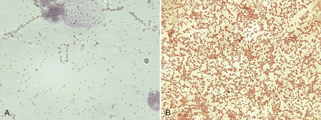
Because brucellosis is the most commonly reported laboratory-acquired bacterial infection, all handling and manipulations of suspected Brucella spp. should be performed in a class II or higher biologic safety cabinet. On Gram stain, the organisms are small coccobacilli that resemble fine grains of sand (Figure 36-2). Brucella spp. are catalase and urease positive, and most strains are oxidase positive. Other nonfermentative gram-negative coccobacilli that may be confused with brucellae are Bordetella, Moraxella, Kingella, and Acinetobacter spp. Brucella spp., however, are nonmotile, urease and nitrate positive, and strictly aerobic. The most rapid test for presumptive identification of Brucella spp. is the particle agglutination test with anti–smooth Brucella serum (Difco Laboratories, Detroit, Michigan).

Brucella spp. are differentiated by the rapidity with which an organism hydrolyzes urea, its relative ability to produce hydrogen sulfide (H2S), its requirements for CO2, and its susceptibility to the aniline dyes thionine and basic fuchsin (Table 36-2). For determination of the CO2 requirement, identical plates of Brucella agar or brain-heart infusion agar should be given equal inocula (e.g., with a calibrated loop) of a broth suspension of the organism to be tested. One plate should be incubated in a candle jar and the other plate in air in the same incubator. Most strains of B. abortus do not grow in air but show growth in the candle jar. Presumptive identification can be reported based on the colony’s morphology and a positive catalase, oxidase, urease, and slide agglutination reaction (see next section). Brucella isolates should be sent to state or other reference laboratories for confirmation or definitive identification, because most clinical laboratories lack the necessary media and containment facilities.

TABLE 36-2

Characteristics of Brucella spp. That Are Pathogenic for Humans

        INHIBITION BY DYE
Species CO2 Required for Growth Time to Positive Urease H2S Produced Thionine* Fuchsin*
B. abortus ± 2 hr (rare 24 hr) + (most strains) +
B. melitensis 2 hr (rare 24 hr) +
B. suis 15 min ± + (most)
B. canis 15 min +

image

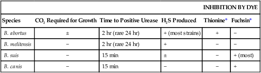
+, >90% of strains positive; − >90% of strains negative; ±, variable results.

*Dye tablets (Key Scientific Products, Round Rock, Texas).

Subtyping of biovars may be performed using a variety of molecular techniques, including pulsed-field electrophoresis, random amplification of polymorphic DNA, amplified fragment length polymorphism, various PCR techniques, and multilocus sequence typing.

Serodiagnosis

Because isolating brucellae is difficult, a serologic test is widely used (e.g., serum agglutination test [SAT] or microplate agglutination [MAT]). This technique detects antibodies to B. abortus, B. melitensis, and B. suis; however, the SAT does not detect B. canis antibodies. An indirect Coombs’ test is performed after the SAT. This test detects nonagglutinating or incomplete antibodies in complicated and chronic cases of brucellosis.

The serology associated with Brucella infection follows the classic antibody response: IgM appears initially, followed by IgG. A titer of 1 : 160 or greater in the SAT is considered diagnostic if this result fits the clinical and epidemiologic findings. The SAT can cross-react with class M immunoglobulins with a variety of bacteria, such as Francisella tularensis and Vibrio cholerae. Enzyme-linked immunosorbent assays (ELISAs) also have been developed. Purified LPS or protein extracts are primarily used in ELISAs. However, currently no reference antigen exists; therefore, it is important to identify the antigen in the commercial antigen when evaluating test results. In patients with neurobrucellosis, ELISA offers significant diagnostic advantages over conventional agglutination methods.

Additional serologic assays are commercially available, including a lateral flow dipstick for screening outbreaks and an immunocapture agglutination method. The immunocapture assay demonstrates sensitivity and specificity similar to a Coombs’ test and is less cumbersome to perform. The dipstick test has a high degree of sensitivity (greater than 90%).

Prevention

Successful vaccines against Brucella infection have been developed for livestock. However, the development of human vaccines has met with serious medical contraindications and low efficacy. The prevention of brucellosis in humans depends on elimination of the disease in domestic livestock.