Chapter 545 Breast Concerns
Abnormal Development
Precocious Puberty
Premature thelarche is usually an isolated condition but it may be the first symptom of precocious puberty. Precocious puberty occurs in 14-18% of girls with premature thelarche (Chapter 556). Serial examinations, with particular emphasis on growth velocity, secondary sex characters such as pubic hair, pigmentation of the labia or areola, or vaginal bleeding are imperative to identify precocious puberty. Unless there are associated signs of precocious puberty, the parents should be reassured and the child should be followed.
Amastia
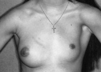
Complete absence of the breast, or amastia, is rare and is thought to occur from lack of formation or obliteration of the milk line. Amastia is usually unilateral and can be congenital or associated with systemic disorders (e.g., malnutrition, Crohn disease) or endocrine disorders (e.g., congenital adrenal hyperplasia, gonadal dysgenesis, hypogonadotropic hypogonadism). It can be associated with anomalies of the underlying mesoderm, such as abnormal pectoralis muscles seen in Poland syndrome (aplasia of the pectoralis muscles, rib deformities, webbed fingers, and radial nerve aplasia) (Fig. 545-1). Amastia or hypomastia can also be iatrogenic, resulting from injuries sustained during thoracotomy, chest tube placement, radiotherapy, severe burns, and inappropriate biopsy of the breast bud. Treatment is surgical correction.
Polymastia and Polythelia
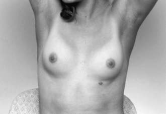
Supernumerary breast tissue (polymastia) and accessory nipples (polythelia) occur in approximately 1-2% of the population (Fig. 545-2). The abnormally placed tissue can be seen anywhere along the milk line but is usually noted on the chest, upper abdomen, or just inferior to the normally positioned breast. An association has been made between polythelia and anomalies of the urinary and cardiovascular system. Surgical excision of the accessory breasts or nipple is not usually needed. Resection of accessory tissue may be warranted if the patient has pain or for cosmetic reasons.
Juvenile or Virginal Hypertrophy
Spontaneous massive growth of the breasts during puberty and adolescence is thought to be the result of excessive end-organ sensitivity to gonadal hormones. The underlying cause, if any, should be determined and removed (Table 545-1). When growth is extreme it is termed macromastia or gigantomastia. It is more commonly bilateral, often occurs over a brief period, and most commonly affects adolescent girls. Physical and psychologic problems can affect posture and quality of life. Strong emotional support should be provided as this can affect an adolescent’s self-esteem at a vulnerable time in her psychologic development. Management should be individualized and may range from reassurance or the use of supportive brassieres to reduction mammoplasty. Surgery should be delayed until late adolescence to allow complete breast development. Medical therapy is available to slow down breast growth in extreme cases until surgery can be performed. Surgical intervention often necessitates relocation of the nipple, which can result in decreased sensation and altered lactation.
Table 545-1 DIFFERENTIAL DIAGNOSIS OF MACROMASTIA
From Desilva N, Brandt M: Breast disorders in children and adolescents. In Sanfilippo J, Lara-Torre E, editors: Clinical pediatric and adolescent gynecology, London, 2008, Taylor & Francis.
Infections
Mastitis is the most common infection of the breast. Though it is most common in lactating mothers, it can occur in young infants and adolescents. Neonatal mastitis is an infection that usually occurs in term or near-term infants. It should be treated aggressively to reduce the risk of forming abscesses. Adolescents can develop nonlactational mastitis or a breast abscess for unknown reasons, as a result of irritation of the skin (through shaving or nipple stimulation), trauma, a foreign body (e.g., piercing), ductal abnormality (such as ductal ectasia), or infection of an epidermal cyst. The initial therapy of all breast infections is antibiotics and analgesics. Staphylococcus aureus (Chapter 174.1) is the offending organism is almost all cases. Owing to the potential for breast abscess, the neonatal population should be treated with parenteral antibiotics guided by gram stain, when available. Adolescents may be initially treated with warm compresses and oral antibiotics. Abscesses should be surgically evaluated (with ultrasound guidance if necessary) and drained as necessary. If incision and drainage is performed, a small, periareolar incision is indicated.
Nipple Discharge
Nipple discharge must be carefully evaluated and a distinction made among galactorrhea (milky white discharge), blood, or other discharge (Table 545-2). A careful history and physical examination directed at the possible etiologies of galactorrhea will help the practitioner determine the etiology. Examination of the discharge assists in diagnosis. Benign conditions are usually associated with a milky, sticky, thick discharge; infection is associated with a purulent discharge; intraductal papilloma and cancer are associated with a serous, serosanguineous, or bloody discharge.
Galactorrhea
To test for galactorrhea, obtain cytologic evaluation of the discharge fluid by Sudan (fat) stain. Serum pregnancy testing and prolactin and thyroid levels are obtained to rule out the presence of a thyroid abnormality, a pituitary prolactinoma, and pregnancy (in the postpubertal adolescent). If the prolactin level is elevated, visual field studies and a head MRI might reveal presence of a pituitary adenoma (Chapter 554). Treatment is directed by results of history, physical exam, and lab studies. Patients should be instructed to avoid nipple stimulation and stop any offending drugs. Hypothyroidism should be treated and prolactin tumors managed with appropriate medical or surgical care. Treatment of galactorrhea (not thyroid related) consists primarily of dopamine agonists such as bromocriptine or cabergoline. Surgical intervention, usually transsphenoidal hypophysectomy, is rarely required.
Breast Masses
A mass in the developing breast can be of concern to the adolescent and her family.
Common Adolescent Breast Masses
The differential diagnosis for breast masses in the adolescent patient is seen in Table 545-3. The patient should be questioned about the variation in symptoms with the menstrual cycle, associated symptoms such as nipple discharge, recent trauma to the breast, family history of breast masses or cancer, and history of chest radiation or malignancy. Because breast cancer in the adolescent is extremely rare, masses can be expectantly managed for extended periods with little concern for malignancy in this population.
Table 545-3 BREAST MASSES IN THE ADOLESCENT GIRL
BENIGN
MALIGNANT
Data from Dehner LP, Hill DA, Deschryver K: Pathology of the breast in children, adolescents, and young adults, Semin Diagn Pathol 16:235–247, 1999; Simmons PS: Diagnostic considerations in breast disorders of children and adolescents, Obstet Gynecol Clin North Am 19:91–102, 1992; and Laufer MR, Goldstein DP: The breast: examination and lesions. In Emans SJ, Laufer MR, Goldstein DP, editors: Pediatric and adolescent gynecology, ed 5, Philadelphia, Lippincott Williams & Wilkins, 2005, pp 729–759.
Malignant Masses
Primary breast cancer is extremely rare in adolescents. Surveillance Epidemiology and End Results (SEER) data from 1975-2006 lists no incident cases of breast cancer in situ in girls and women <20 yr and publishes an incidence of invasive breast cancer of 0.2/100,000 for females up to age 19 (http://seer.cancer.gov/csr/1975_2006/results_merged/sect_04_breast.pdf). Although malignancy is rare, lesions with suspicious imaging findings or progressive growth should undergo cytologic or histologic examination. A small study in Austria noted a 4.7% malignancy rate among palpable masses in teens. Additional research in this area is needed to assess the validity of the findings of the latter study.
American College of Obstetricians and Gynecologists. ACOG Committee opinion no. 350, November 2006: Breast concerns in the adolescent. Obstet Gynecol. 2006;108:1329-1336.
American College of Obstetricians and Gynecologists. Fact sheet: plastic surgery. Tool kit for teen care, ed 2. American College of Obstetricians and Gynecologists, Washington, DC, 2009.
American Society of Plastic Surgeons. Policy statement: breast augmentation in teenagers (PDF), http://www.plasticsurgery.org/d.xml?comp=x983. Accessed April 1, 2010
Centers for Disease Control and Prevention. Idiopathic granulomatous mastitis in Hispanic women—Indiana, 2006-2008. MMWR. 2009;58(47):1317-1321.
Chung EM, Cube R, Hall GJ, et al. From the archives of the AFIP: breast masses in children and adolescents: radiologic-pathologic correlation. Radiographics. 2009;29:907-931.
Dixon JM, Khan LR. Treatment of breast infection. BMJ. 2011;342:484-489.
Greydanus DE, Matytsina L, Gains M. Breast disorders in children and adolescents. Prim Care. 2006;33:455-502.
Henley DV, Lipson N, Korach KS, et al. Prepubertal gynecomastia linked to lavender and tea tree oils. N Engl J Med. 2007;365:479-485.
Laufer MR, Goldstein DP. The breast: examination and lesions. In: Emans SJ, Laufer MR, Goldstein DP, editors. Pediatric and adolescent gynecology. ed 5. Philadelphia: Lippincott Williams & Wilkins; 2005:729-759.
Ries LAG, Melbert D, Krapcho M, et al. SEER Cancer Statistics Review, 1975–2005 (website), http://seer.cancer.gov/csr/1975_2005/. Accessed April 1, 2010
Sadove AM, van Aalst JA. Congenital and acquired pediatric breast anomalies: a review of 20 years’ experience. Plast Reconstr Surg. 2005;115:1039-1050.
Stricker T. Mastitis in early infancy. Acta Paediatrica. 2005;94:166-169.
Tea MM, Asseryanis E, Kroiss R, et al. Surgical breast lesions in adolescent females. Pediatr Surg Int. 2009;25:73-75.
Tiryaki T, Senel E, Hucumenoglu S, et al. Breast fibroadenoma in female adolescents. Saudi Med J. 2007;28:137-138.


