Brain Tumors

Published on 13/03/2015 by admin

Filed under Neurosurgery

Last modified 22/04/2025

Print this page

rate 1 star rate 2 star rate 3 star rate 4 star rate 5 star
Your rating: none, Average: 0 (0 votes)

This article have been viewed 1912 times

CHAPTER 96 Brain Tumors

An Overview of Current Histopathologic Classifications

The current World Health Organization Classification of Tumors of the Central Nervous System, fourth edition (WHO 2007), lists more than 120 types of brain tumors. Classification of brain tumors is an evolving process, with obsolete entities being discarded and newly recognized tumors added with each successive revision. In the past, classification has relied heavily on recognition of morphologic patterns and immunohistochemical identification of differentiation antigens, but with the discovery a decade ago of the association between the translocation and subsequent deletion of chromosomal arms 1p and 19q and the responsiveness of anaplastic oligodendroglioma to treatment, a new era of molecular classification of brain tumors began. Current advances in molecular methodologies, particularly in the fields of genomics, transcriptonomics, and proteomics, have revolutionized brain tumor classification, and although the present classification remains based on morphology, histology is increasingly being complemented by genetic characterization of neoplasms. In this chapter we present an overview of the current histopathologic classification of brain tumors based on WHO 2007 criteria.

Astrocytomas

Brain tumors that exhibit purely astrocytic differentiation can be separated into two major categories: diffuse and circumscribed. Complete surgical resection is rarely attained with diffuse astrocytoma because of the inherently infiltrative nature of the neoplastic cells, which extend well beyond the apparent gross margin of the tumor.

Diffuse Astrocytomas

Diffuse astrocytoma (WHO grade II) is a mildly hypercellular infiltrative neoplasm composed of well-differentiated, spindle- to stellate-shaped astrocytes with minimal nuclear atypia and separated by a loosely fibrillary (and sometimes microcystic) background. Nuclei are characteristically irregular, angulated, and hyperchromatic. Mitotic figures are either absent or rare, and there is no vascular proliferation or necrosis. Glial fibrillary acidic protein (GFAP) may be highly or weakly expressed. Two morphologic subtypes are recognized: protoplasmic and gemistocytic. Protoplasmic astrocytomas are composed of small tumor cells with scant GFAP expression and are frequently associated with a myxoid or microcystic background. Gemistocytic astrocytomas consist of a predominant population of large rounded neoplastic astrocytes with a copious amount of glassy eosinophilic cytoplasm that is strongly GFAP-positive and has an eccentrically located nucleus. Despite their benign morphologic appearance, diffuse astrocytomas have an intrinsic tendency to recur, spread extensively, and undergo anaplastic progression to a higher grade. The latter propensity is especially true of the gemistocytic variant. The time to recurrence and progression after initial clinical evaluation varies from case to case but ranges from months to several years.

Anaplastic astrocytoma (WHO grade III) is a hypercellular glioma composed of poorly differentiated astrocytes consisting of a mixture of pleomorphic fibrillary and gemistocytic cells with significant nuclear atypia and prominent mitotic activity. These malignant tumors may develop as a result of anaplastic progression from a preexisting, low-grade diffuse astrocytoma or may arise de novo. The mean age of patients with anaplastic astrocytoma at initial diagnosis is approximately 41 years, which falls between the age means for patients with low-grade diffuse astrocytoma and glioblastoma. Anaplastic astrocytomas typically show anaplastic progression to glioblastoma after an average of 2 years.

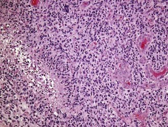
Glioblastoma multiforme (GBM; WHO grade IV) is unfortunately both the most common glioma and the most malignant primary brain tumor arising in adults. Most commonly, GBMs are solitary tumors of the cerebral hemispheres but may develop at almost any site within the neuraxis, including the cerebellum and the spinal cord. In many cases they infiltrate across the corpus callosum or arise directly within it, with bilateral extension (butterfly tumor). Multifocal tumors are observed in about 2% of patients and are often mistaken for metastatic disease on preoperative neuroimaging studies. The necrotic tumor mass may be partially delineated on gross examination, but infiltrating glioma cells can easily be identified microscopically well beyond the apparent gross tumor boundaries. The cellular morphology of GBM cells is highly variable, with the spectrum ranging from small, tightly packed, round or elongated cells to giant bizarre and multinucleated forms, all of which are frequently encountered within a single tumor. Mitotic figures are typically readily identified, and corresponding proliferation marker indices, such as the Ki-67 antigen, show elevated levels. Positive immunostaining for GFAP is characteristic but highly variable and can be absent in some instances. Microvascular proliferation and foci of necrosis are histologic hallmarks of GBM (Fig. 96-1). The presence of necrosis is not required for a diagnosis of GBM; vascular proliferation, in conjunction with pleomorphism and increased mitotic activity, is sufficient according to WHO 2007 criteria.

The typical peripheral, ring-like zone of contrast enhancement seen on computed tomography (CT) and magnetic resonance imaging (MRI) corresponds to the highly vascularized peripheral area of the neoplasm. Vascular proliferation is defined as the presence of blood vessels with multilayered vessel walls (more than two cell layers thick). So-called glomeruloid vascular proliferation constitutes the most obvious and characteristic example of the florid microvascular proliferation seen in GBM.

Two subsets of GBM, primary and secondary, have been described based on clinical and genetic data. Primary GBM arises de novo (i.e., without evidence of a preexisting lower grade astrocytoma), typically in patients in the sixth decade of life and beyond. Epidermal growth factor receptor (EGFR) gene amplification occurs in about 40% of primary GBMs. In contrast, secondary GBM tends to develop in younger adults (<45 years old) by malignant progression from a diffuse astrocytoma of WHO grade II or III. The genetic hallmark of secondary GBM is TP53 mutation. Large areas of necrosis are typical of GBMs, which also frequently exhibit multiple small serpiginous zones of necrosis surrounded by densely crowded tumor cells in a pseudopalisading pattern. The hypoxic perinecrotic apoptotic cells strongly express vascular endothelial growth factor (VEGF), which in turn induces the characteristically prominent vascular proliferation.

Gliomatosis cerebri (WHO grade III) has historically been considered to be a unique glial tumor entity sui generis, but mounting evidence over the last decade, including support from molecular studies, favors conceptualization of gliomatosis cerebri as a pattern of exceptionally diffuse involvement of the central nervous system (CNS) that can be seen with any of the diffuse glioma subtypes, including oligodendroglioma.

Circumscribed Astrocytomas

Pilocytic astrocytoma (WHO grade I) typically arises in children and young adults. Pilocytic astrocytomas are macroscopically circumscribed and grow very slowly. In contrast to diffuse astrocytomas, pilocytic astrocytomas exhibit very little tendency for anaplastic progression. Pilocytic astrocytomas preferentially affect the cerebellum, brainstem, optic nerves, and third ventricular region. The tumor is often manifested as a mural nodule within a cyst. Invasion of the overlying leptomeninges is common, but this feature does not constitute a negative prognostic factor. Microscopically, many pilocytic astrocytomas exhibit a biphasic architectural pattern consisting of compacted areas of elongated, piloid (hair-like) cells alternating with loosely textured and microcystic areas populated by scattered stellate cells. The neoplastic pilocytes of pilocytic astrocytoma are GFAP positive. Rosenthal fibers, which are brightly eosinophilic compacted masses of glial intermediate filaments with entrapped cytosolic proteins and eosinophilic granular bodies (EGBs), are histologic hallmarks of pilocytic astrocytoma, although they are neither a constant nor a pathognomonic feature. Hyalinized and glomeruloid vessels are frequently observed. Vascular proliferation is responsible for the contrast enhancement seen on preoperative MRI studies and can occasionally cause histologic diagnostic confusion with high-grade glioma, particularly in small biopsy samples. In pilocytic astrocytomas, mitotic activity, vascular proliferation, and necrotic foci do not have the same ominous prognostic significance as when present in diffuse astrocytomas. In favorable anatomic locations, such as the cerebellum, surgical resection of pilocytic astrocytoma has the potential to be curative.

Pilomyxoid astrocytoma (WHO grade II) is a newly recognized variant of pilocytic astrocytoma that has been found to be clinically more aggressive than ordinary pilocytic astrocytoma. It occurs most commonly in the hypothalamic/chiasmatic region. Distinctive histologic features include a monomorphic population of neoplastic pilocytes in a prominent myxoid background stroma. Unlike the situation with most pilocytic astrocytomas, in pilomyxoid astrocytoma, Rosenthal fibers and EGBs are either very inconspicuous or completely absent.

Pleomorphic xanthoastrocytoma (PXA; WHO grade II) is another circumscribed variant of astrocytoma that typically arises in a superficial cerebral location in children and young adults with a history of seizures. The temporal lobe is a favored anatomic site. Histologically, PXA mimics GBM in its strikingly pleomorphic tumor cells, but the paucity of mitotic figures and absence of vascular proliferation and necrosis are clues to the correct diagnosis, and the characteristic presence of EGBs, which are also seen in other circumscribed tumors (such as pilocytic astrocytoma and ganglioglioma), is an even stronger signature of PXA. The pleomorphic, giant, and often multinucleated cells may display a variable xanthomatous change in their cytoplasm because of intracellular accumulation of lipids. Most of these pleomorphic cells are GFAP-positive, but neuronal marker proteins can also be detected in some tumors, indicative of dual differentiation. TP53 mutation is not a characteristic of PXA. In addition to prominent EGBs, dense intercellular and pericellular reticulin and perivascular lymphocytic infiltrates are also typical microscopic features of PXA. As with pilocytic astrocytoma, invasion of the overlying meninges is characteristic and does not constitute a negative prognostic factor. PXAs generally exhibit indolent behavior; however, approximately 15% will recur and undergo anaplastic progression to high-grade diffuse astrocytoma.

Subependymal giant cell astrocytoma (WHO grade I) is an intraventricular tumor composed of large cells that resemble gemistocytes or ganglion cells, or both, morphologically and immunohistochemically. This tumor is almost invariably associated with tuberous sclerosis, although this condition is often not known at initial evaluation. Surgical resection is curative.

Desmoplastic infantile astrocytoma (WHO grade I) is a rare tumor of infancy with morphologic and clinical features that overlap those of another rare tumor termed desmoplastic infantile ganglioglioma. Both lesions share a superficial cerebral cortical location, large size, circumscribed growth pattern, and development during infancy. A marked desmoplastic meningeal reaction is characteristic. Surgical resection is the treatment of choice, and the prognosis is generally more favorable than would otherwise be suggested by the typically very large size of most of these tumors at diagnosis.

Oligodendroglial and Oligoastrocytic Glial Tumors

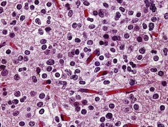
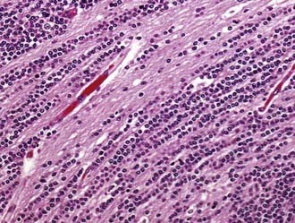
Oligodendroglioma (WHO grade II) is a well-differentiated, diffusely infiltrating tumor composed of cells resembling normal oligodendroglia. Most oligodendrogliomas arise in adults in the fourth and fifth decades. Their preferential location is the white matter of the cerebral hemispheres, from which tumor cells typically infiltrate the overlying cortex. As viewed macroscopically and on neuroimaging studies, oligodendrogliomas often appear somewhat more circumscribed than astrocytomas. They are composed of uniform round cells with cleared cytoplasm surrounding a central spherical nucleus (fried egg appearance). The perinuclear halo is a diagnostically useful fixation artifact present only in formalin-fixed, paraffin-embedded tumor tissue (Fig. 96-2). Mitotic activity is inconspicuous. A branching network of small delicate blood vessels (chicken wire pattern) is a classic histologic feature of many oligodendrogliomas. Microcalcifications are also common. Subpial tumor infiltration, perineuronal satellitosis, and perivascular satellitosis of tumor cells (secondary structures of Scherer) are characteristically seen in oligodendrogliomas that infiltrate gray matter. No oligodendroglioma-specific immunohistochemical markers are currently available. Oligodendrogliomas generally recur locally and ultimately undergo anaplastic progression.

Anaplastic oligodendroglioma (WHO grade III) is an oligodendroglioma in which high-grade features, such as increased mitotic activity, microvascular proliferation, or necrosis (or any combination of such features), are present. Some tumors show florid vascular proliferation and necrosis that mimic what is seen in GBM. Varying numbers of cells with eosinophilic GFAP-positive cytoplasm that resemble miniature gemistocytes (minigemistocytes) or astrocytes (gliofibrillary oligodendrocytes) may be present. The hallmark genetic signature of oligodendroglioma (low grade and anaplastic) is combined whole-arm deletion of chromosomes 1p and 19q, which arises secondary to an initial translocation event and constitutes an independent prognostic marker, with 1p or 19q loss being associated with improved outcome regardless of the specific therapeutic regimen.

Oligoastrocytoma (WHO grade II) and anaplastic oligoastrocytoma (WHO grade III) are currently regarded as mixed diffuse gliomas with astrocytic and oligodendroglial components. There is no consensus on the minimal percentage of each component required for diagnosis of a mixed glioma. By WHO 2007 criteria, the diagnosis is based entirely on morphologic features, as assessed on hematoxylin and eosin–stained tissue sections, without consideration of molecular genetic characteristics, and is very subjective with high interobserver variability. In general, anaplastic oligoastrocytoma exhibits high-grade features such as increased mitotic activity and often microvascular proliferation, but otherwise it has the same subjective diagnostic criteria as oligoastrocytoma. According to WHO 2007 criteria, the presence of tumor necrosis in anaplastic oligoastrocytoma mandates upgrading, and such lesions have been given the designation GBM with an oligodendroglioma component (WHO grade IV). Anaplastic oligoastrocytomas that exhibit deletion of both 1p and 19q are considered to have a favorable genetic signature, whereas those with intact 1p/19q status and p53 immunopositivity are considered genetically closer to astrocytomas.

Ependymal Tumors

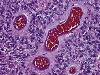
Ependymoma (WHO grade II) is a slowly growing neoplasm of children and young adults that originates from the ependymal lining of the cerebral ventricles. An infratentorial location is the most frequent in children, whereas in adults most of these tumors are supratentorial. Ependymomas may occur outside the ventricular system in the brain parenchyma and also in the spinal cord. Ependymomas are grossly characterized by a sharply demarcated edge. As seen histologically, classic ependymomas are moderately cellular tumors composed of oval cells with monomorphic nuclei and tapering eosinophilic cytoplasm. Some ependymomas have a more glial appearance, whereas others are more epithelioid. Some ependymoma variants (cellular, tanycytic) mimic other primary tumors, although others (papillary, clear cell) may mimic secondary tumors. The histologic hallmarks of ependymoma are the perivascular pseudorosette (perivascular collars of radiating tumor cell cytoplasmic processes) and, more elegantly but less frequent, the true ependymal rosette (tumor cells surrounding a central lumen) (Fig. 96-3). GFAP and epithelial membrane antigen (EMA) immunopositivity is a frequent finding in ependymoma. GFAP reactivity is often strongest in the perivascular pseudorosettes, and EMA positivity takes the form of cytoplasmic dot-like and ring-like staining. Electron microscopy may be required in some cases to identify the ultrastructural features associated with ependymal cell differentiation (intercellular lumina filled with microvilli and sometimes cilia and prominent intercellular junctional complexes).

Anaplastic ependymoma (WHO grade III) typically shows increased mitotic activity and microvascular proliferation.

Myxopapillary ependymoma (WHO grade I) is a distinct low-grade variant of ependymoma that arises almost exclusively from the caudal portion of the spinal cord of adults in the conus medullaris/filum terminale region. The tumor is usually well circumscribed and covered by an outer layer of investing leptomeninges (capsule). Layers of cuboidal or spindled tumor cells surround myxoid microcysts. Collars of myxoid material also surround blood vessels. Surgical resection of intact tumors can be curative in some cases; however, the presence of micrometastases, which are not visible to the unaided eye, or frank capsular breaching by the tumor can lead to diffuse dissemination among the nerve roots of the cauda equina and ultimately result in significant morbidity.

Subependymoma (WHO grade I) is an indolent intraventricular glioma of adults (rare cases arise in the spinal cord). Asymptomatic lesions are often discovered only as incidental findings at autopsy, but subependymomas occasionally produce ventricular obstruction of the lateral or fourth ventricles. Surgical resection is curative. As seen microscopically, the tumor is composed of clusters of small, uniform, benign-appearing tumor cell nuclei separated by extensive cell-free areas of finely fibrillary matrix. Lateral ventricular examples are prone to prominent microcystic change that can obscure the characteristic multinodular architecture. Cases of mixed subependymoma and classic ependymoma are rare but well documented; the clinical course of such tumors is similar to that of WHO grade II ependymoma.

Choroid Plexus Tumors

Choroid plexus papilloma (WHO grade I), atypical choroid plexus papilloma (WHO grade II), and choroid plexus carcinoma (WHO grade III) originate from the epithelium of the choroid plexus of the cerebral ventricles. The lateral ventricles are more often involved in patients younger than 20 years. As viewed both macroscopically and microscopically, choroid plexus papilloma closely recapitulates the papillary architecture of normal choroid plexus, but the tumor cells tend to be more crowded and columnar, as opposed to the cuboidal morphology of normal choroid plexus epithelium. Most intraventricular choroid plexus papillomas are benign and can be cured with surgery, but dissemination via cerebrospinal fluid (CSF) can occur in up to 20% of cases.

In contrast, choroid plexus carcinomas are aggressive tumors that arise more frequently in childhood and show histologically frank features of malignancy, including effacement of the papillary pattern by solid areas of tumor, increased cellularity, nuclear pleomorphism, brisk mitotic activity, necrosis, frequent invasion of the adjacent brain parenchyma, and CSF dissemination. Choroid plexus carcinomas express cytokeratins, but the staining characteristics of S-100 protein, transthyretin, and GFAP, which are usually positive in choroid plexus papilloma, are inconstant and thus less helpful in diagnosing this tumor. Some cases of choroid plexus carcinoma require ultrastructural identification of the characteristic features of choroid plexus differentiation (microvilli, cilia, and intercellular junctional complexes).

Atypical choroid plexus papilloma is a recently codified entity that differs from choroid plexus papilloma in exhibiting increased mitotic activity. Curative surgery is still possible, but the probability of recurrence appears to be significantly higher.

The choroid plexus may also be involved by a variety of other neoplastic and nonneoplastic mass lesions, most prominently intraventricular meningioma, metastatic carcinoma (especially renal cell carcinoma), and xanthogranuloma (which is a reactive mass lesion characterized by cholesterol clefts and attendant multinucleated giant cell reaction).

Other Neuroepithelial Tumors

Chordoid glioma of the third ventricle (WHO grade I) is a unique glial neoplasm of adults, with a highly characteristic anatomic site of origin in the rostral third ventricle. The tumor consists of cords and clusters of strongly GFAP-positive epithelioid cells set in a prominent basophilic myxoid background. Chordoid glioma is hypothesized to originate in association with the specialized ependymal cells of the lamina terminalis circumventricular organ (organum vasculosum of the lamina terminalis). Prominent lymphoplasmacytic infiltrates are highly characteristic. The tumor is well circumscribed, but its anatomic origin, close to the hypothalamic region, often precludes total resection.

Angiocentric glioma (WHO grade I) is a newly identified glial tumor that arises most frequently in the frontoparietal or temporal cortex of children and young adults who have a history of refractory epilepsy. As seen microscopically, angiocentric glioma is characterized by an infiltrating population of monomorphous bipolar cells, a prominently angiocentric growth pattern, and immunoreactivity for EMA (cytoplasmic dot-like pattern), GFAP, S-100 protein, and vimentin. The histogenesis of angiocentric glioma is uncertain, with the differentiation features overlapping those of both astrocytic and ependymal tumors.

Astroblastoma is a rare glioma, viewed as a subtype of ependymoma by some, that primarily arises in children and young adults. This compact tumor exhibits a solid growth pattern, with the GFAP-positive cells forming back-to-back perivascular pseudorosettes that differ from those of typical ependymoma in that the cell processes are broad and stout rather than delicately fibrillary. In fact, astroblastoma typically does not exhibit any fibrillar matrix at all. Areas with prominent vascular mural thickening and hyalinization are very characteristic. Patients with low-grade tumors have a good prognosis after undergoing gross-total resection. The behavior of high-grade tumors is less certain; some, with only increased mitotic activity, may also portend a good patient outcome after resection, whereas others display a range of high-grade features that at the high end, merge with those of GBM, and for these, the diagnosis and prognosis are less confidently determined.

Neuronal and Mixed Neuronal-Glial Tumors

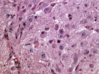
Gangliocytoma (WHO grade I) and ganglioglioma (WHO grade I or III) are well-differentiated tumors composed of neoplastic mature-appearing neurons alone (gangliocytoma) or neoplastic ganglion cells combined with glioma cells (ganglioglioma). They frequently occur in the temporal lobe. Ganglioglioma is the most common tumor associated with chronic temporal lobe epilepsy (40% of tumor-associated temporal lobe epilepsy cases). Both tumors are grossly circumscribed, may be solid or cystic, and frequently contain calcifications. At the microscopic level, gangliocytoma is composed entirely of clusters of dysmorphic mature ganglion cells (Fig. 96-4), whereas the ganglion cells in ganglioglioma are accompanied by glioma elements (usually astrocytoma). The glial component may include cell types resembling pilocytic astrocytoma (with Rosenthal fibers and EGBs), fibrillary astrocytoma, or rarely, oligodendroglioma. In the latter two cases, ganglioglioma has the potential to undergo anaplastic progression. Ganglioglioma must be differentiated from the cortical invasion of diffuse astrocytoma with entrapped neurons. Lymphocytic perivascular infiltrates are common. Immunopositivity for neuronal markers, such as synaptophysin and NeuN, in a subpopulation of tumor cells is characteristic. Another helpful marker is the oncofetal CD34 antigen, which is expressed in the neural component of gangliogliomas but not in normal brain.

Dysplastic gangliocytoma of the cerebellum (Lhermitte-Duclos disease; WHO grade I) is a distinctive clinicopathologic entity that is characterized by a cerebellar location, gross enlargement of the folia on MRI, and a disorganized cerebellar cortical histology in which large ganglion cells predominate. A layer of myelinated axons in the outermost part of the molecular layer just beneath the pia is also a distinctive feature. An association with Cowden syndrome is observed in 50% of patients. Complete surgical resection is curative.

Dysembryoplastic neuroepithelial tumor (DNT; WHO grade I) is a low-grade quasihamartomatous tumor that occurs in children and young patients with a history of long-standing resistant seizures. Neither mass effect nor peritumoral edema is observed on neuroimaging studies. Characteristic features of DNT include a multinodular architecture and a predominantly intracortical location. The temporal lobe is the preferred location, but DNT can arise in any part of the supratentorial cortex. The rostral septum pellucidum/head of the caudate nucleus/frontal horn of the lateral ventricle region is an additional rare but well-recognized site of occurrence. The specific glioneuronal element is considered the histologic hallmark of DNT and consists of prominent clusters of oligodendroglial-like cells with interspersed neurons that often appear to float in the cystic spaces of the loose background stroma. DNT closely mimics low-grade oligodendroglioma. Foci of cortical dysplasia may be identified in the adjacent peritumoral cortex. Resection is curative, and even partial resection usually stops the seizure activity.

Central neurocytoma and extraventricular neurocytoma (WHO grade II) are low-grade neoplasms of young adults, composed of remarkably monomorphous round cells with immunohistochemical and ultrastructural evidence of neuronal differentiation. Neurocytomas are typically located in the lateral ventricles or third ventricle, or both, with an attachment to the septum pellucidum. Neoplasms with similar histopathologic characteristics and biologic behavior occur outside the ventricular system. Diffuse positivity for synaptophysin is the rule. Surgery can be curative with small lesions, but local recurrence results with partially resected tumors. Increased mitotic activity and vascular proliferation may rarely be seen but are not generally associated with a poor prognosis.

Atypical neurocytomas are defined by an increased Ki-67 antigen (MIB-1 antibody) labeling index of greater than 2%, and these tumors have an increased likelihood of recurrence. Rare tumors consisting of neurocytic cells with focal areas of lipomatous differentiation resembling mature adipose tissue typically occur in the cerebellum of adults and are designated cerebellar liponeurocytoma (WHO grade II).

Paraganglioma of the filum terminale (WHO grade I) is an uncommon neuroendocrine tumor that arises from the conus medullaris/filum terminale region. As seen histologically, paragangliomas of the filum terminale can mimic ependymoma, with perivascular pseudorosette formation. Strong tumor cell positivity is seen for neuronal markers such as synaptophysin. Most tumors are encapsulated by an investing layer of leptomeninges and may be cured by total excision.

Papillary glioneuronal tumor (WHO grade I) and rosette-forming glioneuronal tumor of the fourth ventricle (WHO grade I) are both rare mixed neoplasms that have recently been codified as new entities by the WHO. Papillary glioneuronal tumor is a supratentorial lesion (often temporal) histologically characterized by pseudopapillary structures of cuboidal glial cells surrounding hyalinized vessels, with the intervening zones filled with neurocytic elements. The cells in direct contact with the vessels are GFAP-positive, whereas cells without vascular association stain for synaptophysin.

Rosette-forming glioneuronal tumor of the fourth ventricle is a tumor of children and young adults. The neuronal component consists of neurocytes that form rosettes with eosinophilic, synaptophysin-positive avascular cores, and the glial component typically exhibits features of pilocytic astrocytoma. Both papillary glioneuronal tumor and rosette-forming glioneuronal tumor of the fourth ventricle are clinically indolent and surgically curable.

Pineal Region Tumors

Pineocytoma (WHO grade I) is a circumscribed slow-growing neoplasm of the pineal area that develops mainly in young adults. The tumor is composed of well-differentiated, uniform, mature-appearing pineocytes arranged in lobules and often forming large rosettes with solid fibrillar cores (pineocytomatous rosettes). Approximately 20% of pineal parenchymal tumors exhibit higher cellularity, nuclear atypia, occasional mitoses, and absence of pineocytomatous rosettes. These tumors are potentially aggressive and have been designated pineal parenchymal tumors of intermediate differentiation (WHO grade II or III).

Pineoblastoma (WHO grade IV) is a highly malignant primitive neuroectodermal tumor (PNET) with a predilection to occur during childhood. Flexner-Wintersteiner rosettes (small rosettes with a central lumen) or fleurettes, which are indicative of retinoblastic differentiation, may be seen. Pineoblastomas are immunoreactive for neuronal markers such as synaptophysin and retinal S-antigen. Like other embryonal neoplasms, pineoblastoma has a tendency to metastasize via CSF.

Papillary tumor of the pineal region (WHO II or III), a recently described rare tumor of the pineal region of children and adults, is an epithelial-appearing tumor with papillary features. Papillary tumors of the pineal region are large, well-demarcated tumors. Mitotic activity is highly variable from case to case. Tumor cells are positive for cytokeratins, EMA, neuronal markers, and GFAP. As viewed ultrastructurally, tumor cells exhibit features of ependymal differentiation. Local recurrence is common, but because of the rarity of the lesion, the biologic behavior and histologic grading of papillary tumor of the pineal region remain to be defined.

Germ Cell Tumors

Germinoma of the CNS (WHO grade III) has a proclivity to affect midline structures, particularly the pineal gland and third ventricular region. Germinoma characteristically exhibits a biphasic cell population, very large malignant cells resembling primitive germ cells and small reactive lymphocytes. A prominent granulomatous response is occasionally present and, in such cases, can overshadow the tumor cell component. Germinoma cells are strongly immunopositive for c-kit and OCT4 and usually positive, albeit sometimes very weakly, for placental alkaline phosphatase. In some cases, β-human chorionic gonadotropin immunostaining identifies the presence of isolated syncytiotrophoblastic cells. Any positive marker may be clinically useful for monitoring response to treatment and tumor recurrence through measurement of the specific marker or markers in serum. Pure germinoma is highly radiosensitive. Patients may be treated with radiation therapy, chemotherapy, or a combination of both.

Teratoma (mature and immature), yolk sac tumor, embryonal carcinoma, choriocarcinoma, and mixed germ cell tumor are histologically similar to their systemic counterparts and usually arise in midline structures of the CNS in a manner similar to germinoma, but they are more prevalent at sites other than the pineal region, suprasellar region, and basal ganglia. Accurate histologic subtyping of these tumors is critical for planning treatment and predicting outcome. After germinoma, teratoma is the most common member of this group to occur as a pure (nonmixed) tumor. The remaining germ cell tumors are most frequently encountered as mixed forms. The prognosis of nongerminomatous germ cell tumors is generally poorer than that for germinoma and largely dependent on the extent of surgical resection.

Embryonal Tumors

Medulloblastoma (MDB; WHO grade IV) is the most common intracranial embryonal tumor and by definition arises exclusively in the cerebellum. The tumor develops primarily in children, with a peak incidence at 7 years, but there is a second incidence peak in adulthood in the 21- to 40-year-old age group. Most childhood MDBs arise in the midline (vermis), whereas involvement of the cerebellar hemispheres is more frequent in adults. One third of patients are initially found to have leptomeningeal dissemination. The gross appearance of most MDBs is as a relatively circumscribed mass. As seen microscopically, an MDB is composed of sheets of densely packed malignant small cells with round to oval or carrot-shaped nuclei and scant cytoplasm. Neuroblastic (Homer-Wright) rosettes are typical, but they are present in no more than 40% of cases. Mitotic figures are usually abundant. Vascular proliferation and necrosis with pseudopalisading are uncommon.

A nodular pattern superficially resembling lymph node tissue, with reticulin-free neurocytic islands (pale islands) surrounded by densely packed pleomorphic cells producing abundant reticulin fibers, is observed in some cases and is designated the desmoplastic/nodular variant of MDB. This variant arises predominantly in the cerebellar hemispheres of adult patients and has a more favorable prognosis than classic MDB.

A closely related variant is the recently described MDB with extensive nodularity, which is rare, is seen only in infants, and displays a unique multinodular appearance that is apparent on preoperative imaging studies. As seen microscopically, this variant is characterized by large lobules of neuropil-like tissue with prominent streaming of neoplastic neurocytes and little or no extranodular tissue (Fig. 96-5).

Anaplastic MDB and large cell MDB are two aggressive MDB variants with overlapping morphologic features. Anaplastic MDB is characterized by marked nuclear pleomorphism, nuclear molding, and cell-cell wrapping. In contrast, the large cell variant is composed of a more monomorphous population of large cells with round vesicular nuclei and prominent nucleoli. Both variants exhibit an increased mitotic rate and abundant apoptosis. The most common type of differentiation in MDB, morphologically and immunohistochemically, is neuronal (immunopositivity for synaptophysin and other neuronal markers). In addition, any MDB variant can display focal glial differentiation (GFAP immunopositivity in bona fide neoplastic cells), myogenic differentiation (in a subtype previously termed medullomyoblastoma, as demonstrated by desmin, myoglobin, and myosin immunopositivity), or melanotic differentiation (in a subtype previously termed melanocytic MDB). In contrast to the atypical teratoid/rhabdoid tumor, which is another malignant neoplasm that is sometimes mistaken for MDB, nuclear INI1 gene expression is retained in all MDB variants. Prognostic indicators of poor clinical outcome include age younger than 3 years, CSF dissemination at diagnosis, partial surgical resection, large cell or anaplastic variant morphology, and the presence of amplification of MYC.

CNS primitive neuroectodermal tumors (CNS supratentorial PNET; WHO grade IV) are a heterogeneous group of rare embryonal neoplasms that may occur at any extracerebellar site of the CNS and are composed of undifferentiated or poorly differentiated neuroepithelial cells that may display divergent differentiation along neuronal, astrocytic, ependymal, muscular, or melanocytic lines.

CNS supratentorial PNETs with exclusively neuronal differentiation are termed cerebral neuroblastoma or ganglioneuroblastoma, depending on the degree of ganglion cell differentiation present.

Medulloepithelioma is the designation reserved for tumors that recapitulate the features of the primitive neural tube. PNETs that exhibit exclusively ependymal differentiation in the form of profuse numbers of ependymoblastic rosettes are termed ependymoblastoma. All subtypes of CNS supratentorial PNETs arise primarily in children and adolescents and display aggressive clinical behavior. CSF dissemination is present in up to a third of patients, and extraneural metastases may also occur.

Embryonal tumor with abundant neuropil and true rosettes is an unusual PNET associated with a poor prognosis and characterized histologically by prominent well-formed true rosettes within a neuropil-like background. CNS supratentorial PNETs are unrelated genetically and pathobiologically to peripheral PNETs, which arise in peripheral nerves, soft tissues, or bone.

An additional recently characterized highly malignant embryonal CNS tumor briefly mentioned earlier is atypical teratoid/rhabdoid tumor. Atypical teratoid/rhabdoid tumor is a rare neoplasm of childhood characterized by a unique combination of rhabdoid and primitive neuroectodermal cells, and it can show divergent differentiation along epithelial, mesenchymal, neuronal, or glial lines (or any combination of such lines). The posterior fossa is the most frequent location for atypical teratoid/rhabdoid tumor (75% of cases), followed by the supratentorial compartment (25% of cases). The hallmark rhabdoid cells, which exhibit eccentrically located nuclei and abundant eosinophilic cytoplasm, may rarely fill the entire tumor (CNS rhabdoid tumor), but they more often constitute only one component of a highly pleomorphic neoplasm; in some cases they may be very inconspicuous. Loss of immunohistochemical staining for the INI1 protein is diagnostic of atypical teratoid/rhabdoid tumor. Loss of INI1 nuclear staining is associated with inactivation of the INI1/hSNF5/SMARCB1 tumor suppressor gene (often referred to simply as INI1). Pure rhabdoid tumors outside the CNS, such as those arising in the kidney, share the same genetic alteration as atypical teratoid/rhabdoid tumor. However, the so-called composite rhabdoid tumors, in which rhabdoid morphology is present in other tumor types, do not have INI1 alterations. Germline mutations of INI1 result in rhabdoid tumor predisposition syndrome, in which affected patients are predisposed to the development of CNS and systemic rhabdoid tumors in infancy.

Tumors of the Meninges

Meningiomas (WHO grade I) are slowly enlarging neoplasms that originate from meningothelial (arachnoidal) cells and are thus typically firmly attached to the inner surface of the dura. The meninges covering the cerebral convexities, the falx cerebri, and the skull base are the most frequent sites of origin. Meningiomas may originate in any location where arachnoidal cells are present, including the choroid plexus (intraventricular meningioma). Meningiomas are common tumors and account for an estimated 13% to 26% of primary intracranial neoplasms. They arise most frequently in women in the middle decades of life. Most meningiomas are well-defined, lobulated, firm masses that compress the underlying brain. Gross-total resection is usually curative. Invasion of brain parenchyma is associated with a greater likelihood of recurrence. In contrast, invasion through the dura into the overlying cranial bone is quite common in low-grade (WHO grade I) meningioma.

As seen microscopically, meningiomas can exhibit multiple morphologic patterns. There are nine benign morphologic subtypes that have no associated prognostic significance and four variants that are characterized by more aggressive clinical behavior. The meningothelial, transitional, and fibrous subtypes are most common in low-grade meningioma. Intranuclear pseudoinclusions, nuclear grooves, whorl formation, and psammoma bodies are classic features of meningioma, but they may not be present and are not required for diagnosis. Meningiomas show positive immunostaining for EMA. Occasionally, electron microscopy may be required to identify the characteristic complex interdigitating cell processes and profuse numbers of desmosomes that are present in all morphologic subtypes of meningioma. Based on their histopathologic features and the correlation with the tendency of these lesions to recur and pursue an aggressive clinical course, meningiomas are subdivided into three grades: benign (WHO grade I), atypical (WHO grade II), and anaplastic (WHO grade III).

Any of the benign meningioma subtypes that show either increased mitotic activity (four or more mitoses per 10 high-power microscope fields) or a minimum of three of five specific morphologic features (increased cellularity, small cell formation, prominent nucleoli, sheet-like growth, micronecrosis) warrant upgrading to atypical meningioma (WHO grade II). In addition, two histologic subtypes, the clear cell and chordoid variants, are associated with a more aggressive course and also correspond to WHO grade II. Brain-invasive meningiomas are equivalent to atypical meningiomas in terms of a greater likelihood of recurrence and, accordingly, are classified as WHO grade II.

Anaplastic (malignant) meningiomas (WHO grade III) either have a mitotic rate of 20 or more mitoses per 10 high-power microscope fields or exhibit histologic features of frank malignancy (resembling sarcoma or carcinoma). The papillary and rhabdoid variants characteristically follow the most aggressive clinical course of all of the morphologic subtypes and are thus classified as WHO grade III lesions.

Solitary fibrous tumor and meningeal hemangiopericytoma form a spectrum of mesenchymal nonmeningothelial meningeal tumors referred to as the solitary fibrous tumor/hemangiopericytoma family, with solitary fibrous tumor on the benign end of the spectrum and hemangiopericytoma on the malignant end. Both may resemble fibrous meningioma. Solitary fibrous tumors are typically paucicellular lesions with spindle cells dispersed between prominent eosinophilic bands of collagen. They display strong, diffuse immunoreactivity for CD34 (and, in contrast to meningioma, lack of reactivity for EMA).

Meningeal hemangiopericytomas (WHO grade II or III) are highly cellular, monotonous tumors with a characteristic branching staghorn vascular pattern and dense reticulin deposition that surrounds individual tumor cells (pericellular reticulin). Mitotic activity in meningeal hemangiopericytoma is prominent. Invasion of adjacent brain tissue and bone is commonly seen. Meningeal hemangiopericytomas are classified as grade III tumors (anaplastic hemangiopericytoma) according to WHO 2007 criteria if they exhibit five or more mitoses per 10 high-power microscope fields or necrosis, or both, plus at least two of the following: increased cellularity, high nuclear atypia, and hemorrhage. Meningeal hemangiopericytomas are immunopositive for vimentin, CD34 (typically patchy rather than diffuse as in solitary fibrous tumor), factor XIIIa, and Leu-7 and (as with their low-grade solitary fibrous tumor counterparts) are usually negative for EMA. Recurrence is not unusual, even after ostensible gross total resection followed by irradiation of the tumor bed.

Meningioangiomatosis is another rare meningeal lesion that is not dura-based. It is characterized by a plaque-like proliferation of mesenchymal cells in the subarachnoid space that penetrate the underlying cerebral cortex as perivascular cuffs surrounding blood vessels. The immunophenotype of the tumor cells varies from fibroblastic to meningothelial. Meningioangiomatosis typically arises in childhood as a seizure disorder. The disease may be sporadic or associated with neurofibromatosis type 2. The main differential consideration is brain-invasive meningioma with a perivascular pattern of dissemination. Instances of meningioma associated with meningioangiomatosis have also been reported.

Hemangioblastoma (WHO grade I) is a benign tumor of uncertain histogenesis that commonly affects young adults and consists of neoplastic vacuolated stromal cells amid a dense capillary vasculature. Stromal cells are negative for EMA and other epithelial antibodies. This immunohistochemical profile helps in differentiating hemangioblastoma from metastatic renal cell carcinoma. Hemangioblastoma is commonly sporadic, but in 25% of cases it is associated with von Hippel-Lindau disease. Hemangioblastomas produce erythropoietin, which may cause secondary polycythemia. Hemangioblastoma is one of a number of CNS tumors that frequently appears on preoperative neuroimaging studies as a circumscribed cyst with a mural nodule and that can potentially be cured by surgical resection alone.

Tumors of the Sellar Region (Excluding Pituitary Adenoma)

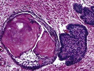
Craniopharyngioma (WHO grade I) is an epithelial tumor of the sellar and suprasellar regions, presumably derived from Rathke pouch remnants, that occurs predominantly in children and young adults. Two morphologic subtypes are distinguished: adamantinomatous, which is the most common, and papillary. The adamantinomatous subtype consists of cords and broad strands of squamous epithelium with peripheral palisading of basal cell nuclei, loosely adhesive areas of epithelium (stellate reticulum), and plump nodules of wet keratin that are prone to calcify (Fig. 96-6). Additional findings are cholesterol clefts, macrophages, and cyst formation. The cystic cavities characteristically contain a dark, viscous fluid that has been likened to machinery oil. Pilocytic astrocytosis with prominent Rosenthal fiber formation is frequently found in the compressed neuropil of the surrounding brain parenchyma. The papillary variant occurs only in adults, is usually well circumscribed, and is exclusively composed of nonkeratinizing well-differentiated squamous epithelium. The extent of surgical resection is the most significant factor associated with survival of patients with craniopharyngioma. Residual tumor will recur.

Granular cell tumor of the neurohypophysis (WHO grade I) is a rare tumor composed of large cells with granular, eosinophilic cytoplasm that arises in the neurohypophysis or infundibulum. The tumor may clinically simulate a nonfunctional adenoma of the anterior pituitary. Cytoplasmic granules are periodic acid–Schiff positive and ultrastructurally correspond to lysosomes and autophagic vacuoles. Granular cell tumors of the neurohypophysis are usually well circumscribed and may be cured by surgical resection.

Pituicytoma (WHO grade I) is a rare, low-grade, spindle cell glial tumor of adults that originates in the neurohypophysis or infundibulum, probably from the specialized glial cells of the region. Pituicytomas show a compact architecture with interlacing fascicles of bipolar glial cells and no Rosenthal fibers or EGBs, which is important for the differential diagnosis with pilocytic astrocytoma. Surgical resection is curative.

Spindle cell oncocytoma of the adenohypophysis (WHO grade I) is another relatively recently recognized entity of the sellar region that is defined as an oncocytic, nonendocrine tumor of the adenohypophysis. It may be macroscopically indistinguishable from a nonfunctioning pituitary adenoma. As viewed ultrastructurally, the cytoplasm of cells of spindle cell oncocytoma of the adenohypophysis is filled with mitochondria. Desmosomes and other intercellular junctions are seen, but secretory granules are lacking.

Lymphoma

Both primary and secondary lymphomas may involve the CNS. Primary lymphoma of the CNS is a tumor of adults that has shown an increase in incidence over the last several decades in both immunocompromised and older immunocompetent patients. Primary lymphomas of the CNS may vary widely in MRI appearance, including having a superficial cortical or deep periventricular location, appearing as solitary or multiple lesions, or mimicking GBM as a butterfly lesion of the corpus callosum.

In contrast, secondary lymphomas preferentially involve the leptomeninges. Stereotactic biopsy is currently the method of choice to establish the histologic diagnosis because surgical resection has not been demonstrated to be of benefit. As viewed histologically, primary lymphomas of the CNS are composed of noncohesive neoplastic lymphocytes that diffusely infiltrate the neural parenchyma and show a characteristically prominent vascular mural infiltration. Most primary lymphomas of the CNS are of the large cell B-cell type and express CD20 and other pan–B cell markers such as CD79a and PAX5. A distinctive variant of systemic lymphoma with a tendency to involve the CNS is angiotropic lymphoma (intravascular lymphoma), which is also predominantly of B-cell lineage. Primary lymphoma of the CNS is highly sensitive to steroid administration, often decreasing dramatically in size on interval imaging studies, but the response is temporary. Steroid therapy can make histologic diagnosis of primary lymphoma of the CNS extremely difficult, with posttreatment biopsies often yielding only gliosis and inflammatory reaction. With current therapeutic regimens of radiotherapy and chemotherapy, the median survival rate is as high as 70% at 2 years and can reach 45% at 5 years in immunocompetent patients. Patients with acquired immunodeficiency syndrome do worse, with a median survival time of approximately 13.5 months with multimodal therapy.

T-cell lymphomas rarely involve the CNS. Low-grade B-cell lymphomas of the mucosa-associated lymphoid type occasionally involve the dura.

Metastatic Tumors of the Central Nervous System

Brain metastases are most often located at the gray-white matter junction of the cerebral and cerebellar hemispheres, but they may affect any part of the CNS. The most common primary sites of origin are the lung, breast, skin (melanoma), kidney, and gastrointestinal tract. Metastasis to the brain may be the initially detected feature of the primary cancer. Carcinomas of the gastrointestinal tract, breast, prostate, and uterus often produce solitary metastasis, whereas multiple brain metastases are frequently associated with cancers arising in the lung, from melanoma, and when the primary cancer site is unknown.

Immunohistochemical markers such as cytokeratins, c-kit (CD117), HMB-45, Melan-A, EMA, hormonal receptors, CD10, and CD56, among many others, may be of assistance in evaluating tumors from an unknown primary and resolving the differential diagnosis problem of whether the lesion is metastatic or a primary brain neoplasm. Epidural metastases are often associated with metastases to the skull from primaries in the breast, prostate, lung, or kidney in adults and from neuroblastoma in children. Breast cancer is frequently the origin of isolated dural metastasis.

Cranial leptomeningeal metastatic infiltration may occur alone or in conjunction with parenchymal metastasis. In most cases, the primary site is well established at the time of subarachnoid tumor invasion. Adenocarcinomas (of the lung, breast, and stomach), hematopoietic neoplasms, and melanomas are the most common primary sources. Single tumor cells may be identified in CSF. Leptomeningeal dissemination can also occur in advanced disease secondary to a primary CNS neoplasm, such as MDB or glioma.

Molecular Classification of Brain Tumors

Classification of brain tumors by microscopy has a long history, and the clinical utility of these time-tested classification systems cannot be overemphasized. However, a major paradigm shift occurred in 1998 with the reported association of deletions involving chromosomes 1 and 19 indicating chemosensitivity and a better prognosis of anaplastic oligodendroglioma. The impact of molecular and genomic approaches to tumor diagnosis and classification has since been ascendant. The development of a wide variety of molecular techniques, including high-throughput genomic technologies, has permitted better understanding of the molecular oncogenesis of brain neoplasms and has improved traditional morphologic diagnosis in tandem with the development of new treatment options such as gene therapy and targeted therapeutics.

Examples within the sphere of neuro-oncology include the assessment of INI1 gene deletion/mutation in atypical teratoid/rhabdoid tumors, O6-methylguanine-DNA methyltransferase (MGMT) gene silencing in GBM, and, most prominently, deletion status evaluation of chromosomal arms 1p and 19q in oligodendroglial tumors.

Two approaches have proved fruitful in preliminary attempts at molecular classification: (1) patient stratification based on the assay of a small number of molecular markers and (2) molecular stratification based on transcriptome profiling, comparative genomic hybridization, or proteomic profiling (or any combination of the three). Contemporary basic and translational molecular biologic research has yielded a number of molecular marker assays with proven diagnostic, prognostic, or therapeutic significance in pediatric and adult brain tumors. Examples of molecular advances that can improve patient stratification for prognosis and treatment are assessment of the deletion status of 1p and 19q in oligodendroglial tumors, MGMT status in GBM, coexpression of EGFR deletion mutant variant III (EGFRvIII) with the tumor suppressor protein PTEN in high-grade gliomas, and INI1 gene inactivation in embryonal and rhabdoid tumors.

The first and most salient of these is deletion testing for markers on chromosomes 1p and 19q in oligodendrogliomas. Combined deletion of chromosomes 1p and 19q, which is mediated by the translocation t(1;19)(q10;p10), is associated with improved prognosis and responsiveness to therapy in adult patients with anaplastic oligodendroglioma and with superior overall survival and progression-free survival in low-grade gliomas, especially gliomas with an oligodendroglial component. Assay of a few additional molecular markers, such as TP53 mutation, can be used to further refine molecular substratification of anaplastic oligodendroglial tumors. Currently, 1p/19q deletion analysis is routinely being used in many reference institutions.

GBM is highly resistant to therapy, largely because of its invasive properties, which preclude surgical cure; the presence of a population of neural stem cell–like tumor cells, that may harbor resistance mechanisms distinct from those of most non-neural stem cell–like tumor cells; and retention of abundant DNA repair mechanisms, that abrogate the effectiveness of chemotherapy and radiotherapy. In 2005, a randomized phase III trial of temozolomide and radiation therapy in patients with newly diagnosed GBM determined that patients whose tumors exhibited methylation of the MGMT gene promoter survived longer after treatment than did those without evidence of promoter methylation. MGMT is a ubiquitous cellular enzyme largely responsible for resistance to the cytotoxic action of DNA alkylating agents such as temozolomide, carmustine, lomustine, and procarbazine. Inactivation of MGMT impairs the ability of the cell to repair DNA damage. To date, MGMT promoter methylation status is the single most important prognostic marker for predicting benefit from chemotherapy with alkylating agents. However, the most frequently used assay technique, methylation-specific polymerase chain reaction (MS-PCR), is technically problematic because of the high dependence on tissue quality and quantity, the specificity of the primers selected, the adequacy of bisulfite treatment, and PCR conditions. Several new therapeutic agents directed against VEGF (or the VEGF receptor), the EGFR, the platelet-derived growth factor receptor, and integrins are in clinical trials for GBM. Testing of single, specific, small-molecule inhibitors of signaling pathways initially proved less effective than expected. However, combined testing for both EGFRvIII and PTEN identifies a subset of patients responsive to the EGFR kinase inhibitors, hence suggesting that combinations of molecular alterations common to GBM may need to be targeted for more effective treatments and that treatment may need to be tailored to the individual tumor. GBMs are among the most angiogenic tumors in the body, and VEGF thus represents a particularly attractive target. Recent studies incorporating anti-VEGF agents have achieved improvements in treatment response and survival in patients with recurrent malignant glioma.

Quantitative information on DNA alterations, mRNA levels, or protein composition, obtained through molecular genomic and transcriptonomic techniques, can potentially provide data useful for tumor molecular classification and novel tumor class discovery. Transcriptome profiling and array comparative genomic hybridization have been used to generate brain tumor molecular classifications, especially for diffuse gliomas. Some conclusions can be drawn from the collective experience accumulated to date: (1) molecular classification can separate tumor types and grades as well as and often better than histopathologic classification, (2) gene expression profiling can identify subgroups within histologic tumor types that are not identifiable by morphologic or immunophenotypic evaluation, and (3) nosologic groups identified by expression profiling have prognostic significance with respect to patient survival. Genes that are found to be highly expressed in tumor tissues might be good candidates for molecular diagnostics or therapeutics. Small sets of only a few genes can constitute strong classifiers and can be used to stratify tumors for various ends, such as prognosis, susceptibility, or resistance to specific therapies, through the use of simple chip sets or, with greater ease, less cost, and broader availability, by using immunohistochemistry. In summary, advances in the molecular classification of brain tumors have already had a major impact and are certain to only increase in importance in the near future.