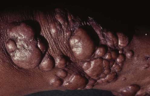
Blastomycosis

(From Lebwohl, M.G., 2003. The Skin and Systemic Disease: A Color Atlas, second ed. Churchill Livingstone, with permission from Elsevier.)
Second-line therapies
Guidelines
Clinical practice guidelines for the management of blastomycosis: 2008 update by the Infectious Diseases Society of America.
 Itraconazole is the drug of choice for the treatment of mild to moderate pulmonary or disseminated infection. The recommended dose is an initial loading dose of 200 mg three times daily for 3 days, followed by 200 mg once or twice daily for 6 to 12 months. Serum levels should be measured after 2 weeks to ensure adequate drug exposure.
 Amphotericin B is the treatment of choice for those with severe pulmonary or disseminated infection, and in the immunocompromised. Either the lipid formulation (3–5 mg/kg/day) or amphotericin deoxycholate (0.7–1 mg/kg/day) is given for 1 to 2 weeks or until improvement is noted. Itraconazole is then recommended as step-down therapy (loading dose and then 200 mg twice daily for 6 to 12 months except in the immunocompromised who require at least 12 months of treatment or until immunosuppression has been reversed).
 Liposomal amphotericin B at a dose of 5 mg/kg/day is the recommended treatment for CNS infection. Treatment should be given for 4 to 6 weeks followed by an oral azole (fluconazole 800 mg daily/itraconazole 200 mg two to three times daily/voriconazole 200–400 mg twice daily), which should be given for at least 12 months and until there is resolution of CSF abnormalities.
 Amphotericin B is the drug of choice for patients who have failed to respond to azole treatment.
 Amphotericin B is the only drug approved for the treatment of blastomycosis in pregnant women.


 Itraconazole
 Amphotericin B
 Ketoconazole
 Fluconazole
 Voriconazole
 Caspofungin