Bioterrorism
Smallpox
Specific investigations


Seward JF, Galil K, Damon I, Norton SA, Rotz L, Schmid S, et al. Clin Infect Dis 2004; 39; 1477–83.
Diagnostic criteria and a CDC algorithm to evaluate and manage suspected cases of smallpox. Three categories (i.e., low, moderate, or high risk of actually having smallpox) dictate subsequent diagnostic strategies. Specific variola laboratory testing is reserved for high-risk persons. An interactive version of the algorithm is available online at http://www.bt.cdc.gov/agent/smallpox/diagnosis/riskalgorithm/index.asp.
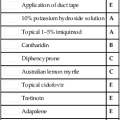
First-line therapies


Anthrax
Specific investigations
 Culture and Gram stain of tissue, blood, or other fluids

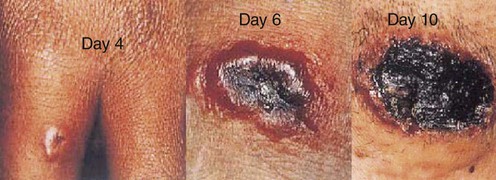
 Punch biopsy fixed in formalin from a papule or vesicular lesion and including adjacent skin
 Biopsies should be taken from both vesicle and eschar, if present
 A photograph, digital image, or diagram indicating the site of each biopsy in relation to the lesion