Published on 19/03/2015 by admin
Filed under Dermatology
Last modified 22/04/2025
This article have been viewed 2444 times
James M. Spencer and Brooke M. Walls
Evidence Levels: A Double-blind study B Clinical trial ≥ 20 subjects C Clinical trial < 20 subjects D Series ≥ 5 subjects E Anecdotal case reports
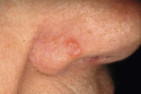
Basal cell carcinoma (BCC) is a slow-growing malignancy originating in the epidermis. It most commonly arises in areas chronically exposed to UV light, especially the head and neck. Although it is very rare for BCC to metastasize, it can produce significant local tissue destruction, including cartilage and bony invasion.
Basal cell carcinoma slowly but relentlessly grows larger and deeper, and therefore therapeutic intervention is geared towards complete eradication of all malignant cells. Local recurrence is the consequence of inadequate therapy. Complete eradication is especially important because recurrent tumors are often larger and more aggressive than the original, incompletely treated primary tumor. Although complete eradication is the primary goal, the therapy chosen should achieve this with the maximal preservation of function and the optimal cosmetic result. Most often, therapy uses destructive techniques such as cryotherapy or curettage and electrodesiccation (C&D); more complex tumors may be treated by excisional surgery, Mohs surgery, or radiation therapy. The decision about which therapy to use is best made by considering four factors: tumor size; location; histology; and history (recurrent vs primary). When assessing a tumor, the clinician may wish to consider each of these four factors and decide whether the patient is high risk or low risk for each, to determine whether to use a simple or complex therapeutic strategy.
Most BCCs are discovered as primary tumors when they are still less than 1 cm in diameter. Generally tumors smaller than 1 cm on the face and 2 cm on the body are low risk.
Histologic growth pattern is a separate risk factor. The cytology of BCC does not vary: that is, all BCCs have well-differentiated, relatively monomorphic cell populations, and these tumors are not graded the way other malignancies are. However, the pattern of growth is variable and makes a large difference in choosing therapy. One must consider whether the tumor has a circumscribed or a diffuse growth pattern. Basal cell carcinoma most typically exhibits a circumscribed, cohesive growth pattern known as nodular. Nodular BCCs may show partial differentiation towards other structures, such as cystic or keratotic, but these variants are without therapeutic significance because the growth pattern is still nodular. Morpheaform, micronodular, infiltrating and superficial BCCs are all variants that exhibit a diffuse growth pattern. These lesions are more likely to recur as a result of subclinical extension or more aggressive tumor behavior, or both. Unfortunately, all too often biopsy reports come back to the clinician and simply state ‘BCC’, with no information about the growth pattern. Inadequately treated nodular BCC often recurs with a more aggressive diffuse growth pattern, such as infiltrating or micronodular.
Location is also an important variable to consider when choosing which therapy to use. Basal cell carcinoma tends to occur in chronically sun-exposed sites, especially the head and neck. Approximately 80% occur on the head and neck, and fully 25% occur on the nose. The central portion of the face, which has the highest incidence of BCC, contains the eyes, nose, and mouth, structures of functional and cosmetic importance highly vulnerable to the destructive effects of BCC. These same structures are also highly vulnerable to the destructive effects of therapy directed against BCC. The center of the face extending onto the area around the ears defines a roughly H-shaped area known as the H zone. Tumors in this zone have the highest recurrence rate and thus deserve special therapeutic attention. This zone also contains the most vulnerable structures and has the highest rate of BCC occurrence. Tumors near the ear canal, in the H zone, are of special concern. Extension down the ear canal provides the tumor with access to the brain and other intracranial structures, and when there is evidence of ear canal invasion particularly aggressive therapy is warranted.
Lastly, tumor history is important to consider. Recurrent tumors are more difficult to treat than primary tumors and require more aggressive methods.
When confronted with a BCC, the clinician may wish to consider these four variables in the context of the individual patient. The patient’s overall medical status, medical history, and age may influence the therapeutic decision making.
Biopsy with adequate dermal component
An adequate biopsy is critical in assessing the tumor. The tumor growth pattern is important information that is impossible to determine if only a superficial fragment is submitted to the laboratory. Deep shave, punch, incisional or excisional biopsy can all give sufficient dermis for such an evaluation. Because metastasis is so rare, no further evaluation is warranted.
A number of non-invasive imaging technologies are being investigated to delineate tumor depth and extent preoperatively and thus guide treatment. These include confocal microscopy, infrared spectroscopy, and ultrasound, but these all remain experimental and are not part of routine care.
Rarely, a BCC may have been neglected and reached a size such that direct bony invasion has occurred. If this is strongly suspected, a preoperative CT scan should be considered.
The possibility that patients with a BCC have an increased risk of developing subsequent internal malignancies has been suggested over the years, and remains controversial. At present there is no recommendation for extraordinary evaluation for internal malignancies beyond routine medical care in patients with a history of BCC.
Milan T, Pukkala E, Verkasalo PK, Koskenvuo M, Pukkala E. Int J Cancer 2000; 87: 283–8.
A total of 71 924 patients with a diagnosis of BCC were followed during the study period. There was a statistically significant increased risk of developing non-cutaneous malignancies in patients who had a BCC.
Bower CP, Lear JT, Bygrave S, Etherington D, Harvey I, Archer CB. J Am Acad Dermatol 2000; 42: 988–91.
A cohort of 13 961 patients diagnosed with BCC between 1981 and 1988 were followed for additional malignancies. There was a significant increased risk of subsequent melanoma, but no increased risk for internal malignancies.
Further complicating the relationship of BCC to other cancers is the argument that vitamin D provides chemoprevention for some visceral cancers. Specifically, it has been theorized that elevated levels of vitamin D lower the incidence of a variety of tumors, including breast, colon, and prostate cancers. As vitamin D is manufactured in the skin following exposure to UVB, it has been suggested that those with high UVB exposure should have a higher incidence of BCC but a lower incidence of breast, colon, and prostate cancers, among others.
Soerjomataram I, Louwman WJ, Lemmens VE, Coebergh JW, de Vries E. Am J Epidemiol 2008; 167: 1421–9.
Patients (n = 26 916) with skin cancer (n = 4089 squamous cell carcinoma, n = 19 319 BCC, and n = 3508 melanomas) from the Netherlands were identified during the years 1972–2002 and analyzed for their incidence of colorectal and breast cancers. SCC, and BCC of the head and neck only, were associated with a lower incidence of colorectal cancer, but not breast cancer. Patients with melanoma had a higher incidence of breast cancer.
The effect of vitamin D on cancer incidence remains controversial.
Silverman MK, Kopf AW, Grin CM, Bart RS, Levenstein MJ. J Dermatol Surg Oncol 1991; 17: 720–6.
This retrospective study of 2314 primary BCCs treated by C&D at a university dermatology clinic reports a 13.2% 5-year recurrence rate following C&D. Further analysis showed that size and location were important variables, with 5-year recurrence rates varying from 9.5% in low-risk locations to over 16.3% in high-risk sites. Similarly, 5-year recurrence rates ranged from 8.5% for tumors 0–5 mm in diameter to 19.8% for tumors 20 mm or more.
Rowe DE, Carroll RJ, Day CL. J Dermatol Surg Oncol 1989; 15: 315–28.
Reviewed literature since 1947, and reported a weighted average 5-year recurrence rate of 7.7% of primary BCCs treated with C&D.
Rowe DE, Carroll RJ, Day CL. J Dermatol Surg Oncol 1989; 15: 424–31.
Reports an almost 40% 5-year recurrence rate of recurrent tumors treated by C&D, emphasizing that this modality is not appropriate for recurrent tumors.
Extensive retrospective studies exist supporting the utility of this simple, rapid, and inexpensive method to treat BCC. However, prospective studies directly comparing C&D with other therapeutic modalities are lacking, and drawing conclusions from retrospective studies not controlled for size, histology, location, and history makes comparisons impossible.
Bernardeau K, Derancourt C, Cambie M, Salmon-Ehr V, Morel M, Cavenelle F, et al. Ann Dermatol Venereol 2000; 127: 175–9.
A retrospective study of 395 BCCs in 358 patients reports a 5-year recurrence rate of 9%, which is in line with other reports, but that the use of a cryoprobe or other temperature-sensing device made no difference to outcome.
Thissen MR, Neumann MH, Schouten LJ. Arch Dermatol 1999; 135: 1177–83.
Meta-analysis of published studies evaluating therapeutic methods for treating BCC. Inclusion criteria were prospective studies of at least 50 patients with primary BCC and at least 5 years’ follow-up. Four studies of cryosurgery filled these criteria, with recurrence rates ranging from 0% to 20.4%.
Several large retrospective reports indicate a greater than 95% cure rate with cryotherapy. However, once again the size, histology, location, and history of the tumors are not defined, and hence such reports are difficult to interpret in a clinically useful way. The authors of such series generally recommend two freeze–thaw cycles to maximize cell death and the use of a cryoprobe to assess tissue temperature achieved: −50°C is generally regarded as sufficiently cytotoxic.
Wolf DJ, Zitelli JA. Arch Dermatol 1987; 123: 340–4.
Detailed histologic examination following excision with various margins revealed that for BCC <1 cm in diameter in low-risk areas, a surgical margin of 4 mm of normal-appearing skin around the tumor gave a 98% histologic cure rate.
Salasche SJ, Amonette RA. J Dermatol Surg Oncol 1981; 7: 387–94.
The average subclinical extension of morpheaform BCC is 7 mm, so a 4 mm margin would be inadequate.
Use the 4 mm margin for primary nodular BCC <1 cm in diameter. Larger tumors, diffuse growth pattern tumors, and recurrent tumors require larger margins or intra-operative histologic control.
Chiller K, Passaro D, McCalmont T, Vin-Christian K. Arch Dermatol 2000; 136: 1327–32.
Preoperative curettage to better delineate surgical margins produced a statistically significant reduction in positive margins following surgical excision, suggesting the utility of curettage immediately prior to surgical excision.
Retrospective analysis of the literature since 1947 reports a weighted average 5-year recurrence rate of 1% when primary BCCs are treated using the Mohs technique.
Retrospective analysis of the literature since 1947 reports weighted average 5-year recurrence rate of 5.6% when recurrent BCC are treated using the Mohs technique.
Both this and the previous study are retrospective rather than prospective, and thus direct comparison with other therapeutic modalities is difficult. However, it is most likely that the Mohs technique was used for higher-risk tumors, whereas simple methods such as C&D or cryosurgery are used for low-risk lesions, so the superior results utilizing the Mohs technique may be greater than these numbers would indicate.
Smeets NW, Krekels GA, Ostertag JU, Essers BA, Dirksen CD, Nieman FH, et al. Lancet 2004; 364: 1766–72.
A randomized, prospective trial comparing Mohs surgery with conventional surgical excision has been initiated. In this preliminary report, 408 primary and 204 recurrent facial BCCs were randomized to surgical excision with 3 mm margins or Mohs surgery. At 18-month follow-up the recurrence rate of primary BCC in the surgical excision group available for analysis was 2.9% (5/171) and 1.9% (3/160) in the Mohs group. The patients with recurrent tumor were seen at 30 months’ follow-up, and the recurrence rate of patients actually seen for follow-up was 3.2% (3/93) for the surgical excision group and 0% (0/95) for the Mohs group.
This preliminary report gives the suggestion that Mohs surgery has a lower recurrence rate than conventional surgical excision, but does not reach statistical significance. It is the intention of the authors to continue this study to 5 years of follow-up, at which time any differences may be more obvious.
Avril MF, Auperin A, Margulis A, Gerbaulet A, Duvillard P, Benhamou E, et al. Br J Cancer 1997; 76: 100–6.
A randomized trial in which 347 primary BCC <4 cm in size were assigned to surgical excision or radiotherapy (RT). The 4-year recurrence rate was 0.7% for surgical excision and 7.5% for RT. More significantly, cosmesis as judged by the patient and blinded judges was significantly better in the surgery group than in the RT group.
This is a significant result because RT is often recommended as an option for those patients who wish to avoid a scar.
The New Hampshire Skin Cancer Study Group. Lichter MD, Karagas MR, Mott LA, Spencer SK, Stukel TA, Greenberg ER. Arch Dermatol 2000; 136: 1007–11.
There is a statistically significant increased risk of the development of BCC in the exposure window following therapeutic RT. The development of subsequent tumors is a significant possible side effect.
Radiation therapy is an effective, albeit expensive and time-consuming option for patients unable or unwilling to undergo surgery. Generally, 3000–5000 cGy are given in six to 20 fractionated doses, so therapy may take weeks. Cure rates have repeatedly been reported to be in excess of 90%. BCCs with perineural invasion have a very high local recurrence rate, and postoperative radiation is a wise precaution.
Cornell RC, Greenway HT, Tucker SB, Edwards L, Ashworth S, Vance JC, et al. J Am Acad Dermatol 1990; 23: 694–700.
Patients (n = 172) with nodular or superficial BCC received 1.5 million units of interferon-α three times a week for 3 weeks and had an 80% histologic cure rate 1 year after treatment.
The injection produces transient flu-like symptoms, which are improved with pre-injection or oral acetaminophen (paracetamol). An 80% cure rate is not comparable to the >90% cure rate attainable with other modalities, but this may be an option for some patients unable to undergo surgery or RT.
Kowalzick L, Rogozinski T, Wimheuer R, Pilz J, Manske U, Scholz A, et al. Eur J Dermatol 2002; 12: 558–61.
A total of 133 BCCs were treated with intralesional interferon-β1a, 1 million units three times a week for 3 weeks. At 16 weeks’ follow-up, 66.9% were clinically and biopsy clear. At 2-year follow-up, 4.5% of those that had cleared had recurred.
Alternative preparations of interferons have not been as successful as the α2a preparation.
Peck GL, DiGiovanna JJ, Sarnoff DS, Gross EG, Butkus D, Olsen TG, et al. J Am Acad Dermatol 1988; 19: 176–85.
Twelve patients with multiple BCCs from varying causes were treated with high-dose oral isotretinoin (mean daily dosage 3.1 mg/kg/day) for a mean of 8 months. Of the 270 tumors monitored in these patients, only 8% underwent complete clinical and histologic regression.
Brenner S, Wolf R, Dascalu DI. J Dermatol Surg Oncol 1993; 19: 264–6.
Four lesions (single patient) were treated with 0.05% topical tretinoin twice a day for 3 weeks, followed by a 3-week rest, and a second treatment cycle. Short-term clinical and histologic evaluation showed initial clearance in all four lesions, but all four recurred within 9 months.
Bianchi L, Orlandi A, Campione E, Angeloni C, Costanzo A, Spagnoli LG, et al. Br J Dermatol 2004; 151: 148–56.
A total of 154 small superficial and nodular BCCs were treated daily for 24 weeks with topical tazarotene. At the end of the treatment period, 70.8% of the lesions showed evidence of regression, but only 30.5% actually resolved clinically.
Retinoids, either systemically or topically, have not shown great efficacy in the treatment of BCC. However, retinoids definitely have an effective role in the chemoprevention of future BCCs in high-risk patients.
Geisse J, Caro I, Lindholm J, Golitz L, Stampone P, Owens M. J Am Acad Dermatol 2004; 50: 722–33.
This paper reports results of 724 subjects who applied the cream daily, 5 to 7 days a week for 6 weeks. Twelve weeks after the treatment period, the area of the tumor was excised and examined histologically. The excised area was tumor free in 82% of the five times a week group and 79% of the seven times a week group.
Vidal D, Matias-Guiu X, Alomar A. Clin Exp Dermatol 2004; 29: 518–25.
Fifty-five BCCs measuring more than 8 mm in diameter with superficial, nodular, or infiltrative histologic growth patterns were treated daily, either three times a week for 8 weeks or five times a week for 5 weeks. Punch biopsies were taken 6 weeks after therapy, and patients were followed clinically for 2 years: 4/4 (100%) of superficial BCCs, 7/8 (88%) of nodular BCCs, and 30/43 (70%) of infiltrating BCCs were tumor free following therapy.
This product upregulates interferons α and γ, and interleukin 12, among other cytokines. It seems to be reasonably effective for superficial BCCs, but less so for other histologic growth patterns.
Wilson BD, Mang TS, Stoll H, Jones C, Cooper M, Dougherty TJ. Arch Dermatol 1992; 128: 1597–601.
A total of 151 BCCs in 37 patients were treated with Photofrin, a systemic photosensitizer that preferentially accumulates in tumors, followed by exposure to 630 nm laser light. Overall, complete response rate by clinical observation at 3 months was 88%.
These authors noted that failures tended to be in high-risk areas such as the nose and high-risk histologic variants (morpheaform). Photofrin is a systemic photosensitizer, and some degree of cutaneous and ocular photosensitivity may last up to 4 to 6 weeks. A variety of other systemic photosensitizers are currently under investigation.
Lui H, Hobbs L, Tope WD, Lee PK, Elmets C, Provost N, et al. Arch Dermatol 2004; 140: 26–32.
Fifty-four patients with 421 non-melanoma skin cancers, including superficial BCC, nodular BCC, and SCC in situ, were treated with intravenous verteporfin followed by varying doses of red light. Treated areas were biopsied 6 months after treatment, and patients were followed clinically for 2 years. At the highest light dose, 93% of treated tumors were clear on biopsy, and 95% were clinically clear at 2-year follow-up.
Like Photofrin, verteporfin is an intravenous medication but has the advantage that patients are photosensitive for only 3 to 5 days.
Topical photodynamic therapy (PDT) with δ-aminolevulinic acid (ALA) and its methylated derivative (mALA) has become more popular, and is commonly used in Europe. Surgical excision remains the gold standard to which other therapies must be compared. Comparison of different therapeutic modalities has been hard because few randomized prospective comparative trials have been performed. One such trial with 5-year follow-up has been completed comparing PDT with mALA to conventional surgical excision.
Rhodes LE, de Rie MA, Leifsdottir R, Yu RC, Bachmann I, Goulden V, et al. Arch Dermatol 2007; 143: 1131–6.
Fifty-three nodular BCC were treated with two to four sessions of mALA PDT, and 52 nodular BCC were treated by surgical excision. At 5-year follow-up there was a 14% recurrence rate in the PDT group versus 4% in the surgical excision group. However, the cosmetic outcome was rated higher in the PDT group than in the excision group.
Multiple sessions with topical mALA gives a lower cure rate but a better cosmetic outcome than surgical excision.
Reymann F. Dermatologica 1979; 158: 368–72.
Ninety-five BCCs were treated with 5% 5-FU ointment. At 10-year follow-up there was a 21.4% recurrence rate.
Epstein E. Arch Dermatol 1985; 121: 207–13.
Forty-four thin BCCs were treated with 25% 5-FU in petrolatum under occlusion for 3 weeks with weekly dressing changes. The 5-year recurrence rate was 21%.
These older papers suggested that topical 5-FU is not a good option for BCC. However, two more recent papers suggest this area may deserve a second look.
Gross K, Kircik L, Kricorian G. Dermatol Surg 2007; 33: 433–9.
Thirty-one superficial BCC were treated with 5% 5-FU BID for up to 12 weeks. Three weeks after stopping therapy the area of the tumor was excised, which revealed 90% of the treated lesions to be tumor free; 10% had residual tumor.
Horlock N, Grobbelaar AO, Gault DT. Br J Plastic Surg 2000; 53: 286–93.
Use of the continuous-wave CO2 laser to destroy BCCs was examined by post-laser excision and histologic check. Superficial BCCs of the trunk could be reliably ablated, but nodular and infiltrating (a diffuse growth pattern) could not reliably be treated by this method.
Kaplan B, Moy RL. Dermatol Surg 2000; 26: 1037–40.
Intralesional injection of interleukin-2 (3000–1 200 00 IU in one to four weekly doses) in varying doses from one dose a week to four doses a week was given to eight patients with 12 BCCs. A complete response was seen in eight of the 12 (66%) tumors.
Johannesdottir SA, Chang ET, Mehnert F, Schmidt M, Olesen AB, Sorensen HT. Cancer 2012; [Epub, www.wileyonlinelibrary.com; accessed 6/25/2012].
A total of 13 316 BCCs in northern Denmark were identified and matched with 10 population controls each based on age, gender, and county of residence. The use of aspirin, non-selective NSAIDs, or selective COX-2 inhibitors was ascertained through a prescription database. Ever-use was not associated with a reduced incidence of BCC; however, long-term and/or high-intensity use was associated with a 10% to 17% reduced risk of BCC. They also observed an inverse relationship between acetaminophen use and the risk of BCC at sites other than the head and neck in high-intensity or long-term users. There are many limitations to this study, including the data relied on dispensed prescriptions, which may not reflect actual use. As well, there are no data on lifestyle of the cases.
Siller G, Rosen R, Freeman M, Welburn P, Katsamas J, Ogbourne SM. Australas J Dermatol 2010; 51: 99–105.
Application of 0.05% ingenol mebutate gel on days 1 and 2 resulted in the highest clearance both clinically and histologically. This study was designed to study the safety and efficacy of topic ingenol mebutate. They report a favorable safety profile and 71% tumor clearance rate. Further studies are needed for optimal dosage to treat superficial BCC.
Von Hoff DD, LoRusso PM, Rudin CM, Reddy JC, Yauch RL, Tibes R, et al. N Engl J Med 2009; 361: 1164–72.
Inhibition of smoothen to halted the progression of tumorigenesis in 33 patients with inoperable BCCs. Phase I trial in which 18 patients with metastatic disease and 15 patients with locally advanced BCC were treated with either 150 mg/day (n = 17), 270 mg/day (n = 15), or 540 mg/day (n = 1). Eighteen patients had a response, 11 had stable disease, and four had progression of their disease. Eighteen patients showed a response to the drug; two CR and 16 PR. In the other patients, 15 had stable disease while four had progression of disease. While advanced or metastatic BCC is rare outside of those with basal cell nevus syndrome, study of the hedgehog (Hh) pathway and PTCH genetic mutations has led to a greater understanding of basal cell carcinogenesis. Adverse events were common with eight reported grade 3 events including fatigue, muscle spasms, and atrial fibrillation and one grade 4 event – asymptomatic hyponatremia.
The first oral hedgehog inhibitor, vismodegib, is approved in a dose of 150 mg daily.
Tang T, Tang JY, Li D, Reich M, Callahan CA, Fu L, Yauch RL, et al. Clin Cancer Res 2011; 17: 3378–87.
In mice, topical inhibition of skin Hh signaling blocked signaling and shrank existing BCCs. However, topical application failed to translate into similar results in a phase I clinical study with 42 patients in a randomized placebo-controlled study.
Treatment of Skin Disease Comprehensive Therapeutic Strategies 4e
WhatsApp us

 Curettage and electrodesiccation
 Cryosurgery
 Excisional surgery
 Mohs micrographic surgery
 Radiation therapy
 Intralesional interferon
 Retinoids
 Topical imiquimod
 Photodynamic therapy
 Topical 5-fluorouracil
 CO2 laser
 PEG–interleukin 2
 NSAIDs
 Ingenol mebutate
 Vismodegib
 Intralesional interleukin
 Systemic chemotherapy
